
Abstract
Maintaining cell viability within 3D tissue engineering scaffolds is an essential step toward a functional tissue or organ. Assessment of cell viability in 3D scaffolds is necessary to control and optimize tissue culture process. Monitoring systems based on respiration activity of cells (e.g., oxygen consumption) have been used in various cell cultures. In this research, an online monitoring system based on respiration activity was developed to monitor cell viability within acellular lung scaffolds. First, acellular lung scaffolds were recellularized with human umbilical cord vein endothelial cells, and then, cell viability was monitored during a 5-day period. The real-time monitoring system generated a cell growth profile representing invaluable information on cell viability and proliferative states during the culture period. The cell growth profile obtained by the monitoring system was consistent with 3-(4,5-dimethylthiazol-2-yl)-2,5-diphenyltetrazolium bromide analysis and glucose consumption measurement. This system provided a means for noninvasive, real-time, and repetitive investigation of cell viability. Also, we showed the applicability of this monitoring system by introducing shaking as an operating parameter in a long-term culture.
Introduction
T
A prerequisite to meet the goals of tissue engineering is to maintain cell survival and growth within the scaffolds by providing appropriate culture conditions. Sufficient transfer of nutrients and oxygen is essential for cell survival and growth. Insufficient nutrition leads to incomplete cell growth and oxygen deficiency leads to hypoxia and thus cell apoptosis. 8 Moreover, fluid mechanical forces affect the cell growth. For example, an investigation on the effect of shear stress on 3D human mesenchymal stem cell construct development demonstrated that variation in shear stress altered cell proliferation and differentiation. 9 In addition, it has been shown that different aeration strategies should be adopted to achieve the desired proliferation and differentiation of stem cells.10,11 Therefore, to have the best cell survival and growth, a monitoring system is required to assess the effect of different stimuli and feeding and aeration strategies on cell survival and growth. In other words, to obtain efficient engineered tissues, monitoring cell viability is one of the significant difficulties that should be overcome. 12
Colorimetric and fluorometric methods have been used widely in different tissue engineering applications. 3-(4,5-Dimethylthiazol-2-yl)-2,5-diphenyltetrazolium bromide (MTT) metabolic dye-based assay is one of the most famous assays of this kind, in which soluble tetrazolium salt is converted to insoluble blue formazan crystals by reduction reactions in cells. MTT assay has been used to assess cell viability in 3D tissue-engineered constructs.13–15 However, it has been reported that MTT can alter cell shapes and consequently the physiology of the tissue. 16 Furthermore, it may harm cells by puncturing their membrane during exocytosis. 17 So, it is known as an endpoint assay. Resazurin-based assays, for example, Alamar Blue™ and Presto Blue™ are other methods that have been shown to have more sensitivity and lower toxicity. 18 The feasibility of resazurin-based methods to monitor cell viability in recellularized lung scaffolds has been demonstrated. 19 Besides, it has been applied to bioengineered kidney culture 20 and engineered bone tissue based on 3D porous scaffolds.21–23 Nevertheless, it has been proven that resazurin-based dyes have cytotoxicity when they are exposed to cells for an extended time, therefore, eliminating the possibility of multiple time point reading over a period of time.16,24 So, a repetitive, nontoxic monitoring method must be developed to circumvent such obstacles.
As oxygen transfer rate (OTR) depends on cell respiration activity, OTR is an important parameter reflecting the physiological and biological state of the cells during the culture. OTR-based methods have been used successfully in different biological systems. The effect of oxygen limitation on the synthesis of mycosubtilin, a nonribosomal lipopeptide antifungal biosurfactant, was investigated by an OTR-based monitoring system. 25 OTR monitoring for both fast- and slow-growing organisms was performed using a new device based on microtiter plates. 26 The metabolic activity of plant cells was noninvasively measured using an OTR-based system. The OTR-based system identified compounds with defense priming-inducing activity in parsley cell suspension in culture. 27 Furthermore, an OTR-based device was applied to evaluate the consistency of the results gained from an extended technique called “enzyme test bench” for oxygen-consuming enzyme reactions. 28 For animal cell line culture also, an OTR-based device was used to prove the applicability of OTR measurement of cell cultures in shake flasks. In this study, respiration activity of an industrial hybridoma cell line producing the antibody IgG1 was measured. 29 These studies demonstrate that OTR-based methods are a suitable candidate for tissue engineering applications due to their technical characteristics. Being noninvasive, online, real-time, and repetitive are some of these features. However, there is no study demonstrating the possibility and validity of OTR-based methods to monitor respiration activity in cell culture within 3D scaffolds.
In this study, we investigated the applicability of an OTR-based method as a real-time and noninvasive method for monitoring cell viability within 3D scaffolds. Human umbilical cord vein endothelial cells (HUVECs) were seeded on decellularized rat lungs, and culture was performed up to 5 days. A growth profile was achieved by using the OTR-based online monitoring system. Histological staining, MTT assay, and glucose consumption measurement were applied to ensure the consistency of the results. Furthermore, we showed the applicability of this monitoring system when shaking was introduced as an operating parameter.
Materials and Methods
Harvest, decellularization, and sterilization of rat lungs
All animal experimental work was approved by the University of Tehran Institutional Animal Care and Use Committee. All animal care was performed in compliance with the Guide for the Care and Use of Laboratory Animals. Systemic heparinization was applied before harvesting of lungs from 6-month-old male rats (300–500 g). Tracheal and pulmonary artery cannulae were also inserted (pulmonary artery cannulation was performed through the right ventricle of the heart). Decellularization was done using the protocol described by Ott et al. 4 In brief, the lungs were decellularized using heparinized (10 U/mL) phosphate-buffered saline solution (PBS; Sigma) for 15 min, 0.1% sodium dodecyl sulfate (Sigma) for 2 h, deionized water for 15 min, and 0.1% Triton X-100 (Sigma) for 10 min, respectively. Perfusion pressure was constant at 30 mmHg. After decellularization, prepared scaffolds were rinsed with PBS containing 100 U/mL penicillin G (Gibco), 100 U/mL streptomycin (Gibco), and amphotericin B (Sigma) for 72 h.
Bioreactor assembly and properties
The main part of the bioreactor included an air-tight custom-designed glass chamber (500-mL volume) equipped with a silicone cap containing different ports for oxygen sensor and tubing. Silicone tubes and connectors (Raumedic AG, Dietzenbach, Germany) were inserted through the ports in the cap forming the essential connections to the pulmonary artery and trachea for perfusion. A peristaltic pump (Rainin Dynamax, San Diego, CA) was used for perfusing medium into the pulmonary artery at the rate of 5 mL/min. The gas flow (air containing 5% CO2) passed through a polytetrafluoroethylene filter before entering the bioreactor. In addition to the main chamber, two auxiliary reservoirs were employed: one used for initiation of medium perfusion and the other one was put in the way of gas flow to provide humidity. All the assembly was installed inside the custom-designed bioreactor with controllable temperature and shaking rate (Fig. 1).
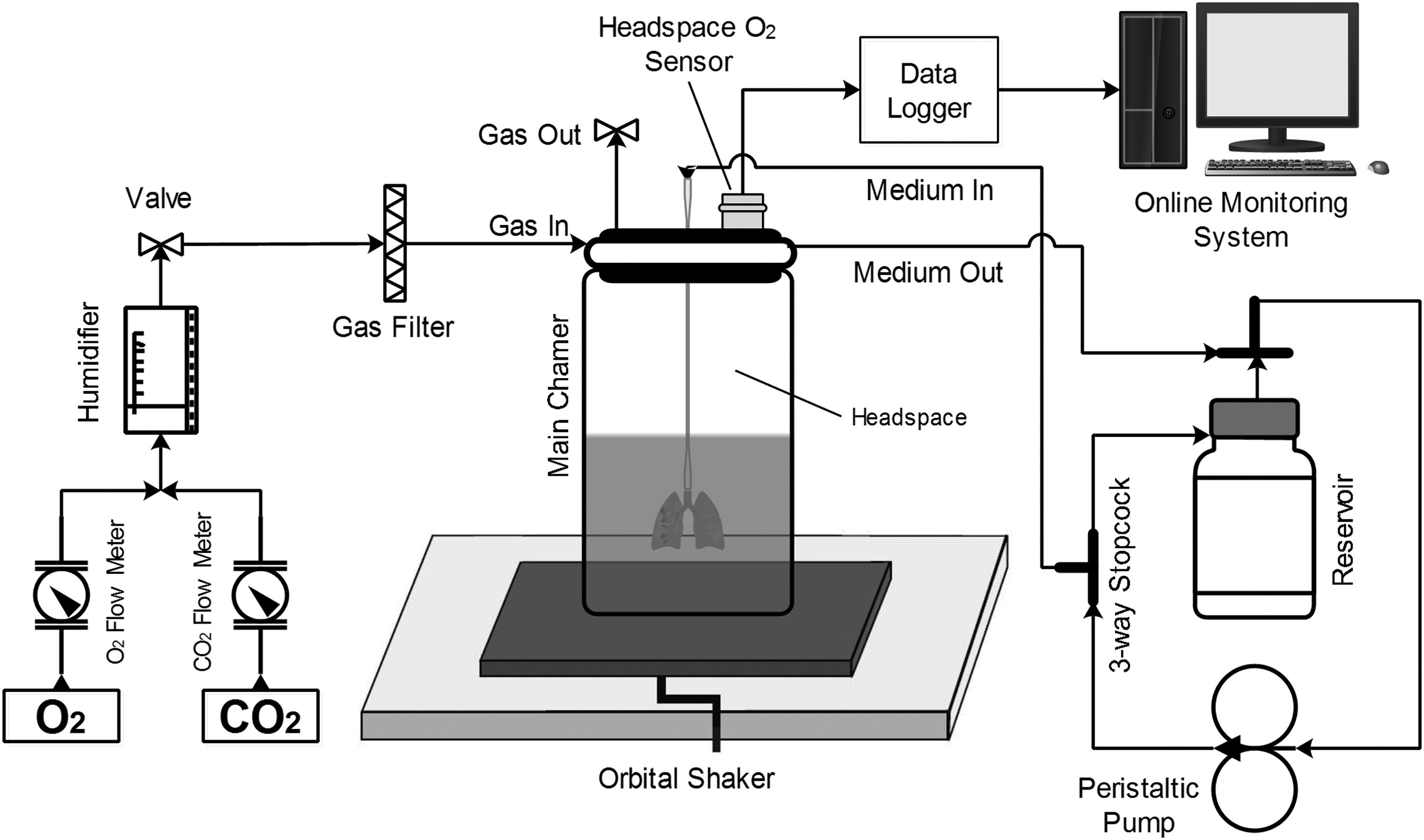
The schematic diagram of the custom-designed bioreactor including the main chamber and online monitoring system. Decellularized lungs are put in the main chamber and HUVECs are seeded into decellularized lungs. The recellularization stage is begun and simultaneously the online monitoring system starts working. First, the valves get opened and the gas flow is let inside. After a user-defined period, the valves get closed and measuring oxygen partial pressure is begun. The measurement process then is continued for a user-defined period. The differences in oxygen partial pressure are used to calculate OTRs from the headspace to the culture medium by a headspace O2 sensor. This process is repeated during the culture time. HUVEC, human umbilical cord vein endothelial cell.
Monitoring of OTR during lung scaffolds recellularization
The method used for monitoring of OTR during the recellularization stage was based on measuring the oxygen partial pressure in the gas phase according to Hansen et al.
30
and Amoabediny et al.
31
At the beginning of the culture, gas inlet and outlet valves of the main chamber were closed. During this period, oxygen was consumed by the cells leading to a decrease in its partial pressure. After a user-defined time period (here it was 20 min), inlet and outlet valves were opened, and gas flowed (air containing 5% CO2) into the main chamber for a user-defined time period (here it was 10 min). This cycle was repeated continuously. The system logged the differences between the initial and the final values of oxygen partial pressure in each cycle. The OTR was then calculated using the following equation:
where ΔpO2 is the variation of oxygen partial pressure (bar), VG is the gas volume (L), Δt is the user-defined time period during which the valves are closed, R is the gas constant (bar·L/mol/K), T is the temperature (K), and VL is the liquid volume (L). For experiments that resulted in growth profiles, OTR measurements were performed during the whole cell culture period (up to 5 days).
Acellular lung scaffolds recellularization and culture in bioreactor
The prepared acellular lungs were mounted in the bioreactor's main chamber for culture at 37°C. Five million HUVECs (National Cell Bank of Iran [NCBI], Pasteur Institute of Iran) were injected into the microvasculature through the pulmonary artery of acellular lungs suspended in 250 mL of culture medium after being counted by hemocytometer. The static culture was done for 18 h allowing cell adherence on the extracellular matrix (ECM). After this period, the dynamic culture was performed using perfusion loop at a constant rate of 5 mL/min up to 5 days (number of experimented samples are presented in Table 1). For cultures in which shaking was used beside perfusion, shaking was applied at the rate of 20 rpm and diameter of 5 cm (with equivalent centrifugal force of 0.0112 g) right after the beginning of the dynamic culture (number of experimented samples are presented in Table 1). The culture medium used in this study was Dulbecco's modified Eagle's medium with 10% fetal bovine serum (Gibco) and 1% penicillin/streptomycin (Gibco). The medium was not exchanged during the culture period. Aeration rate was kept at 250 mL/min for all the cultures.
HUVEC, human umbilical cord vein endothelial cell; MTT, 3-(4,5-dimethylthiazol-2-yl)-2,5-diphenyltetrazolium bromide; OTR, oxygen transfer rate.
Histology
For histological assays, hematoxylin and eosin (H&E; Merck Darmstadt, Germany) staining was performed. First, fixation was done using 10% formalin overnight, and then processing and embedding in paraffin were performed. Next, 5 mm slices were cut from the paraffin-embedded tissues, and after that, routine H&E staining was applied (recellularized lung samples were taken at hour 6 of recellularization).
MTT assay
To perform MTT assay, recellularized scaffolds (n = 2 at each time point, Table 1) were sectioned randomly and put in each well of a 96-well tissue culture plate. MTT reagent (Sigma) was added to culture medium for 4 h, and then, lysis reagent was added overnight. Then, the absorbance was read at 600 nm for 1 s.
Glucose consumption measurement
Glucose consumption measurements of the culture medium were recorded with a GlucCell glucose meter (CESCO BioProducts, Atlanta, GA) based on the manufacturer's protocol. The medium of recellularized lung culture was sampled on hours 6, 24, 48, 72, and 96. Then, glucose consumption was obtained by subtracting two neighboring measurements from each other.
Statistical analysis
The statistically significant differences were indicated using Student's t-tests (two-tail comparisons) and were defined as p < 0.05 and p < 0.005. All data are expressed as “mean ± standard deviation (SD).”
Results
Feasibility of OTR method to monitor cell viability
To show the reproducibility of the results obtained from the OTR method, we applied the method to rat decellularized lungs and HUVEC-seeded lungs at hour 6 of the culture under static condition. As expected, for decellularized lungs, no oxygen transfer was detected, and this showed that there were no oxygen consumers in the chamber. After that, we performed the method for the decellularized lungs seeded with 5 million HUVECs and the OTR value of 0.0025 ± 0.00018 mmol/L·h was observed (p < 0.005) (Fig. 2A). Therefore, the OTR was equal to the cells' oxygen consumption rate. Histological assays of decellularized and recellularized lung are shown in Figure 2B. No cell component was seen in the decellularized scaffolds (up), which confirmed the data obtained from the OTR method. H&E analysis of the recellularized scaffolds (down) confirms that the cells were seeded on the ECM, and adherence to the ECM surface has occurred successfully.

Feasibility of OTR method.
Cell growth profile of recellularized lung scaffolds and correlation between OTR values and number of cells
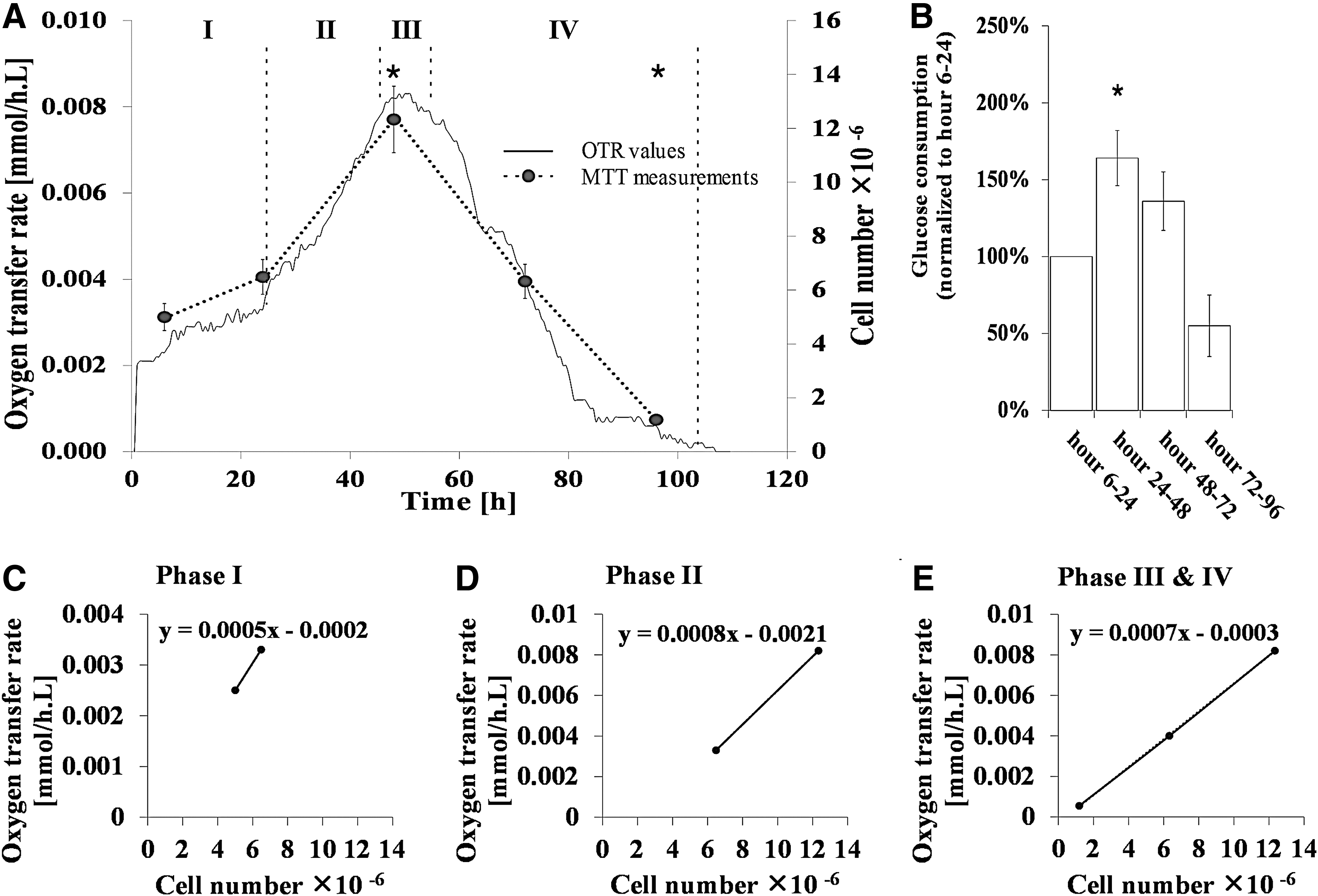
The OTR was monitored online during the recellularization stage. Figure 3A (filled line) shows a cell growth profile when only perfusion was used in the dynamic culture period. First, we performed an 18-h static culture period. Then, we proceeded with the dynamic culture using perfusion. During the static culture period, the OTR values were relatively low (first and last values of OTR in this period were 0.0002 and 0.0031 mmol/L·h, respectively). After the end of the static culture and beginning of the dynamic culture, the OTR values increased intensively. At hour 49, the OTR reached its climax (0.0083 mmol/L·h). Afterward, the OTR values decreased until hour 110. Also, at the top of Figure 3A, different states of the recellularized lungs' proliferation have been illustrated. First, until hour 6, the cells were in their lag phase getting adapted to their new environment (I). After that, propagation of the cells accelerated exponentially until hour 49 (II. exponential growth phase). From hour 49 until hour 53, the growth rate became stationary (III). Then, the cells began to die with higher rates (IV. death phase). MTT assay was used to determine the number of cells corresponding to the OTR values at certain times. Figure 3A (circular points) shows the growth curve of HUVECs in recellularized lung scaffolds assessed by MTT assay at hours 6, 24, 48, 72, and 96. Absorbance was directly proportional to the cell viability demonstrating a significant increase in number of cells until hour 48 of the culture period. The viable number of cells was 5 ± 0.4 million at hour 6 in recellularized scaffolds. Then it reached 6.48 ± 0.65 million at hour 24 (p = 0.108) and 12.33 ± 1.36 at hour 48 (p = 0.015). However, at hour 72, a noticeable decrease was observed in the number of viable cells; it reached 6.32 ± 0.57 (p = 0.114). This implicated that the cells were in their death phase, that is, death rate has surpassed the growth rate. HUVECs did not survive well in the rest of the period. Compared with the number of cells at hour 6, the viable number of cells in recellularized scaffolds decreased to nearly a fifth at hour 96 (1.187 ± 0.095, p = 0.005).
Monitoring OTR of recellularized lung culture.
Glucose consumption measurement was used to validate the changes in cell viability obtained by the OTR method (Fig. 3B). The percentage of glucose consumption over hours 24–48 increased to 164% ± 18% compared with that over hours 6–24 (p = 0.025). After that, the percentage of glucose consumption decreased until the end of the culture period, reaching 136% ± 19% (p = 0.082) and 55% ± 20% (p = 0.06) for hours 48–72 and hours 72–96 compared with hours 6–24, respectively.
To quantify number of cells using the data obtained by the OTR method, a correlation between OTR values and corresponding number of cells was required. To circumvent the errors in the calculation of the number of cells arising from the different metabolic activities of the cells in different growth phases, separate equations were calculated for different growth phases. Figure 3C shows the relationship between the OTR values and the number of cells in the lag phase with the slope of 0.0005 (mmol O2/106 cells·L·h). In the exponential phase, it reached 0.0008 (mmol O2/106 cells·L·h), indicating the highest oxygen consumption rate by the cells (Fig. 3D). Finally, in the stationary and death phase, the slope of the equation was calculated as 0.0007 [mmol O2/106 cells·L·h] (Fig. 3E).
Monitoring of long-term tissue culture in the presence of shaking
The ability of a monitoring method to assess cell viability in a long-term culture without invasivity allows investigation of the effects of different stimuli on the viability of the seeded cells. To show this capability of the OTR method, shaking was added to the dynamic period of the culture, and recellularized lungs were assessed. HUVECs growth profile when shaking was used beside perfusion in the dynamic culture period is illustrated in Figure 4A. Until hour 18, OTR values were the same as those observed for the only-perfusion case. After that, OTR values increased with a higher rate compared with those of the other case. Also, the maximum value of OTR occurred at hour 56, (7 h later than that of the only-perfusion case). The maximum value of OTR was 0.0148 mmol/L·h, which is about 80% more than that of the previous case. After that, the OTR values declined slowly until hour 130.
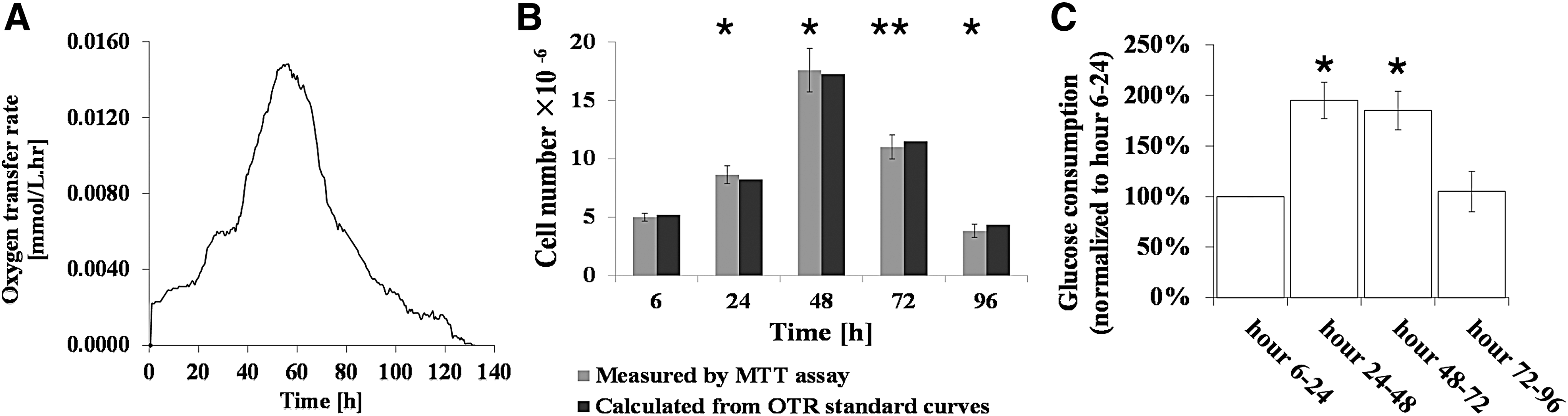
MTT assay of recellularized lungs was performed when shaking was used beside perfusion (Fig. 4B). The viable number of cells was 5 ± 0.34 million at hour 6. At hour 24, the viable number of cells increased to 8.63 ± 0.81 (p = 0.006). The climax was observed at hour 48 with the relative value of 17.58 ± 1.87, which is about 40% more than the only-perfusion case (p = 0.005). At hour 72, the number of viable cells decreased to 11.01 ± 1.03 (p = 0.004). At hour 96, 3.82 ± 0.57 viable cells were observed, which means a threefold increase in comparison with that of the only-perfusion case at the same time point (p = 0.012). To investigate the consistency of the OTR values with the data measured by MTT assay, number of cells at hours 6, 24, 48, 72, and 96 was calculated from the OTR values according to the equations shown in Figure 3C–E. Number of cells in recellularized lungs was calculated as 5.19 million at hour 6. The calculated number of cells reached 8.23, 17.24, 11.49, and 4.35 at hours 24, 48, 72, and 96, respectively. The number of cells calculated by OTR values was not significantly different from those measured by MTT assay (p = 0.435, 0.482, 0.783, 0.504, and 0.249 for hours 6, 24, 48, 72, and 96, respectively).
To confirm the changes in cell viability obtained by the OTR method, glucose consumption measurement was taken (Fig. 4C). The percentage of glucose consumption over hours 24–48 increased to 195% ± 22% in comparison with that over hours 6–24 (p = 0.017). Then, the percentage of glucose consumption decreased until the end of the culture period, reaching 185% ± 23% (p = 0.024) and 105% ± 30% (p = 0.8) for hours 48–72 and hours 72–96 compared with hours 6–24, respectively.
Discussion
Whether the goal is to develop a functional tissue or a disease model for drug screening, maintaining cell viability inside 3D tissue-engineered constructs is a prerequisite. Therefore, monitoring cell viability during the culture period has a great importance since it provides essential information about cell conditions inside the construct. This information is necessary for optimization of process conditions, for example, aeration rate, medium composition, and biomimetic stimuli. One major drawback of conventional methods that rely on the metabolism of a certain dye in cells is that their application is restricted in nonperfusing 3D tissue constructs due to low diffusion of the dye into the tissue-engineered constructs. 32 Also, these assays such as MTT and resazurin-based assays have been proved to be cytotoxic for cells.16,17
Previously, the feasibility of OTR-based methods to monitor respiration activity has been proved in various studies.25–30 Here, for the first time, we demonstrated an OTR-based method to monitor cell viability and proliferative states in recellularized whole rat lungs. The monitoring system was used to measure the rate of transferred oxygen from chamber headspace to the culture medium. Monitoring of decellularized scaffolds with and without cells demonstrated that there was no oxygen consumer in the medium except the cells seeded in the scaffolds (Fig. 2A), which was subsequently confirmed by H&E staining (Fig. 2B).
Owing to the direct proportion between cell oxygen uptake rate and cell population, measured OTR values resulted in a cell growth profile (Fig. 3A, filled line). The profile provided a comprehensive view about the proliferative states of the cells. MTT assay was implemented to determine corresponding number of cells to the OTR values at certain times (Fig. 3A, circular points). Also, further glucose consumption measurements confirmed different trends of the cell viability change obtained by the OTR method (Fig. 3B). Furthermore, to provide the possibility for the translation of the results of the OTR method to the number of cells, correlations between the OTR values and corresponding MTT measured number of cells were obtained (Fig. 3C–E). The slopes of the equations represent specific oxygen uptake rates in different phases. To compare these values with those reported in the literature, we performed a conversion to unify the units. The values obtained here were 2.08 × 10⁻¹⁵, 3.33 × 10⁻¹⁵, and 2.92 × 10⁻¹⁵ mol/cell·min for lag phase, exponential phase, and stationary and death phases, respectively. These obtained values were consistent with reported values of 1.301 × 10⁻¹⁵ mol/cell·min for native rat lung 33 and 3.5 × 10⁻¹⁵ mol/cell·min for HUVECs. 34 Besides, the calculated values for the specific oxygen uptake rates suggest that the cells have had more oxygen uptake rates in their exponential phase than the lag and death phases, which is in agreement with the previously reported values for mammalian cells. 35
To show the applicability of the OTR method to monitor the effect of operating conditions on cell viability over a long-term culture, shaking was added to the dynamic period of the culture, and recellularized lungs were assessed (Fig. 4A). The OTR values were translated to the number of cells using the related equations and were compared with those of MTT measurements, and there was no significant difference to that revealed by MTT measurements (Fig. 4B). Also, the trends of cell viability change obtained by the OTR method were consistent with the values obtained by glucose consumption measurements (Fig. 4C). As our findings showed, applying shaking resulted in improved cell growth. It has been reported that shaking increases OTR to the liquid phase and also causes better oxygen homogeneity in the liquid phase (i.e., culture medium) due to better mixing.36,37 Thus, by enhancing oxygen delivery, the cells grew better inside the recellularized lungs in comparison with the only perfusion case. The ability to monitor the influence of various operating parameters on cell viability using this monitoring system would allow further optimization of the culture conditions.
Oxygen-based monitoring methods are not limited to OTR-based methods. In our previous study, we showed that an online monitoring strategy was applicable over recellularization of an acellular lung tissue using two dissolved oxygen sensors. 38 Also, in another study, a bioreactor was developed that could control oxygen concentration using an optical dissolved oxygen probe, and by this system, oxygen uptake rates of native rat lungs were determined. 33 Nonetheless, the OTR method has two important advantageous as it uses oxygen partial pressure differences in the gas phase: first, the risk of contamination significantly decreases due to the elimination of direct contact of the sensor with the culture medium and second, the data are more reliable because the method measures the overall oxygen uptake rate, so alterations in regional oxygen concentration in the medium do not disturb the measurement procedure.
Although this monitoring system provides an efficient means to monitor tissue growth ex vivo/in vitro, a number of experiments could be made to provide detailed insight into the system capabilities. Examining various ranges of operating parameters including gas-to-liquid ratio, aeration rate, and aeration time, as well as biological parameters such as cell density and metabolic activity is required to be done to determine system's sensitivity more precisely.
Conclusion
This study shows the feasibility of an OTR-based system for monitoring of cell viability during recellularization of decellularized lung scaffolds. Being noninvasive, real-time, and automatic are some of the great features of this system, which allow for repetitive testing without any pause to the culture process. This will help optimize tissue culture conditions, investigate the effect of different feeding or aeration strategies, and screening drug effects.
Footnotes
Acknowledgments
We would like to thank Mr. Hadi Seddiqi and Mr. Seied Ali Safiabadi Tali for their kind helpful remarks. Also, we appreciate the Research Center for New Technologies in Life Science Engineering at the University of Tehran for its scientific and financial support.
Disclosure Statement
No competing financial interests exist.