
Abstract
Corneal endothelium is a single layer of hexagonal cells that maintains the corneal transparency and thickness through its barrier and pump function. For the treatment of corneal endothelial dysfunction, the transplantation of tissue-engineered corneal endothelium and direct injection of cultured corneal endothelial cells were developed because of the severe shortage of donor cornea worldwide. However, the technique difficulty or safety risk still remained. In this study, we report a novel mini-sheet injection for the cultured corneal endothelial cell transplantation and compare with the effects of single-cell injection in rabbit model. Compared with the reported single-cell injection, mini sheets promoted the adhesion and tight junction formation after injection. Rabbit corneal clarity and thickness were rapidly recovered after 7 days of mini-sheet injection, compared with 14 days of single-cell injection. Moreover, typical endothelial morphology was observed as early as 7 days after the mini-sheet injection, whereas until 21 days after the single-cell injection. These results demonstrate that the novel mini-sheet injection of corneal endothelial cells exhibited rapid adhesion, tight junction formation, and corneal clarity recovery, which may represent a more efficient method for the transplantation of cultured corneal endothelial cells.
Introduction
T
The technical difficulty of cultured corneal endothelial cell transplantation is how to insert the fragile cell monolayer into the anterior chamber. Traditionally, the tissue-engineered corneal endothelium was designed with the assistance of carrier substrates, including amniotic membrane, collagen, and vitrigel, which was performed similar to traditional DSEAK or DMEK.8–12 Considering the safety and transparency of the carrier, the preparation of corneal endothelial cell sheet was further tried with the use of temperature-responsive culture dish, 13 but met the difficulty of the intact monolayer detachment and transplantation. Therefore, the direct injection of cultured corneal endothelial cells into the anterior chamber was developed with the confirmed effects in animal models and bullous keratopathy patients.7,14–16 However, the injected cells may increase the risk of abnormal adhesion in other tissues of the anterior chamber, since not all the injected cells were rapidly attached on the Descemet's membrane. 14
The goal of this study was to develop an approach for cultured corneal endothelium transplantation by preparing the mini sheets for the anterior chamber injection. This approach combines the advantages of cell sheet culture and anterior chamber injection while avoiding their shortcomings. The recovery of corneal clarity, thickness, and endothelial cell density was investigated and compared with the single-cell injection in rabbit model.
Materials and Methods
Primary culture
Sixty New Zealand white rabbits (Kangda, Qingdao, China) were housed and treated in accordance with the ARVO statement for the Use of Animals in Ophthalmic and Vision Research. The experimental procedures were approved by the Ethics Committee of Shandong Eye Institute. Rabbit corneal endothelium containing Descemet's membrane was harvested after being sacrificed by euthanasia. The tissues were incubated overnight in the culture media supplemented with 10 μM Y-27632 (Sigma-Aldrich, St. Louis, MO). The isolation and culture of rabbit corneal endothelial cells (RCECs) were modified according to the previous description. 14 The culture media were composed of Dulbecco's modified Eagle's medium (DMEM; Corning, Manassas, VA) supplemented with 10% fetal bovine serum (Gibco, Grand Island, NY), 2 ng/mL human basic fibroblast growth factor (R&D Systems, Minneapolis, MN), and penicillin/streptomycin (Corning). The confluent cells were digested with 0.25% trypsin-EDTA (Sigma-Aldrich) and passaged with 1:3 ratios. The cells within three passages were used for all the following experiments.
Cell viability analysis
Confluent RCECs were dissociated with 3 U/mL Dispase II (Roche), Accutase (Sigma-Aldrich), or 0.25% trypsin-EDTA for 3, 5, 10, and 15 min at 37°C. The cells were harvested, and stained with 0.4% Trypan Blue (Sigma-Aldrich). The percent of viable cells was counted with the TC20™ Automated Cell Counter (Bio-Rad, Singapore).
Single cell preparation
Confluent RCECs were dissociated with 0.25% trypsin-EDTA for 10 min at 37°C and gently triturated into single cell suspension. The density was adjusted into 3 × 10⁵ cells in 250 μL DMEM containing 100 μM Y-27632 for injection.
Mini sheet preparation
Confluent RCECs were dissociated with Accutase for 3 min at 37°C and gently triturated into mini sheet suspension. The mini sheets (obtained from the equal area of 24-well plate with single cell preparation) were subsequently suspended in 250 μL DMEM containing 100 μM Y-27632 for injection.
Anterior chamber injection and evaluation
The rabbits were anesthetized with intramuscular ketamine hydrochloride (40 mg/kg; Gutian, China) and chlorpromazine hydrochloride (20 mg/kg; Shanghai, China). The 20-gauge soft, tapered silicone needle (Inami, Tokyo, Japan) was used to remove the corneal endothelium as described previously. 7 The suspensions of mini sheets or single cells were injected into the anterior chamber of the right eye, with left eye untreated. The rabbits were maintained with the eye-down position for 3 h to allow the rapid attachment. The anterior chamber injection containing Y-27632 after surgery was performed according to previous descriptions. 17 Corneal clarity, endothelial cell morphology and density were evaluated by slit-lamp microscopy (Topcon, Tokyo, Japan) and confocal microscopy (Heidelberg Engineering, Heidelberg, Germany). Corneal thickness was measured with a handy pachymeter (Tomey, Nagoya, Japan).
Immunofluorescence staining
Full-thickness corneal flat mounts were fixed in 4% formaldehyde and blocked with 5% normal serum for 1 h at room temperature. The samples were incubated with the primary antibodies of anti-ZO-1 (Invitrogen, Oregon) and anti-Na+/K+-ATPase (Millipore, Billerica, MA) overnight at 4°C, and subsequently with Alexa Fluor 488-conjugated secondary antibody (Invitrogen) for 1 h at 37°C. The cytoskeleton F-actin staining was performed with the phalloidin (Invitrogen) for 1 h at 37°C. The nuclei were counterstained with 4,6-diamidino-2-phenylindole (DAPI, Beyotime, Shanghai, China). The flat mounts were examined and captured under an epifluorescence microscope (Nikon, Tokyo, Japan).
Statistical analysis
Data in this study are representative of at least three independent experiments and are presented as mean ± standard deviation. Statistical analysis was performed using SPSS 17.0 software (SPSS, Chicago, IL). The differences between the control and experimental groups were tested with Student's t-test. A p-value of <0.05 was considered to be statistically significant.
Results
Preparation of single cells and mini sheets
To prepare the samples for injection, Confluent corneal endothelial cells were dissociated with Accutase, trypsin-EDTA, or Dispase for 3, 5, 10, and 15 min. The results showed that the confluent monolayer was mostly dissociated into single cells with trypsin-EDTA treatment for 3 min, while into cell aggregates of variable sizes with Dispase treatment even for 15 min. Comparatively, a number of aggregates containing 4–10 cells (named as mini sheets for the following injection) were released after the treatment with Accutase for 3 min, and with the extension of digestion time, the shape of mini sheet can still be maintained. The viability of dissociated cells approached or exceeded 90% by Accutase or trypsin-EDTA within 10 min, which was dropped significantly by 15 min dissociation. Therefore, the single cell preparation was fixed with trypsin-EDTA treatment for 10 min, the mini sheets' preparation was performed with Accutase for 3 min. All samples were collected by centrifugation for injection in rabbits (Fig. 1).
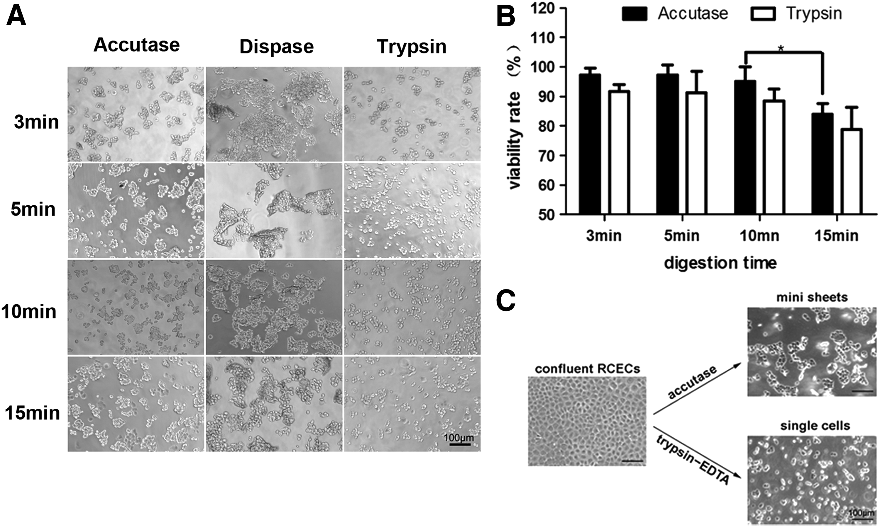
Preparation of single cells and mini sheets. Confluent corneal endothelial cells were dissociated with Accutase, trypsin-EDTA, and Dispase II for 3, 5, 10, and 15 min
Adhesion and tight junction formation after injection
To compare the adhesion and tight junction formation between the single cells and mini sheets, the rabbit corneas were collected at 6 and 48 h after the anterior chamber injection. The F-actin and nuclei staining at 6 h showed that the injected mini sheets adhered rapidly and spread on the surface of Descemet's membrane with the retained cytoskeleton structure (Fig. 2). However, the injected single cells showed the scattered adhesion. According to cell counts, the number of adhered RCECs by the mini-sheet injection was sixfold to sevenfold more than that of the single-cell injection (3025 ± 481 cells/mm2 vs. 415 ± 132 cells/mm2). Moreover, the ZO-1 and Na+/K+-ATPase staining at 48 h showed that the injected mini sheets almost merged into the endothelial monolayer with the formed tight junction and improved expression of endothelial pump functional marker, compared with the injection of single cells (Fig. 2).
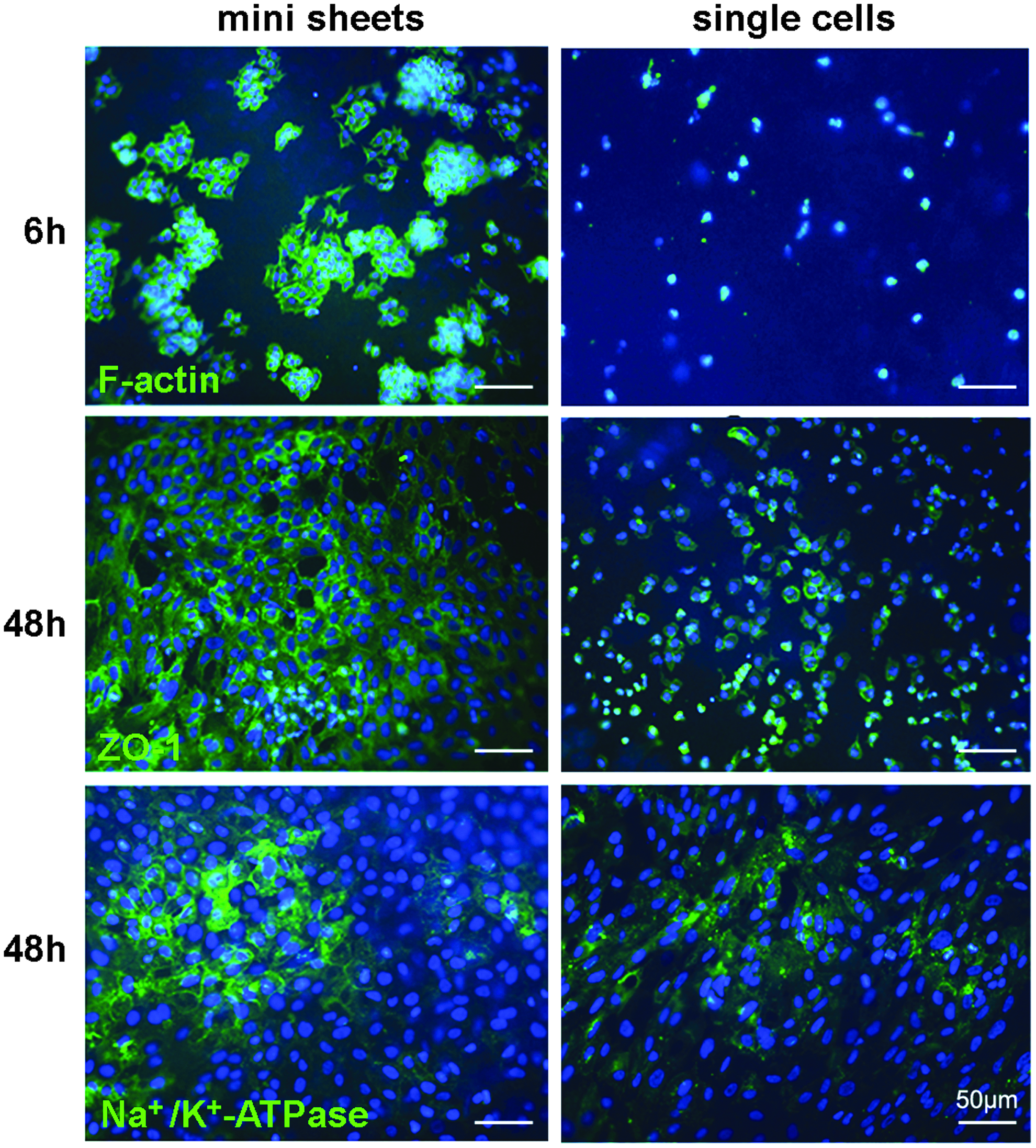
Mini-sheet injection promotes adhesion and tight junction formation. Mini sheets and single cells containing the same number of corneal endothelial cells were injected into the anterior chamber of rabbits with the endothelium removed. The corneas were obtained after 6 and 48 h of injection and stained with the phalloidin (F-actin), and the ZO-1 and Na+/K+-ATPase antibody.
Recovery of corneal clarity and thickness after injection
To compare the functional recovery rates of corneal endothelium between the single cells and mini sheets, the corneal clarity and thickness were examined by slit-lamp microscopy and ultrasound pachymeter. The observation of slit-lamp microscopy showed that the rabbit corneal clarity was rapidly recovered after 7 days of mini-sheet injection, whereas 14 days of single-cell injection (Fig. 3A). According to the analysis of five rabbits every time point, the corneal thickness decreased rapidly and remained stable from 7 days after the mini-sheet injection, whereas the rabbits injected with single cells showed a protracted reduction in corneal thickness and approached the normal level after 14 days (Fig. 3B). There was no increase of intraocular pressure in both groups during the period (data not shown).
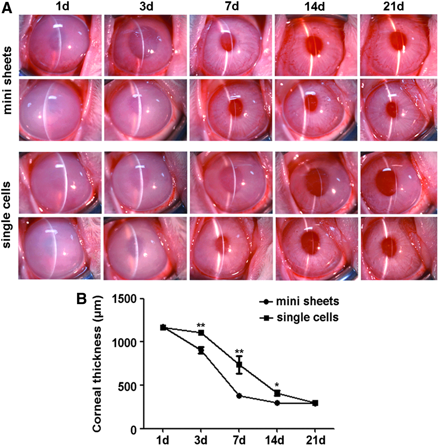
Mini-sheet injection accelerates the recovery of corneal clarity and thickness. Mini sheets and single cells containing the same number of corneal endothelial cells were injected into the anterior chamber of rabbits with the removed endothelium. The corneal clarity
Morphological changes and endothelial cell density after injection
To compare the morphological changes after the injection of single cells and mini sheets, the rabbit corneas were examined by a confocal microscope after 7, 14, and 21 days of injection. Representative results showed that typical morphology and even distribution of corneal endothelium was observed as early as 7 days after the mini-sheet injection, whereas at least 21 days after the single-cell injection (Fig. 4A). According to the counts of endothelial cell density, the injection of mini sheets assumed a higher density than that with the injection of single cells at 14 and 21 days, although both groups had higher than 2000 cells/mm2 (Fig. 4B).
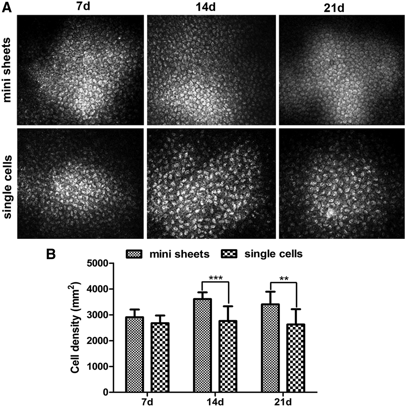
Mini-sheet injection improves the morphological recovery and endothelial cell density. Mini sheets and single cells containing the same number of corneal endothelial cells were injected into the anterior chamber of rabbits with the endothelium removed. The endothelial cell morphology
Discussion
The transplantation of in vitro cultured corneal endothelial cells gained more and more attention for the treatment of endothelial dysfunction. To simplify the preparation of tissue-engineered corneal endothelium and reduce the cell loss after injection into the anterior chamber, in this study, we designed a novel mini-sheet injection for the transplantation of cultured corneal endothelial cells, and compared the effect with the traditional single-cell injection. The results showed that the mini-sheet injection actually promoted the early adhesion and tight junction formation on the Descemet's membrane, as well as the rapid functional recovery in rabbit model.
The sizes of cell sheets for the anterior chamber injection should be controlled. In principle, the larger the sheets injected, the sooner the function appeared. However, oversized sheets may block the syringe needle and accumulate on the posterior surface, which will impair the effective adhesion and function of transplanted cells. Therefore, we compared three dissociation enzymes for the preparation of cell sheets with suitable sizes. Compared with the oversized clumps by Dispase and the single cells by trypsin-EDTA, the treatment with Accutase was preferred with mild dissociation ability and finally acquired the dissociated sheets containing 4–10 cells, as well as some single cells. During the adhesion on the Descemet's membrane, the small amounts of single cells filled the gap between the mini sheets. With the extension of digestion time the shape of mini sheet prepared by Accutase can still be maintained. But the viability of dissociated cells was dropped significantly by 15 min dissociation, so we should control the digestion time within 10 min to improve cell viability rate and achieve better postoperative results. It should be mentioned that additional research need to optimize the preparation of uniform mini sheets and to exclude the single cells for injection, which may further enhance the efficiency of transplanted endothelial cells.
The cell transplantation by injection has been reported for the therapy of several degenerative diseases, such as heart, cartilage, and eye.18–20 The major obstacle is how to promote and ensure the long-term functional integration of transplanted cells in the injection site. For the injection of cultured corneal endothelial cells, the rapid adhesion on the Descemet's membrane plays a critical role in the functional recovery of transplanted cells. 14 Therefore, the transplantation by cell sphere and magnetic assistance had been attempted in animal models.21,22 More recently, the method of corneal endothelial cell injection was developed by the combination with ROCK inhibitor, which promotes the adhesion and proliferation, while inhibits the apoptosis of corneal endothelial cells.7,14 However, the injected cells may be washed off by aqueous humor flow and settled in the iris or angle of the anterior chamber. Accordingly, we tried to prepare the mini sheets to increase the adhesion rate after injection by the Accutase treatment of confluent corneal endothelial cells. Every mini sheet contained 4–10 cells with the remaining adhesion together and the cytoskeleton arrangement. Compared with the single cells after anterior chamber injection, the mini sheets were settled quickly on the Descemet's membrane and hardly washed off by aqueous humor flow. Besides, the tight junction remained in the mini sheets, which accelerated the reconstruction of endothelial barrier function and the recovery of corneal clarity and thickness.
Conclusion
In this study, we developed a novel method of mini-sheet injection for the transplantation of cultured corneal endothelial cells. In comparison to the single-cell injection, the mini-sheet injection improved the adhesion, tight junction formation and functional recovery in rabbit model, which may also provide better choice for cell-based therapy.
Footnotes
Acknowledgments
This work was partially supported by the National Natural Science Foundation of China (81530027, 81700811) and Shandong Provincial Nature Science Fund (JQ201518, ZR2017PH009, ZR2017PH036); W.S. and Q.Z. are partially supported by the Taishan Scholar Program (20150215, 20161059) and the Innovation Project of Shandong Academy of Medical Sciences.
Disclosure Statement
No competing financial interests exist.