
Abstract
The development of an in vitro model resembling the alveolar-capillary barrier might be a highly beneficial tool to study lung physiology as well as the immune response of the lung to infection or after exposure to nanoparticles. This study is based on an in vitro alveolar barrier developed on a basement membrane mimic, composed of ultrathin nanofiber meshes generated via electrospinning using bioresorbable poly(ɛ-caprolactone). As cellular components, NCI H441, resembling the alveolar epithelial cells, and ISO-HAS-1, an endothelial cell line, were used to perform bipolar coculture experiments for a total cultivation period of 14 days. In addition to immunohistochemical and immunofluorescent studies, transepithelial electrical resistance (TER) and transport capabilities of the in vitro model system were investigated. Alveolar barrier function could be clearly determined for the postulated bipolar coculture system on the basement membrane mimic, since TER increased during the course of bipolar cultivation. Furthermore, to gain first insights into possible lung inflammatory reactions in vitro, this coculture model was further expanded by a human leukemia monocyte cell line (THP-1). This triple-culture system was able to maintain adequately the barrier properties of the bipolar coculture, thus making this in vitro model consisting of epithelial, endothelial, and immune cells on a basement membrane mimic a promising basis for further studies in tissue engineering.
Impact Statement
The alveolar-capillary barrier implicates severe requirements and constitutes a difficult challenge for developing adequate in vitro models for possible tissue engineering approaches. During this study, the authors proposed an innovative bipolar cell culture model of the alveolar-capillary barrier consisting of endothelial cells, epithelial cells, and macrophages on a fully synthetic basement membrane mimic as promising basis for more physiological studies of the alveolar-capillary barrier in vitro.
Introduction
T
The specific characteristics of the alveolar-capillary barrier implicate stringent requirements and constitute a difficult challenge for developing and establishing appropriate in vitro models for possible tissue engineering approaches. In general, in vitro cell cultures, mimicking tissues or organs, represent very beneficial model systems to enable analysis of fundamental processes and basic research questions and have the potential to provide important information about cell–cell communication and molecular mechanisms operative in the in vivo situation. Therefore, in vivo-like bipolar cell culture models to mimic approximately the physiological conditions in the lung have been recently developed, especially to analyze cytotoxic effects of different substances or even nanoparticle-mediated modulations.4,5 Nevertheless, bipolar cell culture models usually use commercially available cell culture inserts, in which the basement membrane mimic is an etched poly(carbonate) membrane. Unfortunately, the latter display very limited membrane properties when compared to the natural basement membrane; that is, thickness, lack of interconnected pores, and absent bioresorbability. Hence, the development of basement membrane mimics still remains a scientific challenge, as the engineered biomaterials should be mechanically robust, biocompatible, and biodegradable.
During the present study, an alveolar-capillary barrier model, using a human microvascular endothelial cell line (ISO-HAS-1) and a human lung adenocarcinoma cell line (NCI H441), was developed using a functional nanofiber-based basement membrane mimic. 6 This membrane is composed of ultrathin nanofiber meshes produced using bioresorbable poly(ɛ-caprolactone) (PCL) and bioinert six-armed star-shaped poly(ethylene oxide-stat-propylene oxide) with isocyanate end groups (NCO-sPEG) generated by electrospinning. The human coculture model of the alveolar-capillary barrier was established on this fully synthetic mesh to provide a responsive in vitro model of the air–blood barrier which is physiologically similar to the in vivo situation and might possibly serve as a solid fundamental platform for further lung research in the field of regenerative medicine. In addition, in the course of this work, the coculture model was further expanded by a human leukemia monocyte cell line (THP-1) to gain first insights into possible lung inflammatory reactions in vitro.
Materials and Methods
PCL nanofiber mesh
The 6 wt% PCL solution was dissolved in hexafluoroisopropanol. Ten microliters of 0.2% trifluoroacetic acid solution were added to increase the conductivity of the solution. The polymer solution was filled in 1 mL syringe with 27-gauge flat-tipped stainless-steel as a spinneret was placed in a syringe pump system. A collector with aluminum foil (10 × 10 cm) was connected to a high-voltage generator and the spinneret remained earthed. When a positive high-voltage potential was applied to the collector, the polymer solution was locally charged to produce a jet from a Taylor cone. Spinning was performed using a stationary collector and spinning time for each mesh was kept constant for 5 min, while the syringe pump speed of 0.5 mL/h was maintained. From this, it was calculated that 42 μL (0.5 mL/h × 5/60 h = 0.042 mL = 42 μL) of polymer solution per membrane was spun. To obtain nanofibers and homogeneous membrane voltage of 20 kV, distance of 15 cm and flow rate of 0.5 mL/h were maintained. The obtained PCL fibers were 200 nm in diameter and were 10 μm thick with an average pore size of 1.5 μm and a porosity of 71%. After solvent evaporation, each mesh weighed ∼0.30–0.37 mg. The meshes were ultraviolet sterilized for 1 h. After fixing the PCL meshes in a cell culture insert, the meshes were incubated in 0.01 wt% of bovine collagen I solution (ThermoFisher) for coating (1 h from both sides) and subsequently washed with phosphate-buffered saline (PBS) before cell culture experiments.
Extraction assay: cytotoxicity test
To exclude possible cytotoxic or cell-damaging effects arising from the membranes on the used cell types, an extraction assay was performed before bipolar cell culture experimentation. PCL basement membrane mimics as well as HTS Transwell® filters were incubated in Roswell Park Institute medium (RPMI) 10% fetal calf serum (FCS) for 4 days at 37°C in an atmosphere of 5% CO2 and 95% air. Subsequently, 200 μL of the leached medium was added to NCI H441 (5000 cells/well; ATCC-HTB-174; Promochem, Wesel, Germany) and ISO-HAS-1 (5000 cells/well7,8) that were seeded on 96-well plates 24 h before treatment. The cells were incubated for additional 4 days before analyzing for cell viability using MTS CellTiter 96®AQueous One Solution Cell Proliferation Assay (Promega, Madison) according to the manufacturers' protocol. For this, the leached medium was discarded from the cells. After washing the cells with 0.2% HEPES/BSA, 120 μL MTS diluted 1:6 in fresh medium was added to the cells and incubated for 1 h at 37°C. One hundred microliters of each sample was transferred to a fresh 96-well cell culture plate and the absorbance was measured at 492 nm in a microplate reader (GENios plus, TECAN, Crailsheim, Germany).
Bipolar cell culture system
The epithelial cell line NCI H441 and the endothelial cell line ISO-HAS-1 were used for the bipolar coculture on the PCL nanofiber meshes coated with collagen I. Control bipolar cocultures were additionally performed on HTS 24-Transwell filters (polycarbonate, 0.4 μm pore size; Costar, Wiesbaden, Germany) coated with rat tail collagen type I (12.12 μg/cm2; BD Bioscience, Heidelberg, Germany). ISO-HAS-1 (1.6 × 10 4 /well ≙5 × 10 4 /cm2) were seeded on the lower surface of the inverted filter membrane and incubated at 37°C and 5% CO2 to allow adhesion of the cells on the membrane (Fig. 1). Subsequently, NCI H441 (8.4 × 10 3 cells/well ≙2 × 10 4 cells/cm2) were placed on top of the surface, and bipolar cell cultures were cultivated in RPMI 1640 (Gibco, Karlsruhe, Germany) supplemented with 10% FCS (Gibco, Karlsruhe, Germany) and 1% penicillin/streptomycin (P/S; Sigma, Taufkirchen, Germany). After 4 days of cultivation, 1 μM dexamethasone (Sigma) was added to the apical chamber (H441) and the bipolar coculture further cultivated for 10 days for analysis. Cell culture medium was changed every second day.
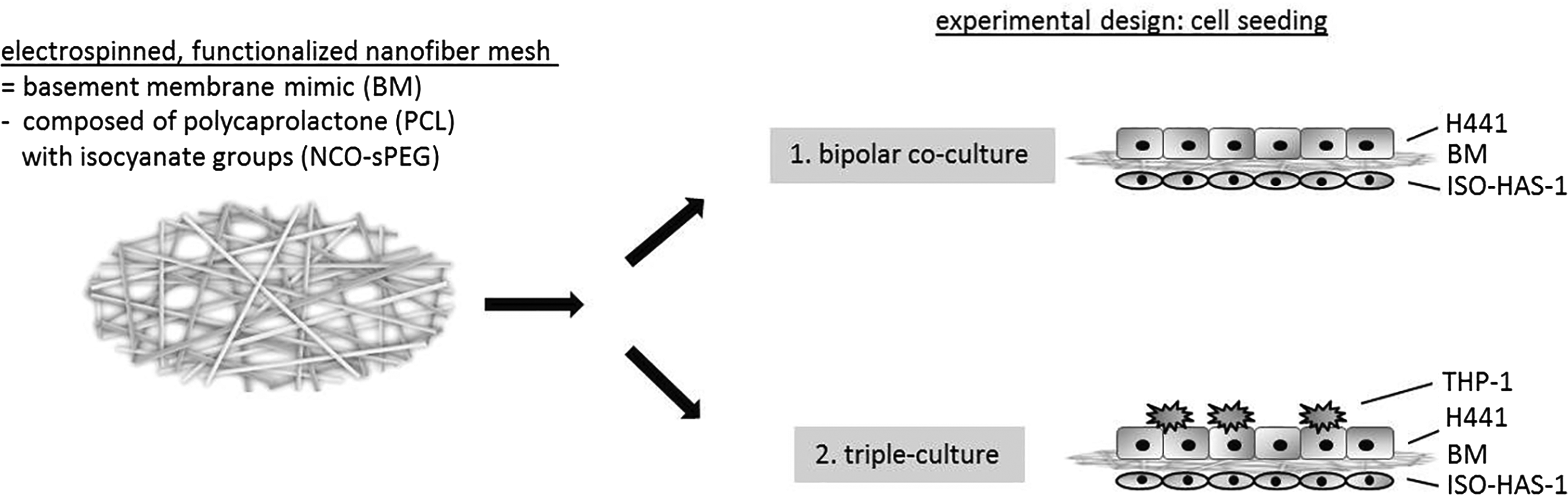
Schematic overview of the experimental setting. Bipolar cocultures consisting of NCI H441 and ISO-HAS-1 as well as triple-culture consisting of NCI H441, ISO-HAS-1, and THP-1 were seeded on an electrospun, functionalized nanofiber mesh as a basement membrane mimic.
Triple-culture conditions
The acute human monocytic leukemia cell line THP-1 was obtained from the ATCC (Manassas, VA). THP-1 cells were grown in suspension at 1.2 × 10 5 cells/mL in RPMI 1640 medium containing 10% FCS (Gibco, Carlsbad, CA) and 1% P/S (Invitrogen, Carlsbad, CA) and maintained at 37°C in an atmosphere of 95% air and 5% CO2. To differentiate the monocytic cell line to macrophages, 5 × 10 5 THP-1 monocytic cells were seeded on fibronectin-coated (5 μg/mL; Millipore, Billerica, MA) six-well plates on a growth area of 9.6 cm2 in 3 mL RPMI medium (Gibco) containing 8 nM PMA (Phorbol-12-myristate-13-acetate; Sigma-Aldrich, St. Louis) for 4 days. Cells were characterized before and after treatment with PMA using immunofluorescent staining for the macrophage marker CD68 (1:100; Dako) and CD105 (1:10; Dako). The PMA treatment induces the proinflammatory (M1) phenotype of the macrophage. Bipolar cell culture consisting of NCI H441 and ISO-HAS-1 was performed on PCL basement membrane mimics as well as on control HTS 24-Transwell filters as described in Bipolar Cell Culture System section. Stimulated THP-1 were seeded on the upper chamber of the transwells on top of H441 cells after 7 days of preseeded bipolar coculture (Fig. 1). THP-1 macrophages were seeded only after the transepithelial electrical resistance (TER) measurement in the bipolar culture (Ω × cm2 > 300). Triple cultures as well as control cocultures were cultivated for further 3 days in RPMI before supernatants were collected for enzyme-linked immunosorbent assay (ELISA) and membranes were fixed for immunohistochemistry. Over the period of triple cultivation, TER was measured every day.
Transepithelial electrical resistance
TER was measured using an EVOM voltohmmeter (Epithelial Voltohmmeter; World Precision Instruments, Berlin, Germany). One electrode of the voltohmmeter was placed in the upper, the other one in the lower compartment of the transwells, and electrical resistance was calculated in Ω × cm2, evaluated as barrier resistance (Ω) multiplied with the total membrane area (0.33 cm2). PCL basement membrane mimics as well as HTS 24-Transwell filters were examined with and without bipolar culture every second day during the whole cultivation time.
Transport experiments
The paracellular transport of sodium-fluorescein was measured across the bipolar culture from apical to basolateral. To achieve this, cell culture medium in the apical chamber of the transwells was replaced with sodium-fluorescein (10 μg/mL; Acid Yellow 73; Sigma, Taufkirchen, Germany) in RPMI with 10% FCS and 1% P/S. Samples from the basolateral chambers were taken after 30, 60, 90, and 120 min and refreshed with the same volume of fresh RPMI. Subsequently, the samples were diluted with 1 mM NaOH, and fluorescence was measured using a fluorescence plate reader (GENios Plus, TECAN, Germany) at a wavelength of 485/530. Permeability coefficients (Papp) were calculated according to the following equation: Papp = (dQ/dt) × (1/(A × c0)).
Immunohistochemistry
For immunohistochemistry, the membranes were fixed in 4% buffered formalin (Roti-Histofix 4% acid free pH 7; Carl-Roth, Germany) and then removed from the transwells before they were dehydrated in an ascending ethanol series using a tissue processor (Leica TP1020) and finally embedded in paraffin blocks. The membranes were cut into histological sections with a thickness of 4 μm using a rotation microtome (Leica RM2255, Wetzlar, Germany). After rehydrating the sections in a descending ethanol series (100%, 95%, 80%, 70%) and pretreating with citrate-buffer at pH 6 for 20 min at 96°C, the sections of the bipolar co- and triple-cultures were immunohistochemically stained with anti-human CD31 (mouse, 1:40, Dako MO 08223), anti-human E-cadherin (mouse) and anti-human CD105 (1:10, Dako M3527) using an Autostainer (Lab Vision Autostainer 360; Thermo Fisher Scientific). Mouse-specific secondary antibody was used (HRP UltraVision Kit; Thermo Fisher) and visualization was detected using DAB (Dako). Histological examination was performed using a light microscope (Nikon Eclipse 80i, Tokyo, Japan) and images were taken with a connected Nikon DS-Fi1/Digital camera and a Nikon Digital sight unit DS-L2.
Immunofluorescence staining
For immunofluorescence staining, whole membranes were fixed in 4% buffered formalin (Roti-Histofix 4% acid free pH 7; Carl-Roth, Germany), permeabilized with 0.5% Triton X/PBS, and washed three times with PBS before incubation with the appropriate antibodies: mouse anti-human CD31 (1:40, Dako MO 08223), mouse anti-human β-catenin (1:50, Santa Cruz Biotechnology), mouse anti-human CD68 (1:200, Dako M0814), mouse anti-human CD105 (1:10, Dako M3527) diluted in a 1% bovine serum albumin/PBS solution for 60 min at room temperature. After washing three times with PBS, the cells were incubated with the secondary anti-mouse antibody Alexa 488 (Molecular Probes, MoBiTec, Göttingen, Germany) diluted 1:1000 in a 1% bovine serum albumin/PBS solution for 60 min at room temperature, protected from light. The cells were mounted with Fluoroshield (ImmunoBioScience Corp., Mukilteo, WA) and examined using a confocal laser scanning microscope (LeicaTCS-NT; Leica Microsystems, Wetzlar, Germany).
Enzyme-linked immunosorbent assay
Culture supernatants from the apical and basolateral transwell chambers of PCL basement membrane mimics and HTS 24-transwell filters were collected separately to gain insights into the potential origin of cell-specific proteins and to compare barrier properties of the bipolar culture on the different membranes. Supernatants were collected after 10 days of cultivation. The concentration of the adhesion molecule endothelial selectin (E-selectin) was measured using DuoSet® ELISA Development Systems according to the manufacturer's protocol (R&D Systems). A streptavidin-HRP colorimetric reaction was used to visualize protein concentrations and the optical density of each well was measured using a microplate reader (Tecan, Crailsheim, Germany) at a wavelength of 450 nm. Results are presented as absolute values as indicated in the relevant figure.
Statistical analyses
All experiments were repeated at least three times, independently. The data are presented as mean values ± standard deviation. Statistical significance was evaluated using the paired student's t-test. Statistical analyses were performed with MS Excel (Microsoft Office, Microsoft) and significance was assessed by *p-value <0.03 or *p-value <0.05, respectively.
Results
Barrier properties of the bipolar coculture seeded on basement membrane mimics
Initially, to exclude cytotoxic effects of the basement membrane mimic composed of the collagen-coated PCL nanofiber meshes, an extraction assay was performed before bipolar cell culture experiments (Fig. 2). Cell viability assays (MTS assay) following cultivation of the cells in monoculture with leached medium obtained from basement membrane mimics incubated without cells in culture medium for 4 days, revealed no negative effects. This was also the case for the cell viability of NCI H441 or ISO-HAS-1 cultivated with control cell culture medium (Fig. 2). Subsequently, bipolar cell culture consisting of NCI H441 (upper compartment) and ISO-HAS-1 (lower compartment) was established on the basement membrane mimics and cultured for 14 days on the membrane, cell culture medium being supplemented with dexamethasone after the first 4 days of bipolar cultivation. In parallel, bipolar cell culture was also performed on control HTS 24-Transwell filters under the same conditions. Figure 3a illustrates the values for TER of the bipolar coculture on basement membrane mimics over the cultivation period (light grey trend), beginning at day 2 following posttreatment with dexamethasone and ending on day 10 ( = day 14 in total), compared to TER measurements of bipolar cultures cultivated on control HTS 24-Transwell filters (dark grey trend). In both cases, TER values increased over the period of cultivation, starting with ∼65 Ω × cm2 evaluated on day 2 and reaching TER values of >300 Ω × cm2 after 10 days of cocultivation posttreating with dexamethasone. This demonstrates the barrier qualities of the postulated bipolar in vitro system on the basement membrane mimic (Fig. 3a). In addition, bipolar cultures on the different membranes were exposed to a fluorescent tracer solution in the apical compartment of the transwells, and the leakage of the fluorescent dye was measured in the basolateral compartment. Hence, the permeability coefficients were calculated in cm/s (Fig. 3b). Transport experiments were performed on day 10 after dexamethasone treatment and showed a decrease of the apical to basolateral permeability in the bipolar cocultures on basement membrane mimics as well as on control HTS 24-Transwell filters when compared to the transport of this fluorescent dye across the membranes alone (controls, without cells), thus confirming the formation of tight monolayers in the bipolar cocultures (Fig. 3b).
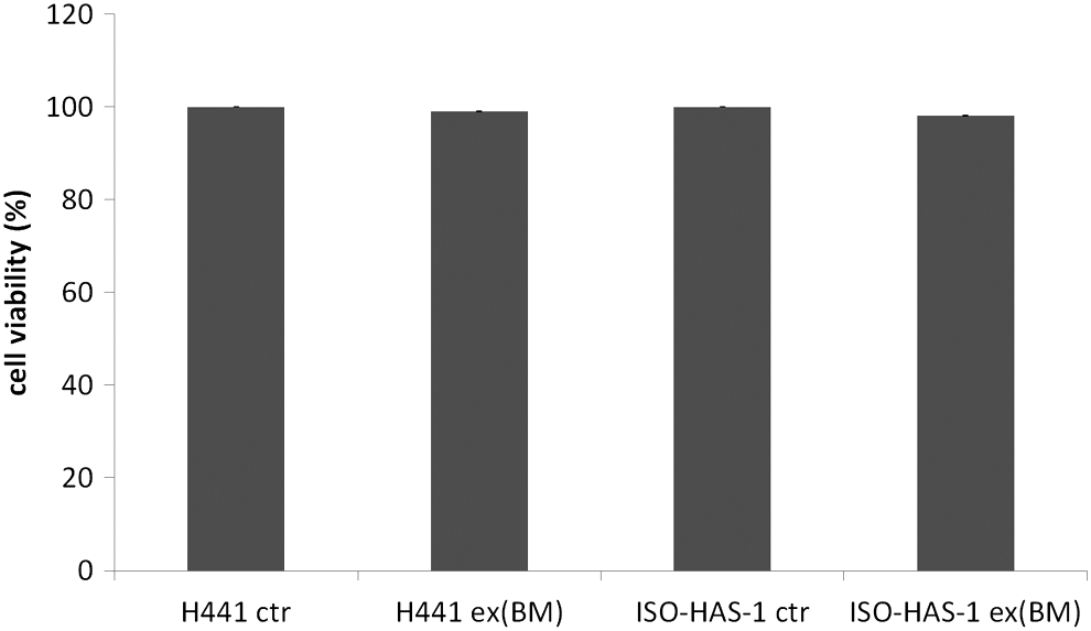
Cell viability (MTS assay). Basement membrane mimics were cultivated in cell culture medium for 4 days ( = leached medium) before monocultures of NCI H441 and ISO-HAS-1 were cultivated in the leached medium for 4 days. To exclude possible cytotoxic effects of the leached medium, mitochondrial activity was measured after 4 days of cultivation. All groups demonstrated excellent cell viability, with no statistically significant differences between groups.
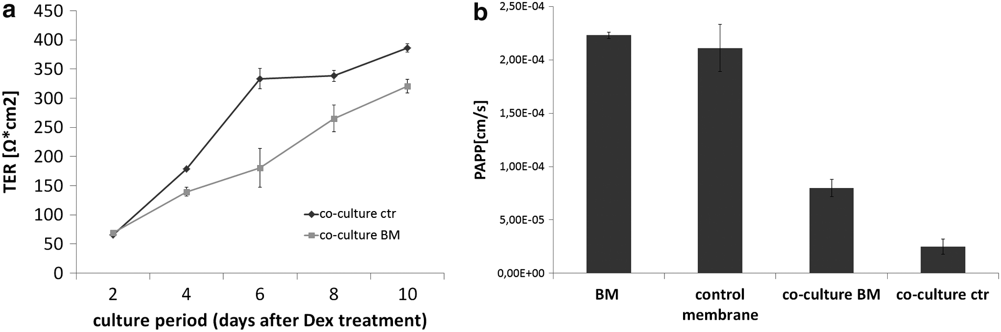
Barrier properties of in vitro alveolar barrier model seeded on basement membrane mimics compared with barrier properties of in vitro alveolar barrier model seeded on the standard system, with a membrane composed of polycarbonate (HTS 24-Transwell® filters).
Cell morphology and expression of cell-specific proteins in bipolar cocultures on basement membrane mimics
To visualize the barrier morphology with NCI H441 on top and ISO-HAS-1 on the bottom of the membrane, membranes cocultivated with NCI H441 and ISO-HAS-1 were fixed, embedded into paraffin, cut into sections of 4 μm thickness, and subsequently stained for cell-specific proteins after 10 days of posttreatment with dexamethasone (Fig. 4a, b). Figure 4a demonstrates the alveolar barrier mimic with NCI H441 on the apical side and ISO-HAS-1 on the basolateral side of the basement membrane mimic, which is visible as a thin and transparent structure between the two cell types. ISO-HAS-1, cultivated on the lower surface of the basement membrane mimic, stained positively for the endothelial marker CD31 (Fig. 4a). As demonstrated in Figure 4a, endothelial cells form a thin monolayer, which is present across the whole basement membrane. In addition, to demonstrate cell morphology of the individual cell types of the bipolar cell culture seeded on the basement membrane mimics, whole membranes were stained for the appropriate, cell-specific marker via immunofluorescent staining (Fig. 4c). Hence, Figure 4c demonstrates the endothelial cell contacts, which are stained positively for CD31, across the monolayer illustrated as top view in this figure. NCI H441, cultivated on the upper surface of the membrane, reveals a flattened morphology with the cells largely organized into a monolayer rather than a bilayer (Fig. 4b). The adherens junction protein E-cadherin is expressed by NCI H441 and was found mainly at the junctions of the epithelial cells when developing a more bilayer structure (Fig. 4b, arrow). β-catenin staining of NCI H441 demonstrates the cell–cell junctions between the epithelial cells, as β-catenin is a subunit of the cadherin complex during adherens junction formation (Fig. 4c). Determination of E-selectin in supernatants of the different transwell compartments of the bipolar coculture seeded on the basement membrane mimics was evaluated using ELISA and revealed a higher concentration of this protein in the basal compartment compared with the apical compartment (Fig. 4d).
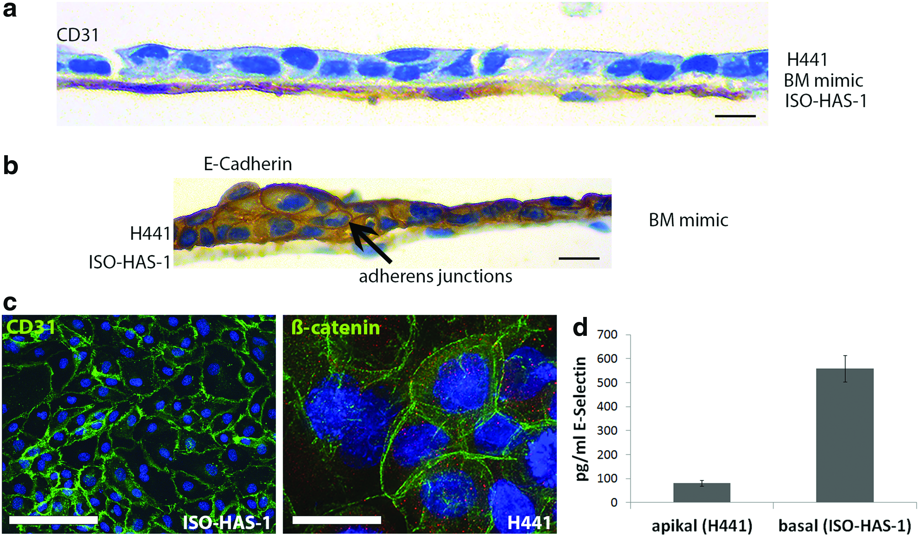
Immunostaining of histological sections of bipolar coculture seeded on basement membrane mimic for the endothelial cell-specific marker CD31
Triple-cultures seeded on basement membrane mimics: incorporation of THP-1
Before triple-culture experimentation, the acute human monocytic leukemia cell line THP-1 was seeded on fibronectin-coated six-well-plates and treated with 8 nM PMA for 4 days to induce macrophage differentiation into the proinflammatory phenotype (M1). Characterization of macrophage-like phenotype was determined via immunofluorescent staining for macrophage-specific marker CD105 and CD68 (Fig. 5a). After 4 days of PMA treatment, adherent THP-1 cells revealed a typical macrophage-like morphology and stained positively for CD105 as well as CD68. Hence, bipolar cell cultures consisting of NCI H441 and ISO-HAS-1 were performed on PCL basement membrane mimics as described, and PMA-treated THP-1 were seeded on the upper chamber of the transwells on top of the epithelial cells after 7 days of precultured bipolar coculture (Fig. 1). Before starting the triple culture, TER needed to be measured (Ωcm2 > 300) for the bipolar culture as an essential prerequisite for seeding THP-1 to the bipolar culture. TER increased during the course of cocultivation from 2 to 7 days, reaching ∼377 Ωcm2 after 7 days of bipolar culturing (Fig. 5b). In response to macrophage treatment on day 7 (Fig. 5b), TER decreased slightly over the period of triple cultivation up to 10 days (∼329 Ωcm2, Fig. 5b). In general, NCI H441 cells of alveolar barrier mimics composed of triple-culture, are organized in a more multilayered structure (Fig. 5c) compared to the barrier composed of the coculture without THP-1, in which the epithelial cells formed a mono- or bilayer structure (Fig. 4a, b). Immunohistochemical staining of the alveolar barrier mimics composed of NCI H441, ISO-HAS-1, and THP-1 for the macrophage marker CD105, revealed the distribution of the differentiated THP-1 cells in the in vitro system (Fig. 5c). Detached CD105-positive cells, demonstrating the macrophage-like THP-1, are incorporated into the NCI H441 cell layer within the alveolar barrier mimic in the triple-culture (Fig. 5c, arrows).
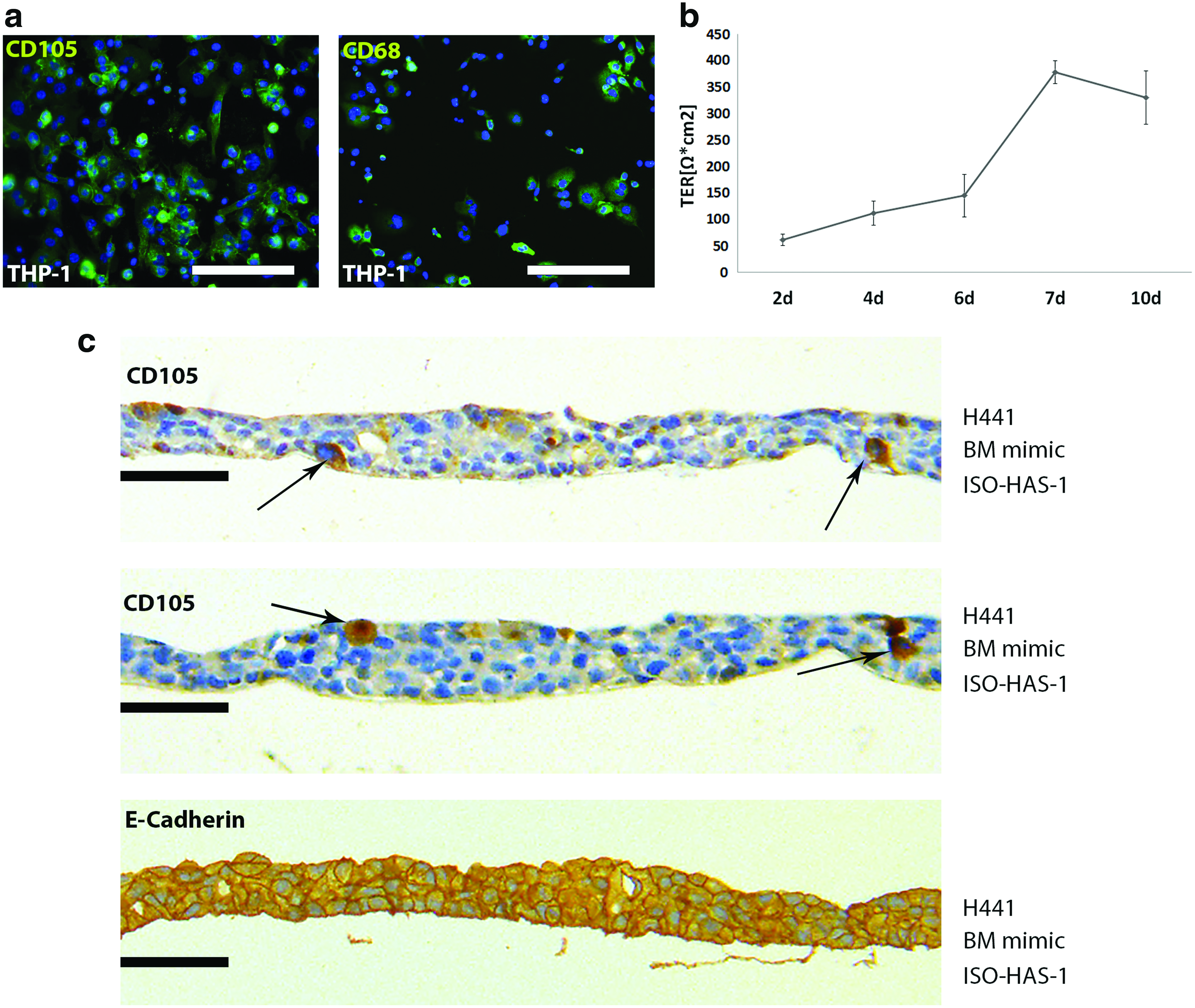
Triple-culture experiments.
Discussion
In this study, an in vitro cell culture model of the human alveolar-capillary barrier was established on an electrospun, functional mimic of a nanofiber-based basement membrane, composed of ultrathin nanofiber meshes. The properties of the human air–blood barrier, especially the characteristics of the natural basement membrane, actually pose a difficult scientific challenge for material scientists. Hence, numerous in vitro alveolar barrier studies are based on bipolar coculture systems seeded on commercially available transwell inserts, composed of poly(carbonate). While the latter might be suitable, and even beneficial, for basic research questions,4,5,9 they poorly represent the properties of the natural basement membrane in the human lung from the clinical point of view. In this study, we were able to postulate a bipolar coculture model of the alveolar barrier on a noncommercially available, ultrathin, nanofiber-based membrane, which can be easily modified in composition, thickness, porosity, and functionality, depending on the experimental purpose. Commercially available transwell inserts that were used in a number of previous studies to separate endothelial cells from epithelial cells did not result in a structure similar to the natural basement membrane with an ultrathin fibrous arrangement and its interconnected pores.
The process of electrospinning enables the fabrication of bioresorbable, ultrafine continuous fibers of small diameters (10 nm to 10 μm), which can be varied in terms of their properties (material composition, diameter, stiffness) and which can be additionally tuned and functionalized with bioactive molecules.6,10 Especially, the possibility to functionalize these membranes with specific ligands for alveolar endothelial cells and epithelial cells ensures long-term cell adhesion and reconstruction of the artificial membrane. For experimental purposes, it is beneficial for the alveolar membrane mimic to be bioresorbable expecting to promote the substitution of the synthetic membrane by extracellular matrix deposition, which is very important to improve cellular junction formation as well as maintaining mechanical stability. Importantly, the degree and time of biodegradability of artificial membranes can be also tuned by selecting suitable polymer for electrospinning. Therefore, during the past few years, electrospinning techniques have become a focus of attention with regard to biomedical applications in the field of tissue engineering. 11 In the present study, an alveolar-capillary barrier mimic was developed using a functional nanofiber-based basement membrane fabricated via electrospinning. This was then used as substratum for the epithelial cell line NCI H441 and the endothelial cell line ISO-HAS-1 as cellular components.
Earlier to bipolar cell culture experiments, cytotoxic effects of the basement membrane mimic itself were excluded following evaluation by an extraction assay. Furthermore, both cell types adhered well to the basement membrane mimic, which was in accordance with the study of Nishiguchi et al. 6 In mimicking the in vivo situation of the alveolar-capillary component in the human lung, a necessary requirement of the proposed in vitro model on the basement membrane mimic was the functionality of the barrier, as barrier function is a conditio sine qua non for physiological function of the lung. The alveolar-capillary barrier is a structural and functional entity, which is composed of three main components: the alveolar epithelium, the capillary endothelium, and the basement membrane in between. Moreover, the barrier must combine high mechanical strength on the one hand and low diffusion resistance on the other hand (Weibel, 1969). The postulated in vitro coculture model should fulfil these requirements and, in addition, should permit cellular crosstalk, leading to the formation of a functional air–blood barrier. In general, the alveolar epithelium plays an essential role during physiological pulmonary function by forming a tight barrier against toxic compounds or bacterial invasion.12–14 During this study, TER was determined for NCI H441 in bipolar coculture seeded on the basement membrane mimic, resembling a tight alveolar epithelial layer. TER values of bipolar cocultures increased during the course of cultivation up to TER >300 Ω × cm2 after 10 days of cocultivation postdexamethasone treatment, reaching approximately the measured TER of the control bipolar cocultures on the transwell inserts. This comparison strongly demonstrates the barrier qualities of our novel bipolar in vitro system on the nanofabricated mimic. In 2004, Hermanns et al. had already developed an in vitro model of an alveolar barrier and reported a strong barrier function for dexamethasone-treated NCI H441 or NCI H441 in coculture with HPMECs. 4 Adherens junction formation, evaluated by immunofluorescent staining of the epithelial cell layer for E-cadherin and β-catenin, additionally confirm epithelial barrier properties of this in vitro model, since cadherin/catenin-based adherens junctions are strongly involved in forming the epithelial barrier, integrating intra- and intercellular signaling and therefore mediating cell and tissue behaviour.15–17 Furthermore, bipolar culture seeded on basement membrane mimics reveals a reduced flux of sodium-fluorescein, thus establishing the paracellular restrictiveness of this in vitro system. The pores in the nanofiber meshes are interconnected, which allows separate cell culture of each layer as well as high permeability of liquid factors, permitting selective transport. Thus, the thin basement membrane mimic allows a sufficiently direct contact between the cells on the upper side of the membrane with the cells on the lower side. This ensures the selected transfer of growth factors and signals from one cell type to the other, while the interconnected pores prevent the penetration of the membrane. In contrast to this, the pores of the control transwell inserts are etched and their low porosity might lead to blocking of the inserts with cells resulting in a lower permeability, which consequently might also suppress cell-to-cell crosstalk. 6 E-selectin, an adhesion molecule, which is expressed by endothelial cells, can be mainly found in the basolateral compartment containing only the endothelial cell layer, suggesting a selective separation of the presented in vitro barrier model. 18
During the course of this study, the discussed in vitro alveolar barrier mimic was further improved by the addition of another important cell type, which can be found in the respiratory system, namely macrophages. This additional cell type is mandatory in the study of possible lung inflammatory reactions in vitro, as it represents a more complex and relevant system by combining endothelial, epithelial, and immune cells.19,20 To obtain a macrophage-like phenotype, the human monocytic cell line THP-1 was treated with PMA for 4 days, resulting in adhesion of the cells on the tissue culture plastic and a typical macrophage-like morphology and corresponding protein expression. In general, triple-cultures of THP-1 macrophages, NCI H441 epithelial cells, and ISO-HAS-1 endothelial cells on the basement membrane mimic leads to a thicker membrane when compared to the bipolar coculture, since the epithelial cells seem to organize into a multilayer when macrophages were added to the system. Macrophages can be found on top of the epithelial cell layer as well as on the bottom, adjacent to the basement membrane mimic, thus indicating a migrating property of the THP-1 in this system. TER decreased slightly after macrophage treatment of the coculture, but still remained at a high level, demonstrating an intact barrier even in response to macrophage treatment. To defend the host against exogenous pathogens, macrophages and alveolar epithelial cells represent the first line of defence. 21 Upon pathogen recognition, a definitive and orchestrated program of defence is activated and involves macrophage-mediated inflammatory cytokine production, which finally triggers the response to the pathogen by activating the epithelial cells. 22 A number of studies focus on the pathogen or nanoparticle-defending role of macrophages in different settings of in vitro alveolar barrier models.19,20,23 However, we have not yet been able to use the described model for the study of possible pathogenic or toxic effects of nanoparticles on the in vitro alveolar barrier system. The purpose of the present experimental work was to establish whether the rather unphysiological polycarbonate membrane of the Transwell system could be replaced by a thinner, nanostructured bioresorbable membrane, and serve as a suitable substratum for double and triple cocultures of the alveolar-capillary barrier. The data clearly demonstrate that suitable cell lines of human epithelial, endothelial, and immune cells can indeed establish and maintain the barrier properties of the air–blood barrier on this novel membrane. It is hoped that this system will serve as a highly beneficial tool to test different pathogenic stimuli and thus provide a basis for better understanding of the physiology and pathology of the lung.
Conclusion
During this study, a beneficial in vitro model system of the alveolar barrier was developed using a functional, electrospun, nanofiber-based basement membrane mimic, composed of ultrathin nanofiber meshes. Bipolar cocultures consisting of epithelial and endothelial cells seeded on this basement membrane mimic yield a tight alveolar epithelial barrier, as documented by increasing TER values, selective transport capabilities and expression of adherens junction proteins. Moreover, this in vitro system was further expanded to a triple-culture by the addition of macrophages as classical immune cells to develop a stable in vitro triple-culture of the alveolar barrier. Such a model system could be used to study the effects of pathogens or toxic factors in the context of a more physiological in vitro alveolar-capillary barrier.
Footnotes
Acknowledgments
The authors thank Verena Hoffmann for her excellent technical assistance. We also gratefully acknowledge the funding (SAW-2015-DWI-2471) from the Leibniz Senate Competition Committee (SAW) under the Joint Initiative for Research and Innovation as well as partial funding from Marie Curie Actions EU project (SNAL 608184).
Disclosure Statement
No competing financial interests exist.