
Abstract
Continuous delivery of monoclonal antibodies (mAbs) at low concentrations may be helpful in the management of several chronic, especially autoimmune diseases. A possible approach to employ mAbs therapy in vivo, in the absence of in vitro manipulations, could be graft of microcapsules containing mAb-secreting hybridoma cells (HY). Sodium alginate (AG) is a polymeric saccharide that permits simple fabrication of microcapsules that are biocompatible and prevent immune recognition of encapsulated cells, upon graft, by the host's immune system. However, at present, AG-based microcapsules are usually impermeable to large molecules. The aim of this study was to engineer the membrane of AG-based microcapsules, to make it permeable to larger molecular weight classes of mAbs. To this end, we have prepared a new AG-based membrane, using standard reagents already in use, but following different coating procedures and molar ratios. In particular, we fabricated a new capsular membrane permeable to IgM synthesized by the HY cell line, G3C. Morphologic structural and ultrastructural analysis of the new membranes before and after intraperitoneal transplant, in conjunction with IgM outflow secretory kinetics underwent both, in vitro and in vivo assessments. While allowing immunoprotection of the enveloped HY, as demonstrated by the absence of any inflammatory response, the microcapsules permitted G3c mAb egress, on a regulated delivery kinetics. HY viability persisted, upon transplant, for long time periods. In summary, the new AG-based microcapsules allow delivery of big molecules out of the capsules, while protecting the enveloped HY from the host's immune system. These microcapsules could apply to implant cells producing fully active large molecules without the need of time- and cost expensive procedures to purify them.
Impact Statement
We have developed a new prototype of alginate-based microcapsules that while enabling permeation of big molecules (i.e., class IgM monoclonal antibodies) delivered by the enveloped cells, retain immunoisolation properties preventing rejection of the encapsulated cells upon transplantation. The new microcapsules reverse the dogma that large molecules, like Ig, should not cross the membrane to avoid immune rejection. We have engineered the classic capsular wall in a different way, with regard to polymeric molar ratios and coating reagent sequence. The increased membrane thickness augmented permeability, but also modified the reagent molecular interplay to preserve the microcapsules' immunobarrier competence.
Introduction
Use of monoclonal antibodies (mAbs) has steadily expanded due to applications to the treatment of a number of chronic, autoimmune and neoplastic disorders. Long-term delivery of biologics, including mAbs, bifunctional antibodies, and fusion proteins may be a way to stimulate/inhibit the immune system, at both systemic and local levels. For instance, long-term delivery of mAbs or fusion proteins could be useful for the local treatment of rheumatoid arthritis 1 or immune-related diseases of the lung2,3 and the central nervous system,4,5 but also for treatment of systemic autoimmune disorders, such as type 1 diabetes (T1D) mellitus. 6
In preclinical studies, the biologics to be tested can be easily obtained in vivo, by growing the hybridoma cells (HY) in the murine peritoneum. 7 Although some authors tested the effects of mAb-containing ascites, 8 purification of the biologics would be more convenient, also practically, should the mAb be delivered continuously by osmotic pumps. However, the purification of fusion proteins and antibodies is a delicate procedure that takes time and is expensive. Moreover, in the long-term delivery, the protein within osmotic pumps may become unstable. That is why it would be much better to use a system based on live cells, enveloped in special microcapsules, associated with high biocompatibility and selective permeability, producing mAb to be synthesized and released in vivo. 9 On this purpose, we have an in-house method for ultrapurification of sodium alginate (AG), a polysaccharide extracted from brown seaweeds that has been used for three decades to fabricate microcapsules. The AG purification process is key to the microcapsules' biocompatibility and their membrane's molecular weight cutoff selectiveness. AG is easy to handle, and suitable for upscaled production of microcapsules containing live cells. 10 Our previous longstanding experience with microencapsulation of pancreatic islet cells11,12 as well as other cells,13–15 and graft of the former in diabetic animal models, and finally in diabetic patients, within pilot clinical trials, 12 allowed us to gain considerable experience in handling cells to be enveloped in AG-based biomembranes. Since we valued the possible role of rat G3C-based, anti-mouse GITR (glucocorticoid-induced tumor necrosis factor receptor-related) targeting mAb in prevention of T1D mellitus very highly, we addressed to develop a biohybrid device, namely G3C in AG microcapsules, to create an in vivo system for delivery of G3C-derived mAbs in preclinical rodent models of T1D mellitus. To accomplish this innovative goal, we redesigned our standard AG-based microcapsules, and re-engineered the membrane's physical chemical properties, underpinning two major targets: (1) to lose membrane's permeability and allow outflow of as big molecules as IgM; and (2) to preserve immunoprotection properties of our new microcapsules to prevent rejection of the embodied nondiscordant xenogeneic G3C grafted in recipient mice.
In the present study, we describe the preparation of microcapsules designed to allow outflow of a mAb produced by HY lodged within the microcapsules. In particular, we prepared multilayer microcapsules containing murine HY producing an IgM (prototype of a very big molecule) and demonstrated that they continuously secreted IgM in vitro and in vivo, without triggering rejection when transplanted in vivo on immunocompetent mice.
Materials and Methods
Hybridoma cells and cell culture
G3C HY cells were a kind gift of Dr. Jun Shimizu. The cells produce G3c mAb, a rat IgM mAb against murine GITR gene.8,16 These IgM mAbs were used to test the new type of microcapsules. Cells are cultured in RPMI medium (GIBCO) with 10% fetal bovine serum. When purification of IgM mAbs was needed, cells were cultured in CD Hybridoma serum-free chemical medium (GIBCO).
AG properties
Powdered alginate was purchased from Monsanto-Kelco featuring the following properties: molecular weight = 120,000–190,000 kDa; mannuronic acid (M) and guluronic acid (G) = M fraction (FM) 61%; G fraction (FG) 39%. It is a “high M” alginate.
Alginate ultrapurification was conducted under Good Laboratory Practice (GLP) conditions, based on patent no. WO 2009093184 A1. At the end of the process, the obtained alginate solution properties were: (1) endotoxin levels, measured by Limulus Amebocyte Lysate (LAL) test, <27.8 Endotoxin Unit (EU)/g (<0.5 EU/mL; any level below 100 EU/g in this test is considered endotoxin-free); (2) protein content <0.45%; (3) viscosity 100–300 cps; (4) heavy metal content below the recommended cutoff and in particular: Ca <100 ppm; Cu <40 ppm; Fe <60 ppm; Hg <40 ppb; Mg <40 ppm; Zn <40 ppm; Pb <50 ppm; Si <10 ppm; Mn <10 ppm; Sr <40 ppm; and As <100 ppb. To prepare our capsules we used a “high M” alginate, subjected to an in house ultrapurification process to render it free of endotoxin, protein and heavy metals. For the gelling process we selected Ca2+ (Ca chloride solution). Our AG-core-based microcapsules underwent coating with poly-
Preparation of alginate microcapsules
Standard microcapsules
Microcapsules were prepared, according to our Standard Operative Procedures (SOPs), starting from 1.8% high-M AG solution, produced as previously described. 10 The same physical/chemical parameters were used for all experiments. Briefly, the alginate solution was continuously aspirated, at a fixed flow rate, by a peristaltic pump and extruded through a microdroplet generator; the resulting microdroplets were collected into a solution containing divalent cations, which immediately made them turn into gel microbeads. The employed gelling solutions were: 100 mM CaCl2, with this salt (Sigma-Aldrich) being dissolved in sterile NaCl 0.9%. After the gelling, the beads were retrieved, washed twice in saline, and sequentially coated with 0.12%, 0.06% PLO (Sigma-Aldrich), degelled with 55 mM sodium citrate, and finally overlayered with an outer coat of 0.06% ultrapurified alginate, to obtain biologically acceptable and functionally performing microcapsules. 10 Sterility and viability tests, the latter using ethidium bromide and fluorescein diacetate (Sigma-Aldrich), under fluorescence microscopy were performed.
IgM-secreting microcapsules
To fabricate microcapsules that would allow for outflow of big molecules (such as an IgM), in principles we had to modify substantially our standard microencapsulation procedure, where the membrane must be impermeable to Ig. Hence, we changed stoichiometric molar ratios of the usual reagents, AG and PLO, employed for microencapsulation. We also changed the composition of the capsule's multilayered membrane, by reframing the layering sequence, so as to fulfill the desired membrane's molecular weight cutoff. The employed gelling solutions were: 100 mM CaCl2, dissolved in sterile NaCl 0.9%. After gelling, the beads were retrieved, washed twice in saline, and were sequentially coated with 0.06% PLO, washed twice in saline, and overlayered with 0.1% ultrapurified alginate, washed twice in saline and coated with 0.04% PLO, washed twice in saline, degelled for 3 min with 55 mM sodium citrate to liquefy the capsules gel core, and finally washed twice in saline and overlayered with an outer coat of 0.05% ultrapurified alginate. A fundamental principle, in cell microencapsulation, is to calculate optimized polymer/cell volume ratios, to avoid too many empty microcapsules or microcapsules that are overloaded with cells. Both circumstances would be detrimental in a transplant setting. This is why we have selected to mix 1.5 × 10 6 hybridoma cells with 1 mL of ultrapurified alginate. This polymer/cell ratio would yield a balanced population of microencapsulated cells.
Transmission electron microscopy
Samples were prefixed in 2% glutaraldehyde, buffered with 0.2 M Na cacodylate, pH 7.4, for 2 h at 4°C, rinsed in the same buffer, postfixed with 2% osmium tetroxide in the same buffer for 2 h, dehydrated in ethanol graded series, and embedded upon Araldite. Ultrathin sections were stained with uranyl acetate and lead citrate and examined in transmission electron microscopy (TEM) 400 T Philips (B Philips) at 60 kV.
Scanning electron microscopy
Samples were prefixed in 2% glutaraldehyde, buffered with 0.2 M Na cacodylate, pH 7.4, for 2 h at 4°C, rinsed in the same buffer, postfixed with 2% osmium tetroxide in the same buffer for 2 h, dehydrated in ethanol graded series, critically point dried, and coated with gold palladium. Examination of the samples was conducted under SEM, Philips Scanning Electron Microscope, B Philips, The Netherlands, at 15 Kv.
Purification of IgM mAb
G3C HY were cultured for 14 days in a serum-free chemical medium (GIBCO). G3c-derived IgM mAb was purified using HiTrap IgM Purification HP column (1 mL size) for affinity purification (GE Healthcare), following the manufacturer's instructions. The column allows the purification of a maximum of 5 mg of IgM per sample. Briefly, the IgM mAb present in the supernatant of the cell culture (150 mL) was concentrated (final volume 1.5 mL) using Amicon ultra-15 columns (Millipore) with a molecular weight cutoff of 100 kDa so that the proteins bigger than 100 kDa were retained. Then, the concentrated supernatant was diluted in 10 mL of ammonium phosphate 1M solution and loaded on the HiTrap IgM Purification HP column, equilibrated with 5 mL of binding buffer (20 mM sodium phosphate and 1 M ammonium phosphate, pH 7.5); the unbound sample was washed out from the column with 15 mL binding buffer, the IgM mAb was eluted with 12 mL elution buffer (20 mM sodium phosphate, pH 7.5). The buffer containing the IgM mAb was exchanged with phosphate-buffered saline (PBS) through dialysis, using Slide-A-Lyzer Dialysis Cassette (10,000 Molecular Weight Cut Off (MWCO); Thermo Scientific). The purified G3c IgM mAb was quantified using the Bradford method. Considering that the culture medium was synthetic and did not contain serum, the protein concentration was considered to apply exclusively to G3c IgM mAb.
Enzyme-linked immunosorbent assay
To quantify the level of G3c IgM mAb produced by G3C cells, we developed an enzyme-linked immunosorbent assay (ELISA). In brief, the microplate wells were coated with 250 ng of GITR Fc protein (Adipogen) or Fc as control (Adipogen) over 16 h of incubation. After washing and blocking the plate with the blocking buffer (0.2% bovine serum albumin, 0.05% sodium azide, 0.04% Tween20 in PBS) for 30 min, samples (cell culture medium or mice plasma, and purified IgM mAb as standard) were added; following 16 h incubation, wells were washed and biotin-anti-Rat IgM Ab (5 μg/mL; BD Pharmingen) was added; after 2 h incubation, the antibody was washed out, and a Streptavidin horseradish peroxidase (HRP) conjugate (dilution 1:1000; BD Pharmingen) was added. Thirty minutes later, the excess HRP conjugate was washed out, and the ABST substrate solution was added. After an appropriate time, the reaction was stopped with 1% sodium dodecyl sulfate (SDS) solution and the plate was read under Tecan microplate reader (405/600 nm). The purified IgM mAb was used for reference. Optimization of ELISA was obtained by testing different concentrations (plate coating ranging from 100 to 400 ng of the GITR-Fc fusion protein, biotin-anti-Rat IgM ranging from 1 to 10 ug/mL, and blocking buffer Tween-20 percentage ranging from 0.01 to 0.1). ELISA allows a good quantification of IgM within a 10–1000 ng range (data not shown).
Histological and immunohistochemical analyses
For histological and immunohistochemical (IHC) examination, the samples were fixed in 10% neutral buffered formalin for 24 h at room temperature, dehydrated, and paraffin embedded. Paraffin-embedded specimens were cut with a rotary microtome. IHC analysis was performed on 3.5 μm slides using primary antibodies, specific for rat IgM. Immunohistochemistry relied on the automated Leica BOND system (Leica Biosystems Newcastle Ltd, Newcastle, UK) on a Leica BOND-III instrument. The slides were counterstained with Hematoxylin.
Animals
The encapsulated hybridoma cells were grafted intraperitoneally, into n = 7 C57Bl6 mice upon general anesthesia. Around 1.5 × 10 6 hybridoma cells in 1 mL of microcapsules per mouse were grafted. Five mice received empty microcapsule grafts and served as controls. All mice were sacrificed at 3 weeks of transplantation (TX). The microcapsules were retrieved by peritoneal lavage with saline and placed in sterile tubes. Upon accurate washing, to discard blood or peritoneal cells, the capsules were resuspended in complete medium and deposited in culture flasks, at 37°C, 95% air/CO2 for further assessment. Upon 24 h of incubation, microcapsule aliquots were tested for viability, while the remaining capsules were fixed in 10% neutral buffered formalin for immunocytochemical examination.
All immunocompetent mice were housed in the Perugia University Veterinary Service Center according to institution-approved animal care guidelines. All procedures were approved by the University of Perugia animal Welfare Committee.
Results
Production of hybridoma containing IgM-secreting microcapsules
Microencapsulation of hybridoma cells involves suspension of the cells in a 1.8% AG solution at a concentration of 1.5 × 10 6 hybridoma cells per 1 mL of ultrapurified alginate. The cells/AG suspension was pumped through an air droplet generator that, using air shears and mechanical pressure, sprays the generated cells/AG microdroplets onto a 1.2% calcium chloride solution. The latter immediately turned the microdroplets into gel microbeads containing the cells. Up to this point, the production procedure of the new capsules did not differ from the standard microencapsulation process. 10
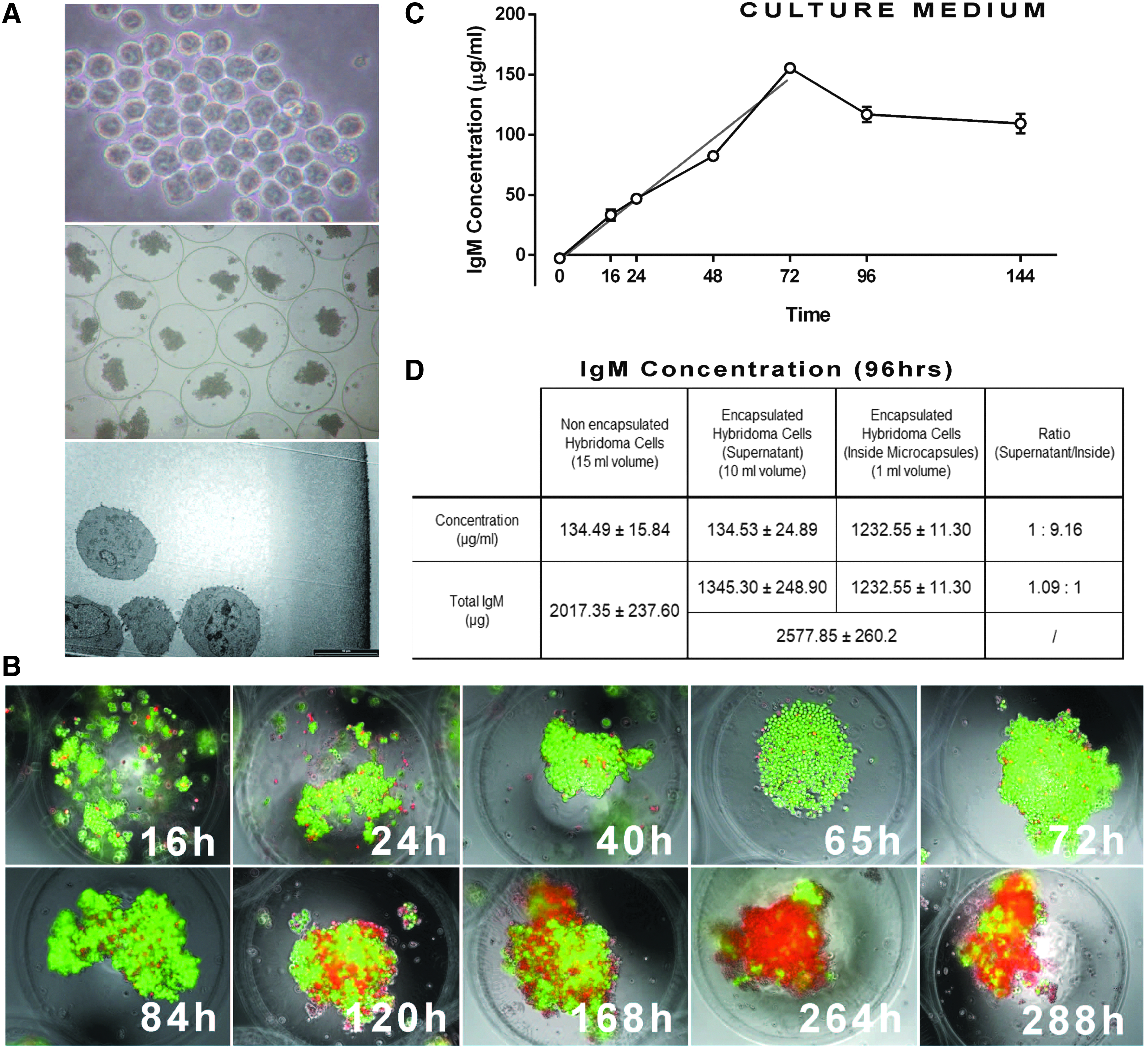
The subsequent procedures to form and stabilize the microbead walls were different from the standard ones and consisted in the coating procedures with PLO and diluted AG described in the Materials and Methods section and summarized in Figure 1A and B. One substantial difference, in comparison with standard alginate microcapsules, lies on the fact that the PLO coatings are differently intertwined with diluted alginate layers. This new technique permits access to microcapsules that macroscopically look the same as usual (e.g., in the mean value of the diameter or the shape), but, when analyzed in detail, they reveal profound differences.

Analysis by TEM and SEM of the new microcapsules
Examination under TEM was associated with considerable differences at the level of the coating layers as compared with the standard microcapsules. First of all, Figure 1B shows that the actual wall thickness was 17 μm as compared with the 10 μm of the standard microcapsules.
Moreover, TEM of the new microcapsules showed different membrane architecture: the gradient layers look homogeneous and continuous, differently from the wall's discontinuous appearance of the standard microcapsules. This seems to reflect different reactivity of osmium with the constituent microcapsule layers in the new versus standard constructs.
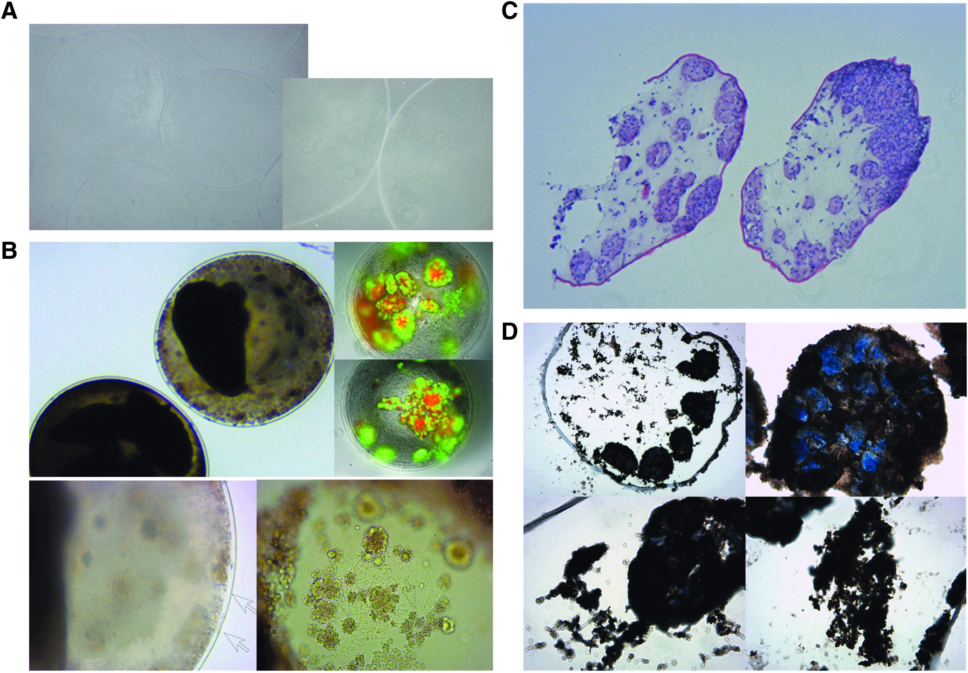
Examination of the new microcapsules under SEM showed that the inner and the outer side of the capsules' membrane were very homogeneous. In the exhibited picture (Fig. 2A), the microcapsules had been cut deliberately. However, when analyzing the uncut samples, the preparations appear to be free of breakages or inhomogeneous surface (Fig. 2B). Hence, the new microcapsules did not show weaker or dented, but just even smoother surface areas.

Microencapsulation of hybridoma cells does not exert adverse effects on cells
After microencapsulation of hybridoma cells, microcapsules were incubated in the culture medium under standard conditions. Figure 3A shows the morphological behavior of hybridoma cells before microencapsulation and after the microencapsulation procedure. TEM analysis confirmed that microcapsules' wall was evenly structured, also in the presence of cells. The latter did not slide through the wall, and looked viable and undamaged by the microencapsulation procedure. In particular, either nuclei and nucleoli or subcellular organelles, such as mitochondria, appeared to be morphologically intact and healthy. At the indicated time, microcapsule aliquots were taken to test cell viability with the ethidium bromide/fluorescein diacetate test. As shown in Figure 3B, the cells grew and retained viability up to 95% throughout 84 h. Moreover, most cells were alive until 7 days of culture maintenance, and some live cells were detectable also after 12 days culture.
Thus, the hybridoma cells secreting IgM mAb can be easily enveloped in microcapsules, remain viable, and eventually implement their growth.
Microencapsulated hybridoma cells produce IgM mAb that can be found outside the capsules
To evaluate the presence of IgM mAb in the medium where microcapsules were present, we employed ELISA.
Figure 3C shows that the immunoglobulin concentration in culture medium increased linearly with increasing culture time for up to 72 h, demonstrating that IgM mAb can exit from the microcapsules. The concentration of IgM mAb reached a plateau after 72 throughout 144 h. We then tested concentrations of IgM mAb in the medium where microencapsulated G3C or control unencapsulated cells were cultured or inside microcapsules containing G3C cells (Fig. 3D). The quantity of the assayed IgM was higher (2577.85 ± 260.20 μg) when G3C were encapsulated, as compared with unencapsulated, monolayered G3C (20,170 ± 237.60 μg). The ratio IgM total quantity in the culture supernatant/capsules equaled 1.09/1, hence identical. However, the ratio IgM concentrations supernatant/microcapsules was 1:9.16, indicating that intracapsular IgM concentrations were far higher.
Microencapsulated hybridoma cells are protected from the graft rejection and survive better in vivo than in vitro
The capsules containing and not containing HY cells were transplanted intraperitoneally into immunocompetent animals, to verify their effective ability to protect the incorporated cells from the recipient's immune system and their ability to release mAb in the animal. After 3 weeks, the capsules were retrieved from the peritoneum and assessed as far as concerns morphology and viability. Moreover, the concentration of IgM mAb in the serum was evaluated.
The new microcapsules did not induce any reaction when transplanted empty into the peritoneum of immunocompetent mice, in fact, their appearance, after recovery, was identical to the pregraft conditions, with no cellular overgrowth, neither inside nor outside the capsules (Fig. 4A). HY cell-containing microcapsules did not elicit any immune reaction. They appeared intact at the time of recovery, and the capsule's wall was clearly detectable (no breakage, dents, or irregular wall patterns) (Fig. 4B). The viability of the encapsulated cells was higher than 60% after 3 weeks of transplantation (Fig. 4B): this value exceeded percent viability of the same preparation under in vitro culture maintenance. Eventually, in vivo conditions help better morphologic/functional preservation as compared with in vitro culture for over a week of follow-up (Fig. 3B). Cells within microcapsules, in some instances, formed aggregates, and color spots were detectable inside the capsules. IHC analysis on the retrieved microcapsules showed IgM mAb within the cytoplasm of hybridoma cells demonstrating that cells were still producing these antibodies, and also within the microcapsules like protein depots (Fig. 4C, D). IgM assay in the plasma of treated animals, confirmed that IgM were both produced by the cells and released outside the new microcapsules (average plasma levels: 0.9 ± 0.2 μg/mL).
Discussion
IgG mAbs are used in clinics to treat patients affected by several disorders, such as tumors, and inflammatory and autoimmune diseases. A number of preclinical studies evaluated the activity of mAbs, fusion proteins, and bispecific antibodies. In this article, we describe a new technology to microencapsulate hybridoma cells. Alginate polymers are very stable and useful for these purposes. They have been employed with a great number of cells producing bioactive proteins, such as hormones,12,17 erythropoietin, 18 angiogenic factors, 19 neurotrophic factors20,21 endostatin, 22 growth factors, cytokines, and immunomodulatory molecules.14,23 Additionally, high biocompatibility of alginate and the benefits associated with cell immobilization systems are the main advantages. Consequently, it is possible to set up an efficient bioprocess system with alginates with care being taken to use suitable materials, with optimized physical/chemical parameters.
Hybridoma cells, microencapsulated by our technology, are protected from graft rejection when implanted into allogeneic/xenogeneic hosts, with full retention of cell viability and function, including an outflow of the synthesized mAbs.
Not only by our longstanding experience in the field of pancreatic islets but also other cell microencapsulation trials using ultrapurified AGs complexed with stoichiometrically suitable aminoacidic polications, we have been able to develop a new prototype of AG-based microcapsules. We especially worked on changing stoichiometric molar ratios of AG and polyaminoacid without losing the capsule's MWCO with no negative interference on the capsule's immunoisolatory properties. The reason why we decided to reduce PLO concentration in both layers was to fabricate a looser membrane (to grant egress to IgM derived from the microencapsulated hybridoma cells). We needed an additional diluted alginate scaffolding in between the two PLO layers to ensure the multilayer stability. The final AG coating layer was essential to bind still free PLO aminoacidic radicals to avoid inflammatory reactions and significantly increase biocompatibility.
The obtained new capsular microenvironment permitted excellent cell survival and function. As a consequence, IgM mAb produced by the G3C HY were stored and slowly released across the modified capsule's membrane. Hence, the newly formulated microcapsules constitute an efficient drug delivery system. This membrane's flexibility is pivotal to deliver variable amounts of mAbs for different time periods. The new microencapsulation system holds the advantage of coupling well-tested basic constituent polymers, whose biocompatibility and physical/chemical stability was observed in hundreds of graft trials from small-size animals to humans, with a completely new chemical setting. The innovation is associated with clear and unprecedented advantages.
Years ago, Dubrot et al. had used AG-based microcapsules to envelop Hybridoma cell lines producing anti-CD137 and anti-OX40 mAb. 24 This mAb was an IgG, hence much smaller in size as compared with an IgM. The employed AG was a 2% G-enriched low-viscosity polymer, and the microcapsules were produced by an electrostatic microdroplet generator. The gel beads were overlayed with polyaminoacids and AG, with a final membrane's MWCO enabling transmembrane crossing of IgG. However, when the capsules were grafted subcutaneously, it elicited substantial inflammatory reaction. 24 Our microcapsules, on the contrary, were associated with retention of the enveloped cell viability throughout the 3 weeks of the intervention, as shown by achieved results, with no induction of any inflammatory response in the host. More recently, cells producing cancer-targeting bispecific antibodies, have been enveloped in alginate-based microcapsules. They have been associated with antibody outflow through the capsule wall, but only in vitro. 25
Microcapsule bioengineering seem to be a straightforward procedure, but several technical skills are required to form a biocompatible, selective, permeable, and mechanically resistant artificial membranes. However, many are the variables of the procedure which need to be complied with, to fabricate functionally performing microcapsules. After long work where cell containing microcapsules were grafted in several mammalians, man included, with very promising results, we turned our attention to producing capsule membranes, associated with the nominally looser cutoff.
This allowed passage of heavy molecules such as IgM but, on the same token, protected the enveloped xenogeneic HY from host's immune rejection. In this study, while representing a novel approach, for bioinnovative molecule delivery, the new microcapsules overcame the dogma of strict membrane's MWCO tightness to grant for retention of the capsules immunobarrier competence.
Footnotes
Disclosure Statement
No competing financial interests exist.