
Abstract
Stem cell therapies are a promising treatment for many patients suffering from diseases with poor prognosis. However, clinical translation is inhibited by a lack of in vivo monitoring techniques to track stem cells throughout the course of treatment. Ultrasound-guided photoacoustic (PA) imaging of nanoparticle-labeled stem cells may be a solution. To allow PA tracking, stem cells must be labeled with an optically absorbing contrast agent. Gold nanoparticles are one option due to their cytocompatibility and strong optical absorption in the near-infrared region. However, stem cell labeling can require up to 24-h incubation with nanoparticles in culture before use. Although stem cell monitoring is critically needed, the additional preparation time may not be feasible—it is cost prohibitive and stem cell treatments should be readily available in emergency situations as well as scheduled procedures. To remedy this, stem cells can be labeled before freezing and long-term storage. While it is well known that stem cells retain their cellular function after freezing, storage, and thawing, the impact of gold nanoparticles on this process has yet to be investigated. Therefore, we assessed the viability, multipotency, and PA activity of gold nanosphere-labeled mesenchymal stem cells (MSCs) after freezing, storing, and thawing for 1 week, 1 month, or 2 months and compared to unlabeled, naive MSCs which were frozen, stored, and thawed at the same time points. Results indicated no substantial change in viability as assessed by the MTT assay. Differentiation, observed through adipogenesis and osteogenesis, was also comparable to controls. Finally, strong PA signals and similar PA spectral signatures remained. Further studies involving more diverse stem cell types and nanoparticles are required, but our data suggest that function and imaging properties of nanoparticle-labeled stem cells are maintained after freezing and storage, which improve translation of stem cell monitoring techniques by simplifying integration with clinical protocols.
Impact statement
Although stem cell tracking techniques are critically needed, stem cells must be labeled with contrast agents in advance of procedures, which is not clinically feasible due to increased procedure time. As a solution, a stock of labeled stem cells could be frozen and stored, ready for immediate use. Results showed that gold nanosphere-labeled stem cells can be frozen and stored long-term without impacting cellular function or photoacoustic imaging contrast, supporting further investigation of other contrast agents and cell types. Creating a bank of nanoparticle-labeled stem cells advances translation and scalability of stem cell tracking methods by improving integration with clinical protocols.
Introduction
Stem cell therapy is a promising solution for many diseases that have limited treatment options and may benefit patients suffering from traumatic spinal cord injuries, amyotrophic lateral sclerosis, type 1 diabetes, heart attacks, and a variety of cancers.1–5 Numerous clinical trials are underway, but translation is inhibited by a lack of in vivo monitoring techniques to guide stem cell therapies.1,2,6–8 Developing tools for intraoperative, real-time image-guidance of stem cell delivery and to provide longitudinal feedback on stem cell therapy progression would improve therapeutic outcomes. 2 A variety of imaging methods are available to address this need. One promising option that has been demonstrated in multiple stem cell tracking applications is ultrasound (US)-guided photoacoustic (PA) imaging.2,9–14
PA signals are produced through the conversion of light to sound.10,15 When an optical absorber is irradiated with a nanosecond pulsed laser, a pressure wave results from localized thermal deposition, which can be detected by an US transducer. 10 By combining the advantages of optical and acoustic techniques, PA imaging provides high-resolution, high-contrast images at a depth of several centimeters.2,10 A variety of exogenous contrast agents are available to label stem cells for noninvasive in vivo imaging and tracking of cells.2,10,16–18 Gold nanospheres (AuNSs) have been previously demonstrated to guide and detect stem cell therapies with US/PA imaging, and their cytocompatibility adds to their appeal.2,10–12,19
One drawback of implementing many stem cell tracking techniques, including US/PA imaging, is that the stem cells must be labeled in culture before use. This requires preparation and an incubation period of at least several hours based on previous studies. 10 This adds substantial procedure time and would require significant changes to clinical protocols, which may not be feasible. Alternatively, stem cells could be harvested, labeled, frozen, and stored well in advance of the surgical procedure. Having a readily available “bank” of labeled stem cells eliminates the additional time required to prepare stem cells for tracking. It may also be possible for companies to label, freeze, and ship stem cells to hospitals or treatment centers when needed, which further minimizes changes to the current clinical environment.
For these reasons, to implement US/PA image-guidance and stem cell tracking, stem cells need to be frozen after being labeled with AuNSs and readily available for immediate use. 20 It is well-established that stem cell function of naive stem cells is retained with freezing and thawing.21–24 However, it is unknown if AuNS-labeled stem cells retain the same function and PA properties as naive stem cells after freezing, storage, and thawing.
The current study seeks to answer these questions. Human adipose-derived mesenchymal stem cells (MSCs) were labeled with AuNSs, frozen, and stored for 1 week, 1 month, or 2 months. At the designated time point, AuNS-labeled MSCs were thawed, and viability, differentiation, and PA detection were evaluated. Results indicated AuNS-labeled MSCs retained functional and PA properties at all time points. Thus, it seems AuNS-labeled MSCs may be prepared in advance of procedures, frozen, and stored until needed.
Our findings have several important implications. First, clinical adoption of US/PA stem cell tracking is more feasible because minimal time is required to prepare the stem cells, which reduces changes to the current clinical protocol. As a result, the overall translatability of stem cell therapies improves because critically needed monitoring techniques can be implemented to improve treatment outcomes. Finally, the current study lays the groundwork to motivate future research on freezing and storage. Many other combinations of contrast agents and cell types should be investigated to create a wide variety of application-specific stem cell tracking techniques.
Materials and Methods
AuNS synthesis
All chemicals were used as received. AuNSs were synthesized following previously established methods.25,26 In brief, glassware was washed with aqua regia (3 parts HCl:1 part HNO3) and rinsed with deionized ultrafiltered (DIUF) water. After preparing glassware, 235 mL of DIUF water was heated to a boil while stirring vigorously at 350 rpm. Next, 2.5 mL of 10 mg/mL HAuCl4 (Alfa Aesar) was added to the flask followed by 12.5 mL of 10 mg/mL sodium citrate tribasic dihydrate (Sigma-Aldrich). Stirring speed was increased to 700 rpm, and the flask was protected from light. Stirring continued until a red color change was observed, indicating the reaction was complete. The solution was cooled to room temperature while stirring. 27
Synthesis was confirmed by UV-vis spectrophotometry (400–900 nm wavelength; BioTek Synergy HT). Hydrodynamic radius was measured with dynamic light scattering (DLS), and zeta potential measured surface charge (Malvern Zetasizer Nano series). DLS data were recorded as the average size from 6 measurements, each consisting of 13 runs. Zeta potential was the average charge from 6 measurements, each consisting of 12 runs. Transmission electron microscopy (TEM) (Hitachi HT7700 TEM) was used to further characterize AuNS size and morphology. Grids were prepared by drop casting and drying overnight.
Cell culture
Human adipose-derived MSCs (Lonza) at passage 8 were plated in T-75 flasks and maintained in alpha-minimum essential media (α-MEM; Corning) supplemented with 1% penicillin streptomycin (Sigma-Aldrich), 20% fetal bovine serum (FBS, Corning), and 2% L-glutamate. Cells were fed every 2 days. Once MSCs reached 80% confluence, they were either labeled with AuNSs or kept as unlabeled (naive) controls.
Stem cell labeling and confirmation
AuNSs were washed twice and concentrated by centrifuging for 10 min at 3000 rcf in 30 kDa filters (Amicon, EMD Millipore) to prepare for cell labeling. MSCs were labeled by incubation for 24 h with AuNSs suspended in media at an optical density (OD) of 1, corresponding to ∼1.5 ng of gold per cell. To confirm labeling with brightfield microscopy, stem cells were grown in six-well plates with a coverslip placed at the bottom of the well. At 80% confluency, stem cells in half of the wells were labeled with AuNSs. After 24 h, AuNS-labeled MSCs were washed three times with 1 × phosphate-buffered saline (PBS) to remove residual AuNSs. The coverslips containing the cells were stained with eosin (VWR). Slides were prepared and imaged with brightfield microscopy (ZEISS Axio Observer). To confirm labeling with UV-vis spectrophotometry (Evolution 220 UV-Visible Spectrophotometer; Thermo Scientific), MSCs were similarly plated and maintained in two T-75 flasks. At 80% confluence, one flask was labeled with AuNSs and similarly washed with PBS. AuNS-labeled MSCs were trypsinized with trypsin EDTA (Corning) and centrifuged at 500 g for 5 min. Centrifugation further size separated residual AuNSs and AuNS-labeled MSCs. The supernatant was aspirated, and primarily pelleted AuNS-labeled MSCs remained. The second flask contained unlabeled, naive stem cells and was similarly prepared. AuNS-labeled MSCs and naive MSCs were suspended in ∼1.5 mL of PBS and transferred to cuvettes for spectrophotometry.
Freezing protocol
AuNS-labeled MSCs and naive MSCs were frozen following conventional slow freezing protocols.21–24,28 Freezing media was prepared and consisted of the same maintenance media supplemented with 10% dimethyl sulfoxide (DMSO; Sigma-Aldrich) as a cryoprotectant. To prepare for freezing, AuNS-labeled MSCs were washed with PBS at least three times to remove residual AuNSs. After washing, AuNS-labeled MSCs were trypsinized, collected, and centrifuged at 500 g for 5 min to pellet the cells. The supernatant was aspirated, and primarily pelleted AuNS-labeled MSCs remained. Naive MSCs were similarly pelleted. Each cell pellet was resuspended at 1 × 10⁶ cells/mL in the freezing media and was rapidly transferred to a 2 mL cryogenic vial (VWR). Each group, AuNS-labeled MSCs or unlabeled, naive MSCs, was separated into several cryovials. Vials were immediately sealed and transferred to a freezing container (Nalgene “Mr. Frosty” Freezing Container; Thermo Scientific). The freezing container was placed in a freezer at −80°C. The cells were cooled at a rate of −1°C/min. After 24 h, the vials of frozen AuNS-labeled or unlabeled, naive MSCs were stored in a liquid nitrogen Dewar in the vapor phase, where they remained frozen for 1 week, 1 month, or 2 months, at which point they were thawed for viability assays, differentiation assays, and PA imaging.
Viability assay
Baseline viability was established from AuNS-labeled MSCs and naive MSCs that were never frozen. A 96-well plate was seeded with 100,000 cells per well. The top six wells contained AuNS-labeled MSCs, the six below them contained naive stem cells for the positive control, and the three below them contained only MSC maintenance media without phenol red for the negative control. The plate was incubated at 37°C in the dark for 48 h. A standard MTT assay (Vybrant, Thermo Fisher) was used to assess cell viability. The standard procedure was followed per the manufacturer's protocol. Assay results were read on a BioTek Synergy HT plate reader at 570 nm. The process was repeated for the designated thawing time point for each sample (1 week, 1 month, or 2 months) using both naive stem cells and AuNS-labeled MSCs. Once thawed, cells were washed and centrifuged before plating to ensure the potentially toxic freezing media was removed.
Data analysis of the viability assessments from the MTT assays was conducted as follows. Absorbance values from blank samples containing media and MTT solutions were averaged and subtracted from each well to remove background absorbance. Average absorbance values were calculated for naive and AuNS-labeled MSCs. The average reading from naive cells was defined as 100% viability and was used to determine relative viability at each time point. Standard deviations were calculated using the replicates for each sample. A two-tailed t-test with a p-value of 0.05 was conducted comparing AuNS-labeled MSCs to naive MSCs for each time point.
Differentiation assay
Baseline differentiation was established from AuNS-labeled MSCs and naive MSCs that were never frozen. Two 6-well plates were seeded with 300,000 cells per well. The top three wells of each plate contained AuNS-labeled MSCs, while the bottom three wells contained naive MSCs. One well of the AuNS-labeled MSCs and naive MSCs was designated for a negative control. All well plates were stored in an incubator at 37°C, 15% O2, and 5% CO2 in the dark.
One plate was fed with purchased adipogenic differentiation media (StemPro; Gibco) every 3 days for 1 week according to established protocols. Control wells were fed with MSC maintenance media on the same schedule. After induction, the media was aspirated, and cells were washed with PBS without Ca2+ and Mg2+. Cells were fixed with 2 mL of 3.8% formalin for 10 min, and washed with PBS again. Oil Red O staining (Sigma-Aldrich) was used to assess adipogenic differentiation. The Oil Red O stock solution was prepared by mixing 300 mg Oil Red O in 100 mL of 99% isopropanol (Merck Millipore). The working solution of Oil Red O was made by mixing three parts Oil Red O stock solution in two parts DIUF water. The solution was kept at room temperature for 10 min before filtering. Once the Oil Red O solution was prepared, cells were incubated with 2 mL of 60% isopropanol for 4 min. The isopropanol was pouted off. Next, cells were incubated with 2 mL of the working Oil Red O solution for 10 min. Samples were then washed with running tap water until the water ran clear. Cells were stained with 2 mL of Harris Hematoxylin counterstain (Sigma-Aldrich) for 1 min. The plate was washed with running tap water in the same manner. Cells were imaged with an LSM 510 VIS Observer Z1 Confocal microscope system (ZEISS) at 20 × .
The second plate was fed with in-house prepared osteogenic induction media every 3 days. Osteogenic induction media consisted of Dulbecco's modified Eagle's medium (DMEM; Corning), 10% FBS, 1% penicillin streptomycin, 100 nM dexamethasone (Sigma-Aldrich), 10 nM beta-glycerophosphate (Calbiochem, Millipore Sigma), and 50 μg ascorbic acid (Sigma-Aldrich). Control wells were fed with MSC maintenance media on the same schedule. After induction, the media was aspirated, and the cells were fixed in the same manner as the adipogenic plate. After the second PBS wash, 2 mL of 5% AgNO3 (Sigma-Aldrich) were added to each well and incubated under UV light for 40 min. AgNO3 reacts with calcium deposited outside osteocytes. Cells were then rinsed with DIUF water, then incubated with 2 mL of 5% sodium thiosulfate (AMRESCO) for 2 min. The plate was rinsed in running tap water. Cells were then incubated with 2 mL of Nuclear Fast Red (Alfa Aesar) for 5 min. The plate was rinsed in running tap water until the water ran clear. Cells were imaged with a LSM 510 VIS Observer Z1 Confocal microscope system (ZEISS) at 20 × .
The process was repeated at the designated thawing time point for each sample. Thawed cells were washed and centrifuged before plating to ensure that potentially toxic freezing media was removed to not impact differentiation results. Differentiation was assessed by comparing adipogenic and osteogenic staining from thawed samples to baseline samples − never frozen AuNS-labeled MSCs and never frozen naive MSCs.
PA imaging
PA imaging experiments were conducted to investigate PA properties of AuNS-labeled MSCs after storage at −80°C. All cell samples were thawed at their designated time points and similarly prepared. Thawed cells were washed to remove free AuNSs and fixed in 10% neutral buffered formalin. The cells were washed again by centrifugation to remove excess formalin, and the cell pellet was resuspended at a concentration of 1 × 10⁶ cells/mL in PBS.
PA imaging studies were conducted using a tissue-mimicking gelatin phantom consisting of a gelatin base with cell inclusions on top. 29 Solutions for cell inclusions were formed by combining equal volumes of the gelatin and cell suspensions in microcentrifuge tubes. These gelatin-cell mixtures were gently pipetted to evenly disperse cells and ensure that no bubbles were produced. A 20 μL volume of the gelatin-cell mixture was pipetted on the solidified phantom base, creating an inclusion—a round gelatin dome containing the cells. The phantom was placed in the fridge at 4°C for 20 min to set. The phantom was then covered with degassed DIUF. The Vevo LAZR US/PA Imaging System (FUJIFILM VisualSonics) was used to acquire combined US and multiwavelength PA datasets (21 MHz transducer, 680–900 nm wavelengths with 5 nm increments). Each imaging frame captured the inclusions containing unlabeled and labeled stem cells. Data were exported to MATLAB (MathWorks, Inc.) for postprocessing.
US images were used to manually define the region of interest, and the average PA signal was calculated throughout the entire inclusion for each wavelength to produce a PA spectrum. A ratio of the PA signal at 700 nm wavelength and 850 nm wavelength was also calculated. These wavelengths were selected because they corresponded to the wavelengths of relatively high and moderate absorption of AuNS-labeled MSCs, while the laser energy at these wavelengths was stable and PA signals had sufficient signal-to-noise ratio. Standard deviations of the PA signal were calculated by dividing the inclusion into at least five smaller regions of interest with the same area. A two-tailed t-test with a p-value of 0.05 was conducted comparing the PA signal ratios of the frozen to never frozen AuNS-labeled MSCs.
Results
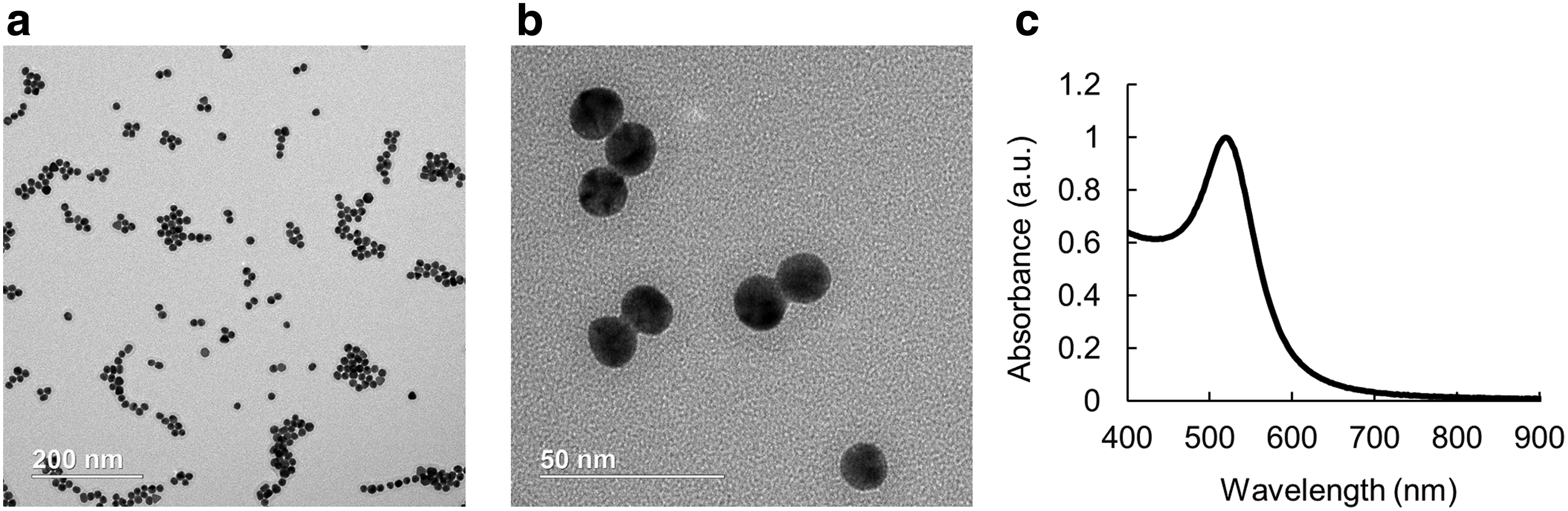
AuNSs for cell labeling experiments were synthesized in-house. TEM confirmed AuNS size and morphology (Fig. 1a, b). Successful synthesis was confirmed via UV-vis spectrophotometry (Fig. 1c). A peak absorption at 520 nm was observed, which was consistent with previous reports. 11 DLS showed an average size of 18.27 ± 6.54 nm, and zeta potential showed an average charge of −42.5 ± 5.66 mV. 27
Characterization of AuNSs.
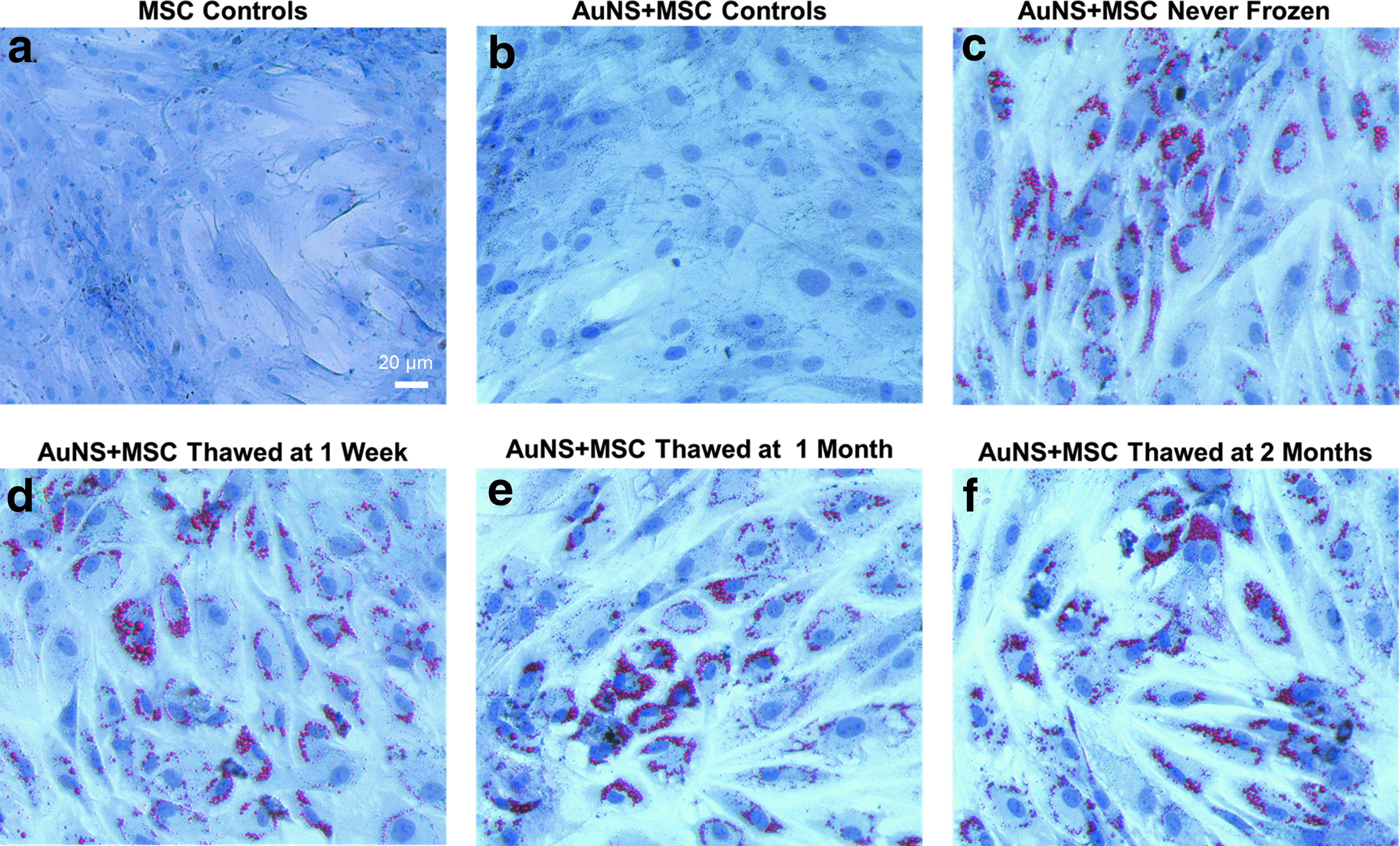
Cellular uptake of AuNSs by MSCs was seen with brightfield microscopy (Fig. 2). Naive stem cells (Fig. 2a, c, e) and AuNS-labeled MSCs (Fig. 2b, d, f) were imaged. AuNS-labeled MSCs had dark blue-purple spots clustered within the cells, which indicated AuNSs (Fig. 2b, d, f). UV-vis spectrophotometry (Fig. 2g) further confirmed successful stem cell labeling of never frozen samples. AuNS-labeled MSCs had a peak at ∼700 nm, which is indicative of surface plasmon resonance coupling of AuNSs upon cellular endocytosis. 18

Characterization of AuNS-labeled MSCs. Cellular uptake of AuNSs by MSCs was visualized with brightfield microscopy.
Frozen AuNS-labeled MSCs were thawed at their designated time points, and the MTT assay was used to confirm AuNS-labeled MSCs remained viable after freezing and thawing (Fig. 3). Initial viability results were compared with two controls that were never frozen: naive (unlabeled) MSCs and AuNS-labeled MSCs. The average percent viability of all AuNS-labeled MSC samples was calculated relative to the control − naive (unlabeled) MSCs that were never frozen. An increase in percent viability was observed for the AuNS-labeled MSCs that were never frozen. Percent viability of AuNS-labeled MSCs frozen for 1 week, 1 month, and 2 months remained relatively stable at ∼85%. Differences between viability were not statistically significant (p > 0.05).
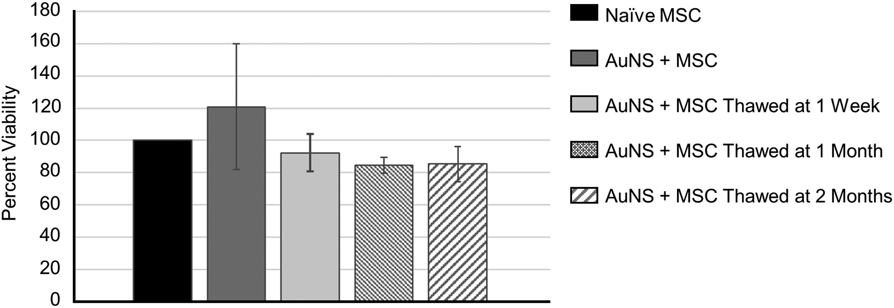
Effect of freezing, storage, and thawing on AuNS-labeled MSC viability. MSCs were labeled with AuNSs at an OD of 1. MSCs were either plated for the MTT assay or frozen and stored to be thawed at a specific timepoint. Results are given as percentages related to naive stem cell controls. Data displayed on the graph are the mean ± standard deviations of the six wells from each timepoint. Differences between viability were not statistically significant (p > 0.05). OD, optical density.
Multipotency of thawed AuNS-labeled MSCs was also assessed to ensure retention of regenerative properties. Thawed samples were plated and subject to adipogenic or osteogenic differentiation. Differentiation was compared with two controls that were never frozen: naive (unlabeled) MSCs and AuNS-labeled MSCs. Brightfield micrographs indicated that stem cell differentiation was unaffected by AuNS labeling, freezing, and storage (Fig. 4). Oil Red O staining was used to assess adipogenic differentiation. The adipogenic-induced AuNS-labeled MSCs (Fig. 4c–f) showed a rounded morphology and prominent red deposits, indicating lipid vesicle formation, compared to controls (Fig. 4a, b), which received standard maintenance media. Silver staining was used to assess osteogenic differentiation (Fig. 5). The osteogenic-induced AuNS-labeled MSCs (Fig. 5c–f) displayed high levels of stacking and linear growth compared to controls (Fig. 5a, b), which received standard maintenance media. The osteogenic-induced cells also showed high levels of calcium, which appeared as brown deposits external to the cells.
Effect of freezing, storage, and thawing on AuNS-labeled MSC adipogenesis. Top row: never frozen

Effect of freezing, storage, and thawing on AuNS-labeled MSC osteogenesis. Top row: never frozen
PA imaging experiments were conducted to ensure AuNS-labeled MSCs produced a PA signal and maintained a similar PA spectrum after freezing, storage, and thawing at each time point. Representative paired US and combined US/PA images from a tissue-mimicking phantom with inclusions containing naive MSCs and AuNS-labeled MSCs thawed at 1 month showed typical phantom morphology (Fig. 6a, b). US images alone depicted each dome, and scattering inside the dome was indicative of cells (Fig. 6a). In the US/PA overlay images, naive MSCs showed no PA signal, but a strong PA signal was observed at the dome containing AuNS-labeled MSCs at 700 nm wavelength (Fig. 6b). PA images for all other domes containing AuNS-labeled MSCs from each time point are displayed in Figure 6c. Qualitatively, AuNS-labeled MSCs retained their ability to produce a PA signal, regardless of freezing time.
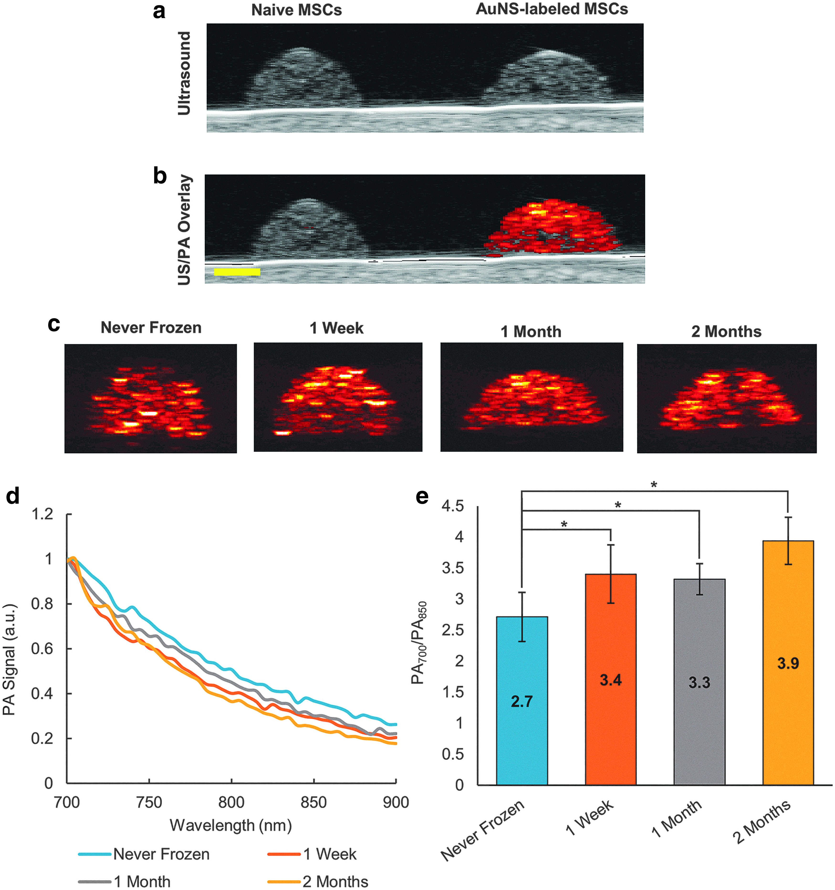
Effect of freezing, storage, and thawing on PA imaging of AuNS-labeled MSCs. Naive stem cells and AuNS-labeled MSCs were frozen and stored for 1 week, 1 month, or 2 months and then thawed to create a tissue-mimicking phantom for US and PA imaging
Multiwavelength PA signals from 700 to 900 nm wavelengths are shown in Figure 6d. The PA spectrum of each cell sample was normalized to best visualize the spectral behavior of the PA signal. Regardless of freezing time, a similar spectral signature was observed. Slight differences in the slope of each spectrum were noted (Fig. 6d), and a ratio was calculated comparing the average PA signal at 700 nm wavelength to 850 nm wavelength (Fig. 6e). The PA700/PA850 ratio increased for the frozen AuNS-labeled MSCs compared to never frozen AuNS-labeled MSCs. Overall PA imaging results indicated that AuNS-labeled MSCs retained their PA signal after freezing and similar PA spectral signatures may support feasibility of quantitative imaging in the future.
Discussion
PA imaging combined with US guidance is an attractive solution to real-time, noninvasive stem cell tracking. To allow US-guided PA imaging of stem cell therapies, stem cells must be labeled in culture with a PA contrast agent before treatment, which takes time and specific facilities. Patients suffering from any acute injury would need immediate treatment, while other patients with chronic conditions may require numerous treatments. Therefore, having frozen stock of labeled cells would be advantageous by eliminating wait time required to prepare cells and allowing immediate use in emergency and regularly scheduled procedures.
In this study, we evaluated a potential clinically translatable solution of advanced labeling of stem cells with AuNSs, followed by freezing, storing, and then thawing AuNS-labeled MSCs. More specifically, MSCs were labeled with AuNSs by incubation in culture for 24 h. AuNS-labeled MSC samples were then frozen and subsequently thawed at 1 week, 1 month, and 2 months. Upon thawing, viability, differentiation, and PA imageability were assessed and compared to naive MSCs and never frozen AuNS-labeled MSCs. The results from all three studies were promising as cell viability and function remained comparable to never frozen cells. In addition, AuNS-labeled MSCs retained their PA properties for imaging.
Viability was assessed using the MTT assay. AuNS-labeled MSCs maintained ∼85% viability when frozen for 1 week, 1 month, or 2 months. The viability spike seen in the never frozen AuNS-labeled MSCs was consistent with our previous studies. 27 Although this was not observed in the frozen samples of AuNS-labeled MSCs, this may be due to an immunostimulatory property immediately following exposure to AuNSs. 30 Alternatively, the viability spike may be due to innate shortcomings of the MTT assay. 31 Results also demonstrated that frozen and thawed AuNS-labeled MSCs had comparable metabolic activity to naive stem cells and AuNS-labeled MSCs that were never frozen. Overall our results agreed with previous reports regarding cell viability upon exposure to gold nanoparticles and also added new information on conservation of viability of AuNS-labeled MSCs after freezing.10,11,27
Stem cell function was further assessed by inducing differentiation. Thawed AuNS-labeled MSC samples were subject to adipogenic or osteogenic differentiation, and multipotency was compared to naive stem cells and AuNS-labeled MSCs that were never frozen. Results indicated that freezing, storing, and thawing AuNS-labeled MSCs had no impact on stem cell differentiation. As differentiation is one of the hallmark characteristics of stem cells, this process is vital to their function as a treatment option for patients. The fact that freezing and thawing the labeled cells did not impact this ability further promotes the clinical application of this strategy as the cells could be labeled and stored for later use. While the studies did not analyze the impact of freezing and thawing on small molecule secretion, based on results from viability and differentiation, we suspect that the secretory profile would also be preserved. 15
Overall results of our studies indicated that stem cells remained viable and multipotent after labeling with AuNSs and freezing for different periods of time. However, it is important to acknowledge that only one freezing protocol was tested in our study and further investigation is needed to confirm that different freezing protocols do not impact stem cell function and PA detection. In this study, MSCs were frozen using a conventional, slow freezing protocol.21–24,28 However, different combinations of cryoprotectant, cooling rate, and storage temperature have been shown to impact stem cell function.24,28 Therefore, it may also be possible that different freezing protocols and storage may impact viability, multipotency, and perhaps even PA properties of AuNS-labeled MSCs. Additional freezing protocols, PA contrast agents, and stem cell types should be studied in the future to best optimize stem cell function and PA trackability after freezing and storage of different combinations of labeled stem cells.
PA activity of the AuNSs allows for noninvasive stem cell tracking. Thus, it was critical that PA properties of AuNS-labeled MSCs remained after freezing, storage, and thawing. Qualitatively, single wavelength PA imaging results (Fig. 6) showed that PA signal was maintained for frozen and thawed AuNS-labeled MSCs. Without the PA activity being maintained after freezing and thawing, the storage of the labeled cells would be useless. However, to allow in vivo detection in the future, single wavelength PA imaging is not sufficient due to high background signals from endogenous absorbers, such as, oxygenated hemoglobin, deoxygenated hemoglobin, and melanin. Thus, multiwavelength PA imaging and spectroscopic analysis will be required.15,32
The critical factors to allow spectroscopic analysis are as follows: (1) the magnitude of the PA signal of AuNS-labeled MSCs must be above the noise floor—this criterion was easily met; and (2) a similar PA spectrum is maintained for each frozen sample of AuNS-labeled MSCs. If the PA spectral signature varied substantially, a different spectrum would always be required for detection, which is impractical. Results indicated the spectral signature was similar for AuNS-labeled MSCs that were never frozen compared to those frozen for 1 week, 1 month, or 2 months. Small variations were observed in the slope of the PA spectra. Ratios of PA700/PA850 were calculated to further analyze differences in the slope. Generally, the ratio increased as a function of freezing, which may indicate some changes in aggregation and, therefore, surface plasmon resonance coupling of AuNSs when frozen within cellular vesicles. 18 Future studies are required to completely analyze this result, and variations in this ratio may indicate an opportunity to noninvasively detect differences in packaging of particles inside cellular vesicles. Despite these subtle variations, the spectral signatures were grossly similar for the purpose of future spectroscopic analysis, detection, and quantification of AuNS-labeled MSCs in vivo after freezing for different periods of time. Overall, our results indicated that PA properties of AuNS-labeled MSCs were maintained after freezing, which is important for clinical implementation of PA detection of labeled stem cells.
Conclusion
Results presented in this study demonstrate an important step in investigating the clinical translation of AuNS-labeled MSCs and implementation of stem cell tracking techniques. Some aspects to be considered moving forward include additional studies on the in vivo imaging performance of the stored AuNS-labeled MSCs. Additional viability studies using multiple assays, such as those that measure ATP production or cellular DNA, should be conducted to confirm MTT results. Prolonged storage of the AuNS-labeled MSCs may also be needed, as this study used 2 months as the longest timepoint. Finally, studies conducted with additional stem cell lines, such as a human embryonic stem cell line (hESCs), are needed to confirm translation to other cell types. It may be worth investigating induced pluripotent stem cells or nuclear clonal cells as well, as they are becoming alternatives to hESCs.
The ability to freeze, store, and thaw AuNS-labeled MSCs has major implications on stem cell therapies. These therapies can benefit from real-time in vivo tracking during and after delivery. The tracking will give valuable insights as to how stem cells initiate a positive response in the patient. The studies conducted here create a paving stone to clinical translation, as stem cells must be easily accessible in large quantities and with minimal preparation to facilitate the implementation of critically needed monitoring procedures to guide stem cell therapies. These investigations can be continued to further demonstrate the power of nanoparticle-labeled stem cells and how they may be translated to clinic.
Footnotes
Disclosure Statement
No competing financial interests exist.
Funding Information
This work was performed, in part, at the Georgia Tech Institute for Electronics and Nanotechnology, a member of the National Nanotechnology Coordinated Infrastructure, which is supported by the National Science Foundation (Grant ECCS-1542174).