
Abstract
Several urological structures, such as the male urethra, have a tubular organization consisting of different layers. However, in severe urethral disease, urologists are limited to replacing solely the epithelial layer. In case of severe hypospadias and urethral stricture disease, the underlying supporting structure (the corpus spongiosum) is either absent or fibrotic, causing suboptimal vascularization and therefore increasing the risk of graft failure. Recapitulating the multilayered architecture of the urethra, including supporting structure with tissue engineering, might minimize urethral graft failure. However, current tissue engineering applications for complex multilayered tubular constructs are limited. We describe a gel casting method to tissue engineer multilayered tubular constructs based on fiber-reinforced cell-laden hydrogels. For this, a multichambered polydimethylsiloxane mold was casted with fiber-reinforced hydrogels containing smooth muscle cells (SMCs) and a coculture of endothelial cells and pericytes. The cell-loaded hydrogels were rolled, with the fiber mesh as guidance, into a tubular construct. In the lumen, urothelial cells were seeded and survived for 2 weeks. In the tubular construct, the cells showed good viability and functionality: endothelial cells formed capillary-like structures supported by pericytes and SMCs expressed elastin. With a graft produced by this technique, supported with subepithelial vascularization, urethral reconstructive surgery can be improved. This approach toward tissue engineering of multilayered tubular structures can also be applied to other multilayered tubular structures found in the human body.
Impact Statement
Recapitulating the multilayered architecture of tubular structures found in the human body might minimize graft failure. Current tissue engineering applications for complex multilayered tubular constructs are limited. Here we describe a gel casting approach based on fiber-reinforced cell-laden hydrogels. A multichambered polydimethylsiloxane mold was casted with cell-loaded, fiber-reinforced hydrogels, with the fiber mesh as guidance, into a tubular construct. A graft produced by this technique can improve reconstructive surgery by providing subepithelial vascularization and thereby can reduce graft failure.
Introduction
Currently there are limited tissue engineering solutions for the regeneration of complex multilayered tubular constructs, especially for urological tissues such as the urethra. The male urethra is mechanically supported and vascularized by the surrounding corpus spongiosum (CS). 1 Major urethral pathologies are hypospadias and urethral strictures. Hypospadias is a congenital disorder through which the urethra and surrounding CS are not correctly formed during embryonic development. 2 Urethral strictures are characterized by constriction of the lumen caused by fibrosis of the urethral epithelium and the CS. 3
Over 300 techniques are known for urethral repair 4 and a wide variety of grafts, including autologous skin and buccal mucosa, have been used for urethral reconstruction. However, these substitutes are inferior to urethral tissue and can lead to stricture formation and/or graft failure. Most complications are related to a suboptimal blood supply due to inadequate vascular embedding of the graft. 5 Especially in case of long defects, the availability of autologous tissue is limited. These limitations have contributed to the growing interest in tissue-engineered grafts for urethral reconstruction.
Most studies on tissue engineering for urethral reconstruction have focused on solely the replacement of the urethral mucosa. In case of a healthy CS, the CS provides a vascular bed for the urethra. 6 However, in urethral stricture disease, the CS is often fibrotic, and in the case of severe hypospadias even absent. Without a healthy CS, using only a mucosal layer is not sufficient for successful reconstruction. By incorporating the CS in the tissue-engineered graft, a better blood supply and mechanical protection of the urethra can be secured. 7
Distinct layers can be identified in the urethra and CS. The urethral epithelium is embedded in a highly vascularized, collagen-rich layer, followed by an elastin-rich layer, thereafter a spongy layer, consisting of veins, arteries, and vascular spaces, and lastly, the tunica albuginea CS. 8 To recapitulate the multilayered architecture of the urethra and CS in tissue-engineered grafts, cell-seeded scaffolds have been combined and rolled into tubular structures.9,10 However, 2D sheets of epithelial cells and fibroblasts lack vascularization. In a 3D setup, endothelial cells and pericytes encapsulated in hydrogels can form microvascular networks, 11 resembling the vascular bed of the subepithelial layer in the CS. Smooth muscle cells (SMCs) can proliferate, express elastin, and display a contractive phenotype when cultured in gelatin-based hydrogels. 12 The hydrogels have been reinforced with an electrospun mesh to enable rolling of the gel into a sutured multilayered construct and potentially meet the mechanical needs during surgery.
The aim of this study was to develop a method for tissue engineering of multilayered tubular structures for reconstructing the distinct layers of the native urethra and CS. 8 In our gel casting approach, we have focused on recapitulating the suburethral layers, with the microvasculature found underneath the urethral epithelium and in the spongy region, as well as the elastin-rich layer in between. A polydimethylsiloxane (PDMS) mold with three chambers was developed and casted with gelatin-based hydrogels, crosslinked with microbial transglutaminase (mTG), containing SMCs and a coculture of endothelial cells and pericytes. The hydrogel supported the growth of urothelial cells. The melt electrowritten mesh, incorporated at the base of the hydrogels, provided mechanical support and enabled rolling of the hydrogels into a tubular construct consisting of three layers resembling the native CS.
Materials and Methods
Hydrogel preparation
mTG (Activa TI®; Ajinomoto) and gelatin (MedellaPro; Gelita, Eberbach, Germany) were used. Prior use, the gelatin solution was set to pH 7.6, dialyzed against distilled water using a cellulose membrane (14 kDa cutoff; Sigma-Aldrich), sterilized, and freeze-dried. Ten percent w/v gelatin and 12.5% w/v mTG stock solutions were made by dissolving in phosphate-buffered saline (PBS) at 50°C for 15 min. The mTG solutions were sterilized using a 0.22 μm filter, coated with bovine serum albumin (BSA; Merck). The stock solutions were mixed with medium and casted into the preferred PDMS mold and incubated for 1 h at 37°C and 5% CO2 in a sealed dish with PBS to prevent evaporation during crosslinking.
Polydimethylsiloxane mold fabrication
PDMS molds were fabricated using the Sylgard® 184 Silicone Elastomer kit consisting of a prepolymer base and a curing agent (10:1). The PDMS was cut according to the design based on patent WO/2013/08540413 with a height of ∼1.5 mm and a width of 5 and 16 mm, respectively (Supplementary Fig. S1A, B). Another mold was made for cylindrical hydrogels with a diameter of 5 mm and height of 3 mm (Supplementary Fig. S1C).
Melt electrowritten microfiber scaffolds
Polycaprolactone-based (PURASORB PC 12; Gorinchem, Netherlands) scaffolds were fabricated using a custom-built melt electrowriting printer, described elsewhere. 14 These square-structured meshes (6 × 6 cm with a height of 150 μm) were fabricated by alternating layer deposition orthogonally, with a fiber spacing of 200 μm and a fiber diameter of 10 μm. Processing manufacturing conditions were set to an acceleration voltage of 5 kV at a spinning gap of 3 mm, a feeding pressure of 3.0 bar, and a heating temperature of 90°C. The meshes were cut into a rectangular shape (length: 5 cm and width: either 7 [Supplementary Fig. S1A] or 20 mm [Supplementary Fig. S1B]), or with a 5 mm cylindrical biopsy punch (Supplementary Fig. S1C), and sterilized with 70% ethanol.
Cell culture
Human urothelial cells (HUCs; ScienCell) were cultured in urothelium culture medium supplemented with urothelium growth cell supplement (ScienCell) and 2% fetal calf serum (FCS; GE Healthcare). Cells were used within eight passages for all experiments. Human umbilical vein endothelial cells (HUVECs; Lonza) with GFP-tag (under the human EF1a promoter) were cultured (37°C and 5% CO2) on gelatin-coated Petri dishes in Endothelial Basal Medium-2® (Lonza) enriched with Endothelial Cell Growth Medium EGM-2® (Lonza Bulletkit™), 1% penicillin/streptomycin, and 1%
Cell encapsulation
The sterile stock solutions were mixed with cell suspensions (in EGM-2) to a final concentration of 3.5% gelatin and 5% mTG. Supplementary Table S1 represents the cell concentrations used in each experiment with the corresponding molds and culturing times. Cylindrical hydrogels were made for investigating cell viability, vascular network formation, elastin expression, and mechanical properties (Supplementary Table S1). For engineering multilayered tubular constructs, the hydrogels were casted in the PDMS molds (Supplementary Fig. S1A, B). An overview of the procedure is given in Supplementary Figure S2. In brief, a mesh was placed on a sterile glass slide, covered with the mold. The small and large chambers were casted with a hydrogel containing a coculture of HUVECs-GFP/pericytes-dsRED and a hydrogel containing SMCs in the middle chamber. After crosslinking, the mold was removed and fresh EGM-2 was added to the hydrogels. All experiments were performed at least in triplicate, and a representative experiment is described.
Rolling of fiber-reinforced hydrogels into a tubular construct
The cell-encapsulated hydrogels were cultured for 24 h, before constructing the tubular construct. Using tweezers, the hydrogel was rolled up starting at the side of the small chamber (Supplementary Video S1) and was sutured to stabilize the rolled-up construct. The constructs were cultured for different periods (Supplementary Table S1). All experiments were performed at least in triplicate, and a representative experiment is described.
Luminal cell seeding
The rolled construct encapsulated with cells (n = 2) was maintained in culture for 7 days before HUCs were seeded into the lumen of the tubular construct, at a concentration of 12.0 × 10⁶ cells/mL in 20 μL. To achieve a desired cell density of 4.0 × 10⁵ cells/cm2, a highly concentrated HUC suspension was pipetted directly into the lumen of the scaffold. After application of the cell suspension to the graft, the cells were allowed to adhere for 15 min before the construct was flipped of 90°. Next, HUCs were added for another round and the construct was flipped for another 90° every 15 min. After 1 h, the rolled construct was cultured in EGM-2 in 5% CO2 at 37°C. After 2 weeks of culture, rolled constructs were fixed and stained for further analysis (n = 2). Measurements from nonluminal seeded grafts were used as controls.
Immunofluorescence staining and analysis
For the live/dead assay, the cell-encapsulated hydrogels were washed with PBS and incubated 20 min with calcein AM and ethidium homodimer according to the manufacturer's instructions (Thermo Fisher Scientific). Hydrogels were stained for alpha smooth muscle actin (αSMA), and elastin in SMCs was stained using anti-α-SMA-Cy3™ (1:500; Sigma), mouse-α–elastin antibody (1:100; Merck), and secondary Alexa 555 goat-α-mouse (1:500; Invitrogen) and Alexa 700 goat-α-mouse (1:200; Invitrogen) antibodies. After luminal cell seeding, HUCs were identified with monoclonal mouse anti-human Cytokeratin 7 (CK7) (1:100, Clone OV-TL; Dako) followed by incubation with Alexa 700 goat-α-mouse (1:200; Invitrogen). In brief, encapsulated hydrogels were fixed with 10% formalin, permeabilized for 15 min using 0.5% Triton X-100/PBS, blocked 1 h with 5% BSA/PBS, and incubated with a primary antibody diluted in blocking solution at 4°C overnight. After washing with PBS-Tween, the gels were incubated for 3 h with a secondary antibody, stained with DAPI (1 mg/L in PBS; Sigma Aldrich), and stored at 4°C until confocal imaging (Zeiss LSM Confocal 700). Vascular network formation was determined with the Angiogenesis Analyzer plugin (ImageJ), in which the branching interval was quantified (total segment length divided by the number of branches).
Mechanical analysis
A dynamic mechanical tester Q800 DMA (TA Instruments) with a compression (40 mm diameter) and tension (film) clamp was used for unconfined compression and tensile testing. For both loading conditions, a static force ramp of 0.1 N/min was used until failure. The (cell-encapsulated) hydrogels and equine urethral tissue (diameter = 5 mm and height = 2 mm) were characterized under compression loading. The compressive tangent modulus (∼10–20% strain) was determined from the acquired stress/strain curves. In addition, the tensile behavior of equine urethra and fiber meshes were characterized (length = 10–15 mm and width = 3–5 mm). The tensile modulus was determined at the initial linear part between 40% and 60% for equine tissue and between 2% and 6% normalized strain for the electrospun mesh.
Statistical analysis
Data analysis was performed by an unpaired t-test or one-way ANOVA with a Tukey's multiple comparison test using GraphPad Prism version 7.0. A p-value of <0.05 was considered statistically significant. The error bars of the bar charts represent the standard deviation.
Results
Human umbilical vein endothelial cell-GFP/pericyte-dsRed coculture forms vascular networks in hydrogels
HUVECs-GFP and pericytes-dsRed showed a round morphology after 24 h (Fig. 1A) and were homogenously distributed throughout the hydrogel (Supplementary Fig. S3A). In the next days, sprouting and network formation by HUVECs-GFP in support of pericytes-dsRed occurred (Figs. 1B, C, and 2A, B). The cells were still distributed throughout the hydrogel after 2 weeks (Supplementary Fig. S3C, D).

Hydrogel-encapsulated cells in the presence
In the fiber-reinforced hydrogels, the cells also had a round morphology and were distributed throughout the hydrogel similar to hydrogels without mesh at day 1 (Fig. 1D and Supplementary Fig. S3D). The cells remained distributed throughout the hydrogel up to 2 weeks (Supplementary Fig. S3E, F). However, less elongation and network formation were observed compared with hydrogels without mesh (compare Fig. 1B, C with E, F). Partial alignment of the branches with the mesh could be observed (Fig. 1E, F, white arrows). Vascular network formation was quantified after 8 (Fig. 2A) and 15 days (Fig. 2B) by determining the branching interval, which showed an increasing trend from 78.7 ± 15.0 μm (day 8) to 123.4 ± 15.1 μm on day 15 (Fig. 2C; p = 0.0643).
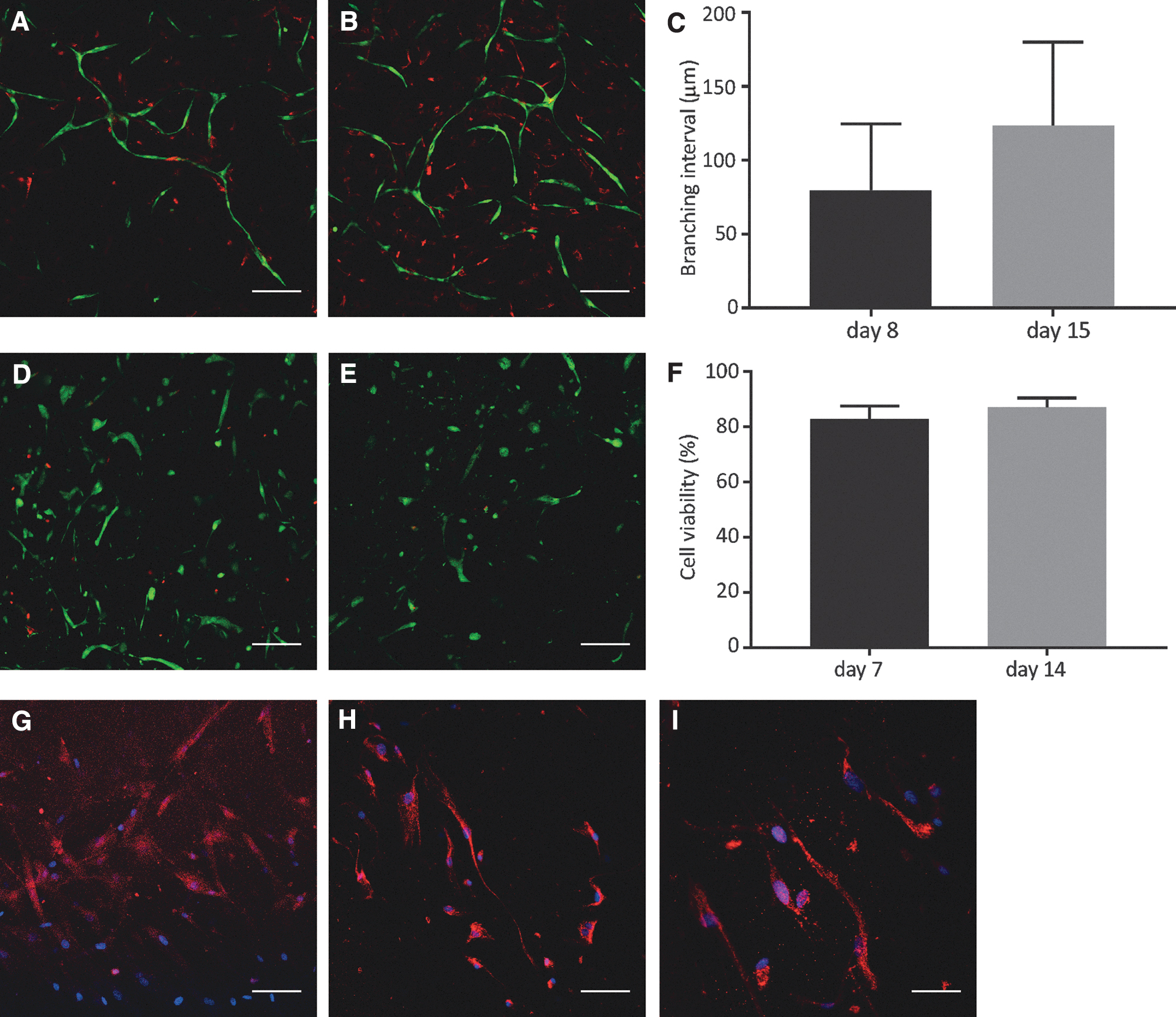
Functionality of cells in gelatin-based hydrogels. Formation of vascular-like networks after 8
Smooth muscle cell viability and functionality
Encapsulated SMCs showed also a round morphology in the first 24 h (Supplementary Fig. S4A). In the next few days, SMCs started to elongate (Supplementary Fig. S4B, C). Overall, >80% SMC viability was achieved up to 14 days (Fig. 2E, F). SMC functionality was confirmed by the expression of both αSMA (Fig. 2G) and elastin (Fig. 2H, I). In 3D cultures of SMCs, the expression of elastin was homogenous throughout the cells and secreted (Fig. 2H, I), whereas in 2D-cultured SMCs, localization of elastin was predominantly perinuclear (Supplementary Fig. S4D). Elastin expression in HUVECs was barely above background (data not shown).
Mechanical properties of the tissue-engineered construct
Native equine urethra had a tensile modulus of 0.176 ± 0.113 MPa (n = 3; Fig. 3A, B). This value was significantly lower than the modulus of the polycaprolactone (PCL) electrospun mesh, which had a tensile modulus of 8.266 ± 1.587 MPa (n = 4, p = 0.0004; Fig. 3B).
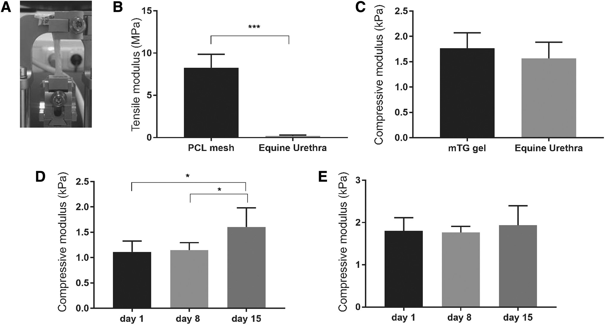
Analyses of the mechanical properties of equine urethra and (cell-encapsulated) hydrogels.
The compressive modulus of equine urethra was 1.567 ± 0.317 kPa (n = 4; Fig. 3C). Hydrogels, in the absence of cells, had a compressive modulus of 1.771 ± 0.301 kPa (n = 7; Fig. 3C), showing no significant difference to the compressive modulus of urethral tissue (p = 0.3176).
In HUVEC-GFP/pericyte-dsRed-encapsulated hydrogels, in the absence of electrospun mesh, an increasing trend could be seen of the compressive modulus over time (Fig. 3D), especially after the formation of vascular networks (Fig. 1). The hydrogels had a mean compressive modulus of 1.115 ± 0.213 kPa at day 1. After day 8, the compressive modulus was similar to day 1. The compressive modulus increased significantly to 1.608 ± 0.375 kPa within 15 days (p = 0.0423 (day 8–15) and p = 0.0297 (day 1–15); Fig. 3D). Meanwhile, more vascular networks had been formed since day 8. This increase of the compressive modulus over time could not be detected in the fiber-reinforced hydrogels (Fig. 3E).
Rolling of the hydrogel into a multilayered tubular construct
HUVEC-GFP/pericyte-dsRed cocultures were effective in forming vascular networks (Figs. 1 and 2) and were used in the inner and outer layer of the construct, to mimic the elastin-rich region of the CS; elastin-expressing SMCs (Fig. 2H, I) were used. The hydrogels could successfully be rolled 1 day postencapsulation around a tweezer and sutured under sterile conditions (Fig. 4A). The hydrogels did not break and sutures successfully maintained the 3D conformation of the construct during culturing (Fig. 4B, C). With this gel casting approach, it was possible to engineer constructs with various lengths; ranging from 5 mm (data not shown) to 16 mm (Fig. 4C).
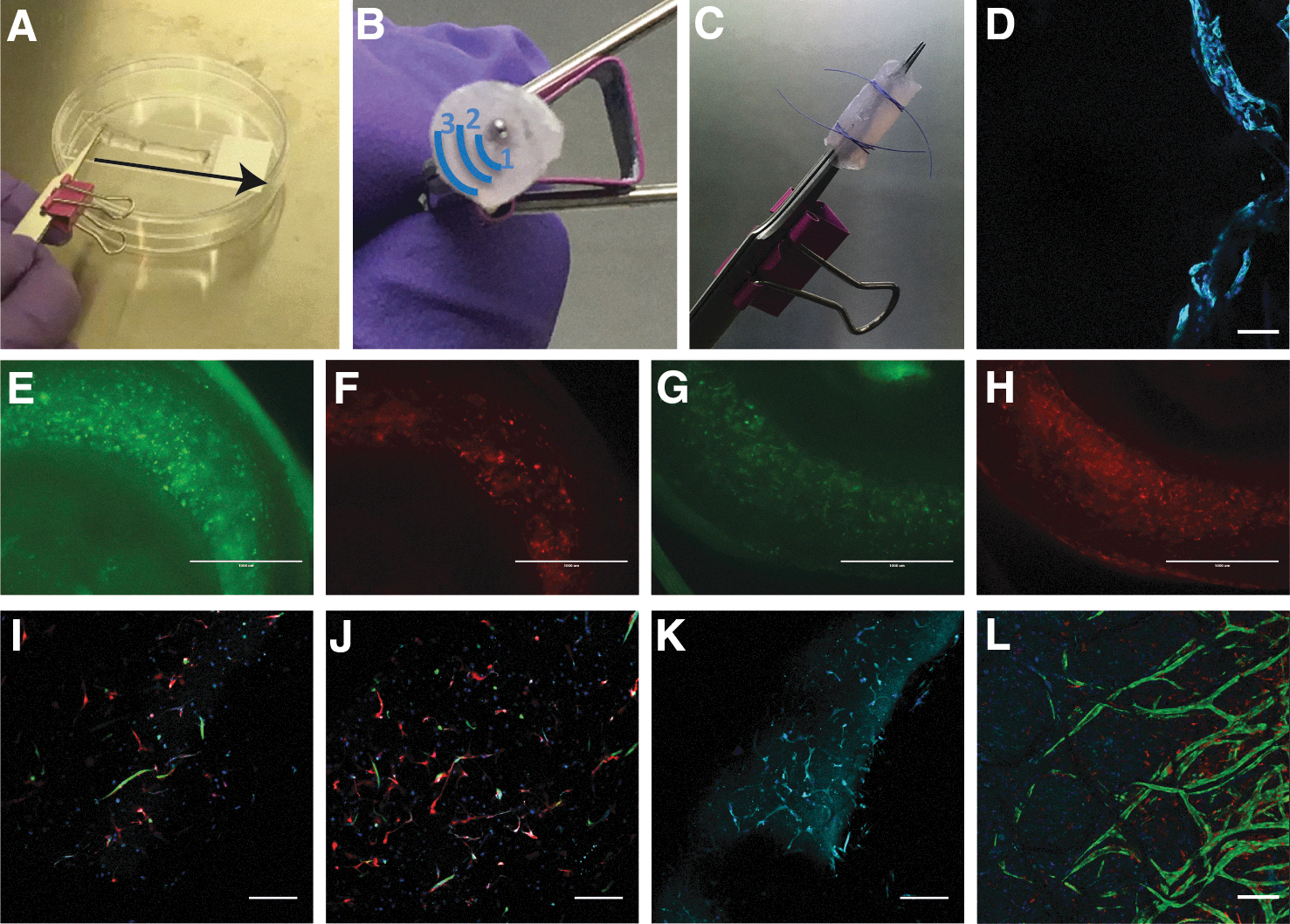
Rolling and cross-sectional view of the multilayered tubular construct.
The constructs showed cell distribution throughout the hydrogel, and survival and functionality were shown by the formation of networks at day 11 (Fig. 4E, F). Increased vascular network formation was observed 14 days postencapsulation (Fig. 4G, H) compared with 11 days (Fig. 4E, F). At day 11, vascular network formation was seen in the outer layer (Fig. 4I) and inner layer (Fig. 4J). Furthermore, SMCs expressed elastin in the middle layer (Fig. 4K). After prolonged incubation for 7 weeks, increased vascular network was observed (Fig. 4L), with visible lumen (Supplementary Fig. S5A). Finally, epithelial cell seeding on the hydrogel tubular structure was tested. Two tubular constructs with a coculture of HUVECs-GFP and pericytes-dsRED as well as SMCs were cultured in EGM-2 for 3–5 days before luminal cell seeding. HUCs were seeded as described in the Materials and Methods section. Fluorescent immunostaining of CK7 showed expression in a monolayer inside the lumen of the tubular construct (Fig. 4D). Cocultures encapsulated in the surrounding gel layers could not been detected (Supplementary Fig. S5B).
Discussion
In this study, we describe a method for tissue engineering of tubular constructs consisting of distinct layers. These initial steps can be further developed to generate grafts that can ultimately be used for urethral reconstruction. As the CS is an integral part of the urethra, all layers of the urethra and CS should be taken into consideration in the tissue-engineered graft.7,8 Two previous studies have focused on tissue engineering of the urethra and CS.15,16 Both studies used acellular CS as scaffold, carrying the problem of shortage of donors and the difficulty of translation to the clinic. In our study, we were able to show elastin expression in the middle layer and the microvasculature found underneath the urethral epithelium with similar mechanical properties to native tissue. Further development of this gel casting approach could potentially result in that the multilayered architecture of healthy urethra and CS can be successfully restored during urethral reconstruction.
Gelatin-based hydrogels, crosslinked with mTG, were used in this study as these were shown to be suitable for encapsulating various cell types.17–23 To our knowledge, this is the first study describing a coculture of HUVECs and pericytes for vascular network formation in mTG-crosslinked hydrogels. This approach can also be used for improving vascularization of tissue-engineered constructs, as vascularization is still a major hurdle in tissue engineering of relevant-sized constructs for clinical application. 24 Overall, the hydrogels seem to provide enough biochemical cues to stimulate both vascular network formation and elastin production. The compressive modulus of the hydrogels is relatively similar to that of native tissue. With the presence of a mesh in the hydrogel, vascular network formation was reduced. Reasons could be alterations in the microstructure and mechanical properties as incorporation of a mesh reinforces and therefore stiffens the hydrogel, 14 resulting in an increased compressive modulus (compare Fig. 3D with E). However, the support of the mesh is required for rolling of the construct and to withstand mechanical forces during surgical handling. As PCL degrades in vivo, the graft will gradually be taken over by neotissue. Future experiments should point out how this influences the mechanical properties of the construct. With this method of gel casting, it is possible to vary the hydrogel composition and the mesh properties, for example, height and spacing of the fibers, as well as the material used to optimally mimic the architecture and native mechanical properties of the tissue-engineered construct.
The results of the mechanical characterization of both native tissue and the tissue-engineered construct should be interpreted with caution. Only a few studies have reported on the mechanical properties of (human) urethra and CS tissue, and therefore, little is known about the variation of the mechanical properties in the different layers of the CS as well as the variation between species.16,25 Another limitation is that, due to limited human tissue availability, we used equine urethra and CS, reported to be less stiff than the human urethra.25,26
Before this construct can be transformed into a functional spongy urethra, the initial steps described here toward a multilayered construct should be further improved. For instance, an epithelial lining of the lumen in the tubular construct is required and the graft should be dynamically cultured in a bioreactor for optimal maturation. We showed viability of urothelial cells in the lumen of the construct (Fig. 4D), yet after luminal seeding, the vascular network in the gel did not survive. Added to that, urothelial cells may not be the ideal cells for a translational approach. Urethral epithelium is different from the urothelium 27 and difficult to harvest. The best alternative is probably oral keratinocytes, as oral mucosa is currently used in urethral reconstruction, 28 as well as in tissue engineering for urethral reconstruction. 29 Moreover, further research is required to optimize the mechanical properties of the tissue-engineered construct as well as optimizing vascular network formation and elastin production in combination with epithelial lining. Finally, innervation of the construct should be addressed, as innervation is of major importance for healthy urethral functioning.
Conclusion
With this gel casting approach, it is possible to tissue engineer multilayered tubular constructs mimicking the elastin-rich region and microvasculature found underneath the urethral epithelium. Our construct has similar compressive mechanical properties to native tissue. Both the gel casting and rolling procedure did not influence the viability and functionality of the cells. This study showed that cocultures of HUVECs and pericytes successfully form vascular networks, and SMCs expressed elastin. This method could also be expanded beyond tissue engineering of the urethra and CS to other multilayered tubular structures.
Footnotes
Acknowledgment
We are thankful for the help of Iris Penning in the production and handling of hydrogels. M.C. would like to acknowledge the strategic alliance University Medical Center Utrecht-Eindhoven University of Technology.
Authors' Contributions
M.J.J.v.V., P.F.C., M.D.C., L.M.O.d.K., and P.d.G. helped to design the study. M.J.J.v.V., R.R., and F.S.Z. performed experiments, collected the data, and performed statistical analysis. P.F.C., M.D.C., J.M., B.J.K., and D.G. gave technical and material support. M.J.J.v.V., B.J.K., P.F.C., M.D.C., and P.d.G. performed the analysis and interpretation of the data. M.J.J.v.V. and P.d.G. drafted the article. All authors discussed the analyses and have contributed to, seen, and approved the article.
Disclosure Statement
No competing financial interests exist.
Funding Information
No funding was received for this study.
References
Supplementary Material
Please find the following supplemental material available below.
For Open Access articles published under a Creative Commons License, all supplemental material carries the same license as the article it is associated with.
For non-Open Access articles published, all supplemental material carries a non-exclusive license, and permission requests for re-use of supplemental material or any part of supplemental material shall be sent directly to the copyright owner as specified in the copyright notice associated with the article.