
Abstract
Inverted colloidal crystal (ICC) hydrogel scaffolds have emerged as a new class of three-dimensional cell culture matrix that represents a unique opportunity to reproduce lymphoid tissue microenvironments. ICC geometry promotes the formation of stromal cell networks and their interaction with hematopoietic cells, a core cellular process in lymphoid tissues. When subdermally implanted, ICC hydrogel scaffolds direct unique foreign body responses to form a vascularized stromal tissue with prolonged attraction of hematopoietic cells, which together resemble lymphoid tissue microenvironments. While conceptually simple, fabrication of ICC hydrogel scaffold requires multiple steps and laborious handling of delicate materials. Here, we introduce a facile route for ICC hydrogel scaffold fabrication using expanded polystyrene (EPS) beads. EPS beads shrink and fuse in a tunable manner under pressurized thermal conditions, which serves as colloidal crystal templates for ICC scaffold fabrication. Inclusion of collagen in the precursor solution greatly simplified preparation of bioactive hydrogel scaffolds. The resultant EPS-templated bioactive ICC hydrogel scaffolds demonstrate characteristic features required for lymphoid tissue modeling in both in vitro and in vivo settings. We envision that the presented method will facilitate widespread implementation of ICC hydrogel scaffolds for lymphoid tissue engineering and other emerging applications.
Impact statement
Inverted colloidal crystal (ICC) hydrogel scaffolds have emerged as a new class of three-dimensional cell culture matrix that represents a unique opportunity for lymphoid tissue modeling and other emerging novel bioengineering applications. While conceptually simple, fabrication of the ICC hydrogel scaffold requires multiple steps and laborious handling of delicate materials with highly toxic chemicals. The presented method for ICC hydrogel scaffold fabrication using expanded polystyrene (EPS) beads is simple, cost-effective, and involves less toxic chemicals than conventional methods, while retaining comparable biological significance. We envision that EPS bead-based hydrogel scaffold fabrication will greatly facilitate the widespread implementation of ICC hydrogel scaffolds and their practical applications.
Introduction
Three-dimensional (3D)
The fabrication of ICC scaffolds involves four steps: (1) preparation of colloidal crystal templates by partial fusion of microspheres with narrow size distribution, (2) infiltration of a hydrogel precursor solution and subsequent polymerization, (3) selective removal of the template microbeads, and (4) if necessary, modification of the pore surface to support stromal cell adhesion 17 (Fig. 1A). The most critical consideration is the selection of template bead material as it determines the possible range of pore sizes and conditions for template preparation and dissociation. Current ICC scaffold fabrication can be categorized into five groups based on template materials (Fig. 1B). The first group is solid polymeric beads synthesized by emulsion polymerization, including poly(methylmethacrylate) (PMMA) and polystyrene (PS).10,12,18–22 ICC geometry was initially developed for nanostructured photonic crystal materials using submicron PMMA and PS beads. By increasing the bead size, ICC structures can be utilized for 3D cell culture experiments. While PMMA and PS beads are commercially available, they are expensive and limited in size selection, with an upper limit of 160 μm due to the decreasing stability of emulsions with increasing size. Addition of crosslinkers allows synthesis of larger beads, but crosslinked beads cannot be dissolved. The second group is polymeric beads prepared by solvent evaporation from emulsion droplets, including polycaprolactone. 23 Polymers dissolved in organic solvents are microfluidically processed into uniform-size emulsion droplets, which in turn solidify as the solvent evaporates. 24 This method increases template bead sizes in a cost-effective manner, but the resultant polymeric beads exhibit a narrow window for thermal and mechanical stability, requiring exceptional care to process into colloidal crystal templates and ICC scaffold fabrication. The third group is hydrogel beads generated by in situ gelation, including alginate and gelatin. Formation and dissociation of alginate beads are exploited by ionic crosslinking and a chelating agent, ethylenediaminetetraacetic acid, respectively.25,26 Synthesis and solubilization of gelatin beads take advantage of their temperature-dependent reversible sol-gel transition.27–30 Although hydrogel beads made from biocompatible materials are removable without using toxic chemicals, their weak mechanical properties make them difficult to process and handle. The fourth group is microbubbles, including air and nitrogen gases.31–34 Microbubbles suspended in a precursor solution generated by a flow-focusing microfluidic device are collected in a confined container and solidified under negative pressure, which opens the closed pores and enables formation of an open porous ICC structure. This method is simple, fast, and essentially nontoxic, but requires significant optimization due to the intrinsic instability of the microbubbles. The last group is glass beads. Glass beads are commercially available with a broad range of sizes and represent high thermal and mechanical stability that facilitate streamlined fabrication of ICC hydrogel scaffolds.14,15,16,35–38 However, requirement of toxic chemicals (i.e., hydrofluoric acid) to dissolve the glass beads has been a critical issue. Currently, there exists no practical and cost-effective method to produce ICC hydrogel scaffolds at high scale under good manufacturing practices. The development of a better fabrication method remains an active area of research.

Summary of the state-of-the-art ICC hydrogel scaffold fabrication methods.
In this study, we introduce a simple and robust method to fabricate bioactive ICC hydrogel scaffolds using expanded polystyrene (EPS) beads, the raw material commonly used for packaging and insulation products. EPS beads are a closed cell foam material composed of 95–98% air, with robust mechanical and thermal stability. 39 We have developed a method to shrink and fuse EPS beads into a free-standing construct that functions as a colloidal crystal template for fabrication of ICC hydrogel scaffolds. Pore size is tunable by adjusting the degree of EPS bead shrinkage. Surface modification for bioactive ICC hydrogel scaffolds was simplified by introducing collagen fibers into the hydrogel precursor solution and polymerizing in a single step. The biological significance of EPS-templated ICC hydrogel scaffolds was demonstrated in vitro by conducting a hematopoietic colony-foaming assay in the presence of bone marrow stromal cells (BMSCs). The ability to form vascularized tissue and attract hematopoietic cells after subdermal implantation was comparatively demonstrated using glass bead-templated ICC hydrogel scaffolds. When compared to existing methods for ICC hydrogel scaffolds, EPS-based fabrication is simple, cost-effective, and involves less toxic chemicals, while retaining comparable biological significance. We envision that the presented method will facilitate usage of ICC hydrogel scaffolds for lymphoid tissue modeling and other emerging novel biological applications.
Materials and Methods
All chemicals and cell culture reagents were purchased from Thermo Fisher Scientific unless otherwise specified. The animal protocol was approved by the University of Massachusetts Amherst Institutional Animal Care and Use Committee.
Pressurized thermal annealing of expanded polystyrene beads
EPS beads (diameter = 1626 ± 195 μm) obtained from Styrofoam Microbead Warehouse (Ebay) were loaded into a perforated stainless-steel mesh container (Houseagain) (diameter = 38 mm and height = 44 mm) to a height of 1 cm. The mesh container was transferred to an elevated rack in a 2.5-quart electric pressure cooker (Chefman) and the cooker was filled with 200 mL of ethanol and water mixture. Using the “High Pressure” setting on the cooker, an annealing time was set between 5 and 30 min. The lid was attached, and the pressure valve was closed to prevent loss of vapor. At the end of annealing, built-up pressure was released before carefully removing the lid and steel mesh containers. Annealed EPS templates were kept at room temperature before further processing. Template images were acquired with an inverted tissue culture microscope. Pore size analysis was performed in ImageJ.
Micro-CT imaging and porosity characterization
Templates fabricated using different annealing conditions and times were scanned using the CT imager on an IVIS SpectrumCT (128201; Perkin Elmer) with a voxel size of 75 μm. Calculation of void fraction based on micro-CT imaging was performed in MATLAB.
EPS bead-templated inverted colloidal crystal hydrogel scaffold fabrication
An acrylamide precursor solution was made with 30 wt% acrylamide monomer (164385000; Acros Organics) and 1.5 wt% bis-acrylamide crosslinker (BP171; Fisher Scientific) dissolved in nitrogen-purged deionized (DI) water. For collagen experiments, nitrogen-purged DI water was displaced with a 4 mg/mL rat tail collagen solution to obtain a final collagen concentration of 2 mg/mL. Before polymerization of the EPS template, a 50 mL centrifuge tube was prepared with laser-cut acrylic inserts (Epilog, Legend Mini 18) to keep the template fully submerged during polymerization. Approximately 10 wt/vol% ammonium persulfate (A3678; Sigma) and N,N,N′,N′-tetramethyl-ethylenediamine (TEMED) (138450500; Acros Organics) were added to the acrylamide precursor solution such that both were at a final concentration of 0.1%. The solution was added to the EPS template-containing centrifuge tube and centrifuged at 4500 g for 20 min at 4°C. Tubes were carefully removed from the centrifuge and left at room temperature to polymerize. Once polymerized, infiltrated EPS templates were retrieved from the centrifuge tubes and carefully scraped on all sides with a razor blade to remove excess polymer. Infiltrated EPS templates were placed in chloroform and left overnight to dissolve EPS beads. A second wash with fresh chloroform was performed to remove residual EPS before thorough washing with water. Hydrogel scaffolds were sterilized by submerging in 70% ethanol (2701; Decon Laboratories) for 30 min. For in vitro and in vivo applications, scaffolds fabricated with 90% ethanol and annealing time of 30 min were used. Initially, large scaffolds were cut down (diameter = 6 mm × height = 2.5 mm) using a 6 mm biopsy punch and a razor blade. Sterile scaffolds were stored in phosphate-buffered saline at 4°C.
Rat tail type I collagen isolation
Type I collagen was isolated from rat tails using serial salt precipitation and dissolution in acetic acid. 15 Briefly, tendons were extracted from rat tails using hemostats and a razor blade, and then dissolved in 3% (v/v) acetic acid (A6283; Sigma) overnight. The resulting solution was passively filtered through a 500 μm mesh and centrifuged for 2 h at 13,000 g, at 4°C to remove debris and impurities. Next, the supernatant was collected and diluted 1:4 by volume in a 30% (wt/v) NaCl (S642212; Fisher Scientific) solution at the rate of 350 mL/h and then left still for 2 h. To collect the collagen, the resulting solution was spun down at 8500 g for 45 min at 4°C, and the precipitated gelatinous collagen harvested. Addition of salt solution and precipitation were repeated until no more collagen was extracted from the solution after centrifugation, upon which all collagen material was combined and resuspended in 0.6% (v/v) acetic acid. The collagen solution was then dialyzed in 1 mN HCl (A144212; Fisher Scientific), and the concentration was determined using lyophilization.
Glass bead-templated ICC hydrogel scaffold fabrication
Soda lime glass beads (diameter = 550 ± 65 μm) (G8772; Sigma) were sorted using an Advantech Sonic Sifter (L3P; Advantech). Beads dispersed in DI water were gradually loaded into a glass vial (8 × 35 mm) to a height of 2–2.5 mm (339130; Fisher Scientific) and were mechanically packed into a lattice structure in an ultrasonic water bath (Branson, B200 Ultrasonic Cleaner). Orderly packed glass beads were dried in a 60°C oven (ThermoFisher, Lindberg Blue M) and then thermally annealed for 4 h in a furnace between 650°C and 680°C. A hydrogel precursor solution composed of 30 wt% acrylamide monomer, 1.5wt% bis-acrylamide crosslinker, 0.2 vol% N,N,N′,N′-tetramethylethylenediamine accelerator, and 0.2 vol% 2-hydroxy-2-methylpropiophenone photoinitiator (405655; Sigma) in nitrogen-purged DI water was prepared immediately before use. Precursor solution (150 μL) was infiltrated into the glass bead template and centrifuged in a microcentrifuge (5415C; Eppendorf) at 8500 g for 15 min and subsequently polymerized under a 15W ultraviolet light (UVP, XX-15S UV Bench Lamp) source for 15 min. Polyacrylamide hydrogel–glass templates were removed from the glass vials the next day to ensure complete polymerization. Excess hydrogel was removed by scraping the glass bead template with a razor blade on all surfaces. Glass beads were selectively dissolved in alternating washes of an acid solution containing a 1:5 dilution of hydrofluoric acid in 1.2 M hydrochloric acid and 2.4 M hydrochloric acid (A513500; Fisher Scientific) (caution: these chemicals are corrosive and must be used in a fume hood with proper protective gear). Washes were completed on a shake plate, and solutions were changed every 4 h until the beads were removed. Scaffolds were thoroughly washed with DI water to remove residual acid and lyophilized. Type I collagen isolated from rat tails was covalently immobilized on the pore surface using Sulfo-SANPAH conjugate chemistry (22589; ThermoFisher) as previously described. 14
Mechanical testing of EPS templates
Mechanical properties were measured using an ElectroForce 5500 (TA Instruments) equipped with a 50 lb load cell. A strain rate of 0.02 mm/s was applied with a 3 mm platen. Ultimate strength was calculated by taking the maximal force before failure.
Multiphoton microscopy imaging of collagen fibers by second-harmonic generation
Collagen fibers on the surface and embedded within hydrogel scaffolds were imaged using a multiphoton microscope (Nikon, A1MP) equipped with a tunable Ti:Sapphire laser source set at an excitation wavelength of 850 nm and a 492 nm short-pass filter.
Primary mouse bone mesenchymal stem cell isolation and culture expansion
A breeding pair of enhanced green fluorescent protein (eGFP) mice (006567; Jackson Lab) was purchased from Jackson Laboratories. A breeding pair of DsRed mice (005441; Jackson Lab) was obtained from Dr. Barbara Osbourne. Femurs and tibias were collected from DsRed or eGFP mice. BMSCs were isolated from the bone by centrifugation and cultured in alpha-Modified Eagle's Medium supplemented with 10% fetal bovine serum (F0926; Sigma) and 1% penicillin-streptomycin (15140122; ThermoFisher). Adherent cells were cultured for a maximum of four passages.
Enzyme-linked immunosorbent assay of mouse IL-6, osteoprotegerin, and receptor activator of nuclear factor kappa-B ligand
EPS scaffolds with and without collagen were seeded with 500,000 adherent eGFP BMSCs and cultured in the wells of a 48-well plate. The culture was maintained until cells spread and covered the scaffold surface. 2D tissue culture plastic controls were seeded at a density of 50,000 cells per well and grown until confluence. Medium was changed every 3 days for the duration of the experiment. At the end of the experiment, conditioned media were collected 48 h after media change and frozen at −80°C. ELISAs were performed on thawed conditioned media samples using ELISA kits acquired from (DY206, DY459, DY462; R&D Systems).
Methylcellulose colony-forming assay
EPS scaffolds were seeded with 500,000 adherent eGFP stromal cells and cultured in wells of a 48 well plate until all surfaces were covered. Freshly isolated bone marrow cells from DsRed mice were resuspended to a concentration of 500 cells/μL in methylcellulose media (HSC001; R&D Systems) (1.27% alpha-Modified Eagle's Medium methylcellulose, 10% fetal bovine serum, and 1% penicillin-streptomycin). Fifty thousand cells were carefully added to the wells containing eGFP BMSC-preseeded EPS-templated ICC hydrogel scaffolds and empty EPS scaffolds. Full-well scans were acquired using a fluorescent microscope (ThermoFisher, EVOS FL) on days 1, 4, 8, 12, and 16. Fluorescent z-stack images were taken by a confocal microscope (Zeiss, Cell Observer) and 3D reconstructed images were generated and animated in an imaging analysis software package (Nikon, Elements AR v5.2.0). FFmpge software was utilized to downsample and compress the 3D reconstructions.
Subdermal implantation of scaffolds in mouse models
Six-week-old Friend Virus B NIH Jackson (FVB/NJ) mice (001800; Jackson Lab) were obtained from Jackson Laboratory. Mice were anesthetized with 1.5% isoflurane (provided by core animal facility) before removing dorsal hair with electric clippers and Nair. Seventy percent of isopropanol wipes (22363750; Fisher Scientific) were used to sterilize the skin. Mice received 2 mg of meloxicam/kg mouse weight (MWI Animal Health) subcutaneously before surgery. Four subcutaneous pockets were formed in mice and EPS and glass-templated ICC hydrogel scaffolds were immediately placed within. Wounds were closed with two 7 mm wound clips. Wound clips were removed after 1 week. Four weeks after implantation, scaffolds were retrieved and flash frozen in Shandon Cryomatrix embedding resin (6769006; ThermoFisher). Tissue blocks were stored at −80°C until use.
Histological characterization
Frozen tissue blocks were sectioned on a cryostat (NX70; ThermoFisher) at a thickness of 20 μm. Hematoxylin and eosin (HXHHEPT, STE0350; American MasterTech) and immunohistostaining procedures were performed as previously described. 40 CD31 (550274; BD), F4/80 (565409; BD), and Ly6G (557445; BD) primary antibodies and anti-Rat AlexaFluor 647 (A21247; ThermoFisher) secondary antibodies were used in this study. Bright-field images were acquired on an EVOS FL Auto microscope and fluorescent images were acquired on a Zeiss Cell Observer SD.
Statistics
Unpaired Student's t-test was performed for comparison of the mean values in all quantitative analysis. Statistical significance was determined if p < 0.05 for a two-tailed analysis. All quantitative data represent mean and standard deviation.
Results
Preparation of colloidal crystal templates using EPS beads
For some PS foam products, EPS beads are subjected to steamed mechanical compression to make a densely packed foam structure without interstitial space. We hypothesized that EPS beads with increased interstitial space could serve as colloidal crystal templates for ICC hydrogel scaffold fabrication. To test this hypothesis, we used a pressure cooker to apply vapor phase water and ethanol solutions to EPS beads without mechanical compression (Fig. 2A). EPS beads (diameters = 1.6 ± 0.19 mm) were placed in a perforated metal container and processed in an electric pressure cooker for 5, 10, 15, and 30 min. The perforated metal container was elevated to prevent contact with the liquid phase at the bottom of the cooker. Pressurized thermal conditions of 0.5 bar at 112°C directed shrinkage and fusion of EPS beads to create a free-standing colloidal crystal-like structure. The shape of the template remained identical, indicating isotropic shrinkage of EPS beads (Fig. 2B). In pure ethanol, beads underwent rapid and uneven size reduction, evident by a wider base compared to top. The overall size reduction was directly correlated to operation time for all ethanol:water solutions. An increasing ethanol concentration was observed to increase the degree of shrinkage. Interestingly a 50:50 mixture had less shrinking ability compared to pure water. Size reduction was greatest at early time points and the rate of reduction gradually slowed down over time (Fig. 2C).
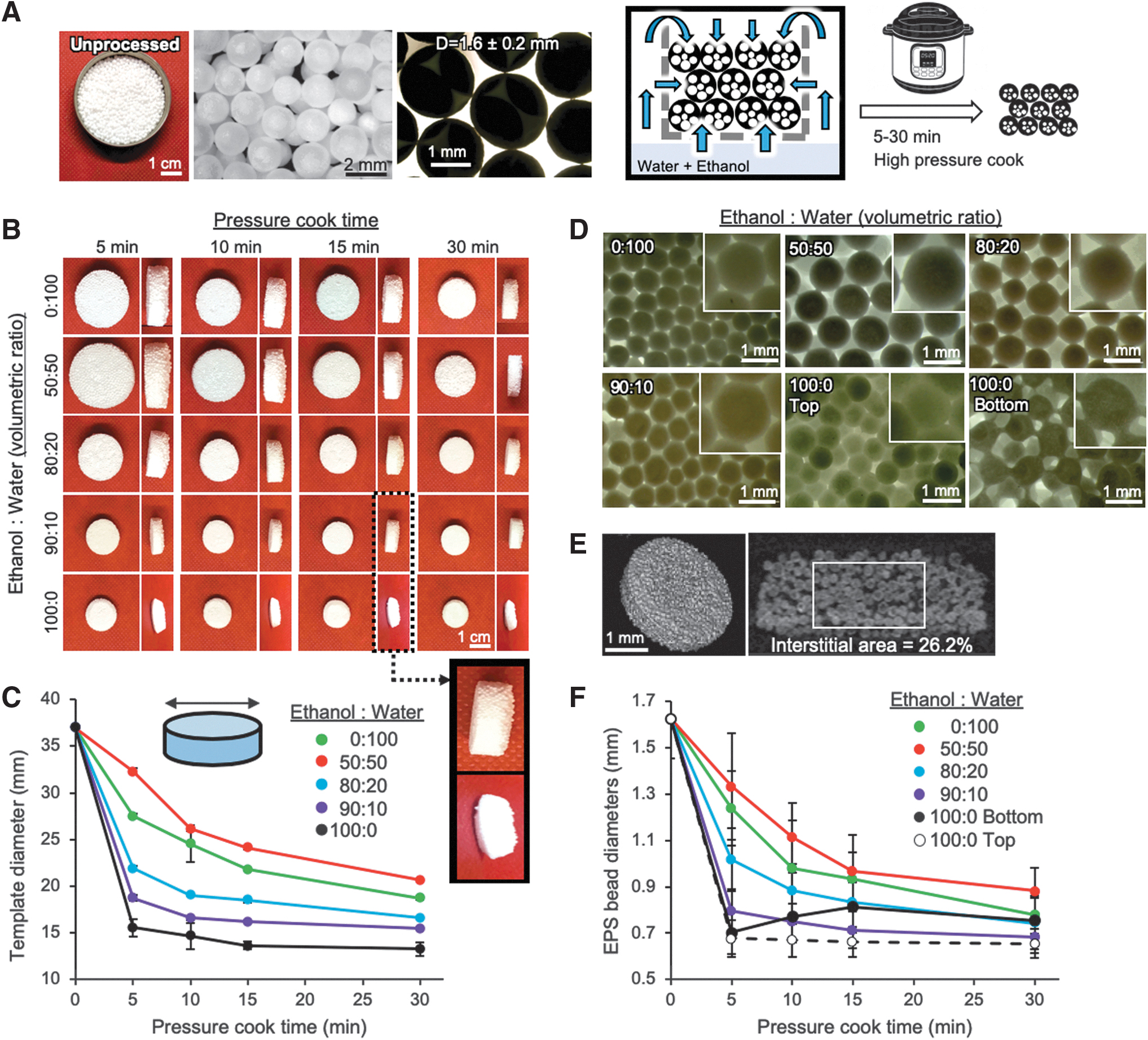
Preparation of colloidal crystal templates using EPS beads.
We next characterized the size and shape change of the individual EPS beads under optical microscopy. Increasing operation time and ethanol concentration reduced the size of EPS beads in an isotropic manner. The interface between the fused beads exhibited elongated necking. In the pure ethanol condition, uneven shrinkage of templates caused different size reduction of EPS beads between the top and bottom surface; EPS beads in the top side showed densely fused morphology, whereas EPS beads in the bottom showed significantly elongated necks (Fig. 2D). The dynamic shrinking of porous EPS beads during bead-bead fusion presents a significantly different annealing process. We further conducted micro-CT scans to visualize the internal packing structure and quantify the interstitial area. While EPS beads have regular sphericity and about 10% of size distribution, their packing density was lower compared to solid beads and microbubbles. The characteristic interstitial area was about 26.2% (Fig. 2E, Supplementary Movie S1). Quantitative analysis of EPS beads substantiated rapid and significant reduction of size with increasing concentrations of ethanol and time (Fig. 2F).
Fabrication of ICC hydrogel scaffolds using EPS bead-based templates
Complete infiltration of hydrogel precursor solution into the interstitial space of colloidal crystal templates requires centrifugation, and the templates should endure the gravitational force. Two critical considerations in using EPS bead-based colloidal crystal templates include flotation of the structure and insufficient mechanical stability. We addressed the flotation issue with a laser-cut acrylic structure designed to fit a standard 50 mL centrifuge tube, consisting of thin rings that support a disk with holes, which are then held down with long bracing pieces that become wedged once the cap is screwed on (Fig. 3A). In this study, we used a hydrogel precursor solution composed of acrylamide monomer and bis-arylamide crosslinker activated by ammonium persulfate and TEMED. After overnight polymerization at room temperature, the polymerized hydrogel and EPS template were taken out from the tube and excess hydrogel was removed. Finally, hydrogel-EPS templates were soaked in chloroform for 4–6 h, during which EPS beads were selectively dissolved, leaving behind an ICC polyacrylamide hydrogel matrix (Fig. 3B). EPS templates prepared in solutions with less than 80% ethanol were broken during precursor infiltration, in which the characteristic ultimate mechanical strength was 1.8 ± 0.2 megapascals (MPa). EPS templates prepared in pure ethanol exhibited the highest mechanical strength (10.3 ± 1.2 MPa), but were difficult to infiltrate with precursor solution due to limited interstitial space (Fig. 3C). When this mechanical correlation was plotted as a function of the overall template size, it turned out that more than 50% size reduction was required to maintain mechanical strength necessary for centrifugation (Fig. 3D). Based on these data, we determined that templates processed with 90% ethanol solution were optimal for fabrication of EPS-templated ICC polyacrylamide scaffolds.
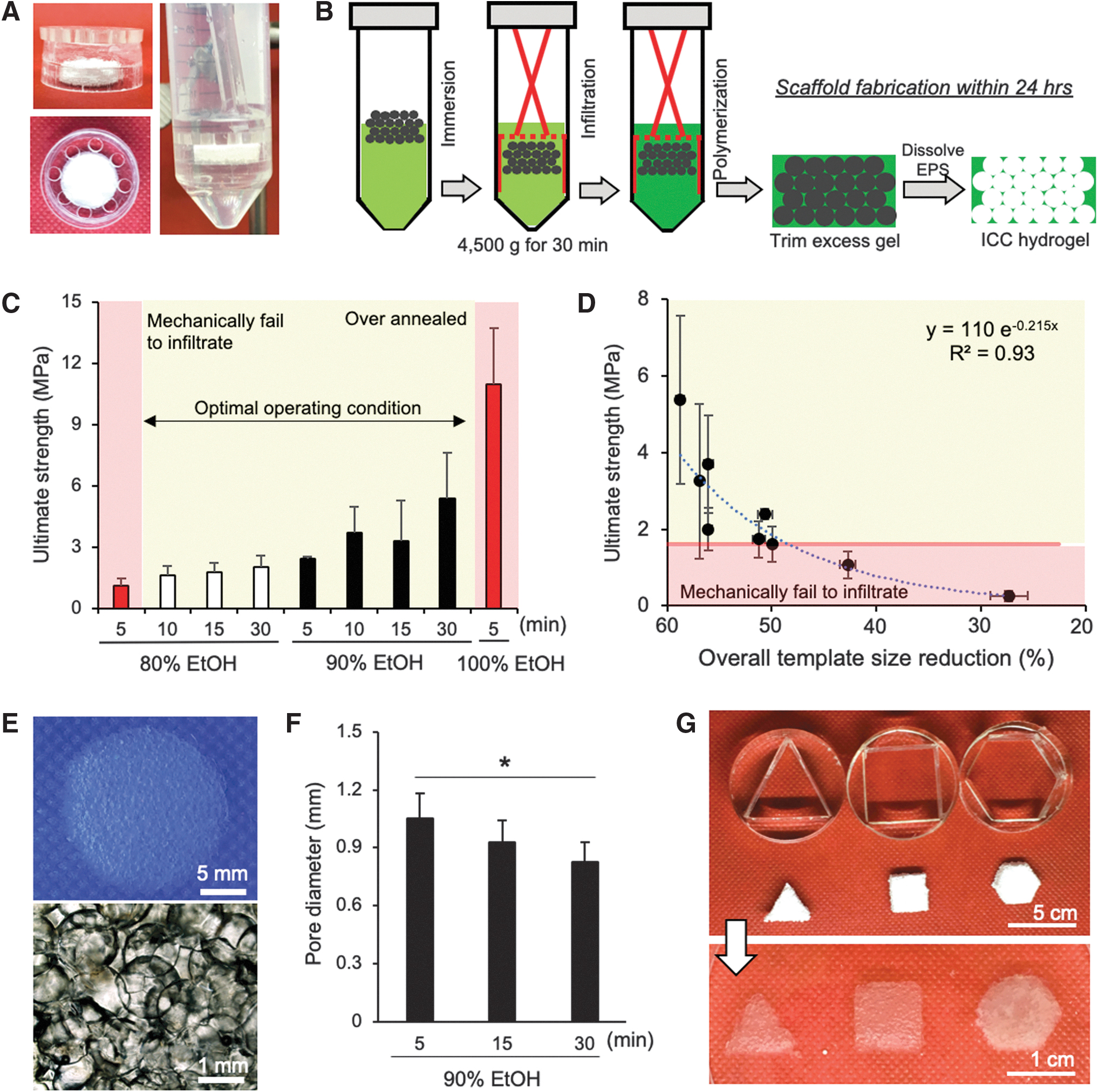
Preparation of EPS-templated ICC polyacrylamide hydrogel scaffolds.
EPS-templated ICC hydrogel scaffolds exhibited spherical pore cavities and optical transparency comparable to ICC hydrogel scaffolds prepared with glass beads of comparable sizes (Fig. 3E). Scaffold pore size was found to be tunable based on the duration of EPS-templated processing. For example, pore diameters were 1.05 ± 0.2 mm, 9.27 ± 0.18 mm, and 8.23 ± 0.15 mm for ICC hydrogel scaffolds fabricated from templates made with 90% ethanol for 5-, 15-, and 30-min operation, respectively (Fig. 3F). The EPS-templated ICC hydrogel scaffold fabrication strategy is flexible and rapid, enabling creation of different prototypes demonstrated by triangular, square, and hexagonal shapes of ICC hydrogel scaffolds by changing the initial mold design (Fig. 3G).
Preparation of bioactive ICC hydrogel scaffolds through type I collagen integration in bulk hydrogel
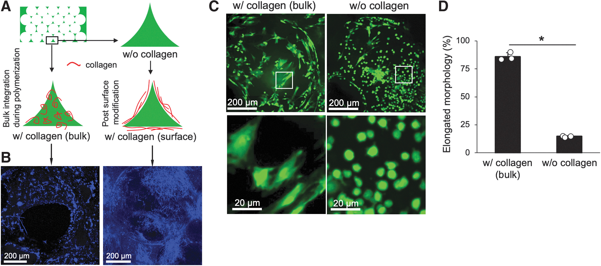
Polyacrylamide hydrogel is not conductive for cell adhesion and requires immobilization of collagen fibers or arginyl glycylaspartic acid peptides to become bioactive. However, reagents for the conjugate chemistry are expensive and this procedure is laborious. Instead, we hypothesized that inclusion of collagen fibers directly in the precursor solution would simplify fabrication of bioactive ICC hydrogel scaffolds. To test this hypothesis, we fabricated ICC hydrogel scaffolds with precursor solution containing type I collagen fibers at a final concentration of 2 mg/mL and compared them to scaffolds prepared by attaching collagen fibers using Sulfo-SANPAH conjugate chemistry (Fig. 4A). Collagen fibers in the hydrogel matrix were visualized through second-harmonic generation in a multiphoton microscope. As expected, the conventional method immobilized collagen exclusively on the pore surface, whereas collagen dispersed in the precursor solution appeared both on the surface and within the bulk hydrogel. Collagen fibers exhibited different morphologies: stretched thin fibers when surface conjugated and coiled bundles when mixed directly in the polymer precursor solution (Fig. 4B and Supplementary Movies S2, S3). Collagen fibers are known to undergo a reversible conformational change depending on the surrounding pH. 41 For example, at low pH, collagen fibers adopt an elongated morphology, whereas at high pH, collagen fibers coil. Our results indicate that the alkaline reaction pH of acrylamide polymerization by addition of ammonium persulfate and TEMED could direct collagen fibers to adopt a more coiled morphology. 42
Hybridization of collagen and polyacrylamide to achieve bioactive hydrogel scaffolds.
We next examined whether the collagen embedded within the polyacrylamide of an ICC hydrogel scaffold could support stromal cell adhesion. Primary BMSCs retrieved from the femurs of eGFP mice were used to easily visualize stromal cells seeded on the scaffold. Fluorescence images taken 3 days after seeding showed that most stromal cells adhered and displayed an elongated, spread morphology on scaffolds with collagen, whereas the stromal cells that adhered on control collagen-free hydrogel scaffolds had a round morphology, indicating poor surface adhesion (Fig. 4C and Supplementary Movies S4, S5). Quantitative analysis of stromal cell morphology revealed 85% had an elongated morphology on collagen scaffolds, whereas only 15% of cells adhered on collagen-free scaffolds (Fig. 4D). These results indicate that direct introduction of collagen fibers in precursor solution promotes stromal cell adhesion comparable to conventional conjugate chemistry.
In vitro modeling of bone marrow tissue microenvironments
For an in vitro functional demonstration, we applied EPS-templated ICC hydrogel scaffolds to model the bone marrow tissue microenvironment focusing on hematopoietic cell activity as a function of BMSCs. We hypothesized that BMSCs would secrete different soluble factors depending on their adhesion pattern on ICC hydrogel scaffolds, which in turn direct hematopoietic colony-forming activity.43–45 A critical issue in characterizing hematopoietic colonies in 3D scaffolds is deciphering stroma from hematopoietic cells and their progenitors using traditional microscopy techniques. To overcome this challenge, we employed cells from eGFP and DsRed reporter mice that facilitate fluorescent monitoring of stromal and hematopoietic cell activity, while preserving the intrinsic phenotype of both cell types (Fig. 5A). We first examined the secretion profile of BMSCs focusing on key factors involved in bone remodeling: IL-6, osteoprotegerin (OPG), and receptor activator of nuclear factor kappa-B ligand (RANKL). Increased RANKL and IL-6 stimulate osteoblasts and osteoclasts to promote bone remodeling activity, whereas OPG secretion suppresses remodeling activity. ELISA data showed that BMSCs secreted significantly more IL-6 and RANKL, and notably lower OPG on collagen-free ICC hydrogel scaffolds than collagen-embedded hydrogel scaffolds (Fig. 5B–D). These results indicate that BMSCs seeded on hydrogel matrix with embedded collagen suppress bone remodeling activity.

In vitro demonstration of functional bone marrow tissue model using EPS-templated ICC hydrogel scaffolds.
We next evaluated if the different soluble environments could direct hematopoietic colony-forming activity. After 1-week culture of eGFP BMSCs on collagen-coated and control ICC hydrogel scaffolds, we introduced freshly harvested femoral bone marrow cells retrieved from DsRed mice. Before loading, bone marrow cells were dispersed in methylcellulose medium without hematopoietic cytokines. Subsequent hematopoietic colony development was monitored with fluorescence microscopy over a 16-day period. DsRed fluorescent colonies gradually emerged in and around BMSC-seeded scaffolds (Fig. 5E). This unconventional 3D colony-forming assay makes it difficult to distinguish specific types of hematopoietic colonies. Instead, we focused on the number of emerging colonies by taking advantage of the endogenous fluorescence. In stromal cell-free control cultures without essential hematopoietic cytokines, no hematopoietic colonies emerged, indicating stromal cells play a vital role in supporting the survival and function of hematopoietic cells ex vivo. Quantitative monitoring of colony number over the experimental period showed an increased number of hematopoietic colonies when cocultured with BMSCs seeded on scaffolds with embedded collagen, whereas the number of colonies stabilized after 1 week of coculture with BMSCs on collagen-free scaffolds (Fig. 5F). 3D confocal imaging at the end of the study revealed the spatial distribution of BMSCs and hematopoietic cells, with some in physical contact with each other (Fig. 5G, Supplementary Movie S6). Collectively, these results indicate that the different soluble milieus produced by BMSCs with and without collagen stimulation can affect hematopoietic colony-forming activity. This also demonstrated that EPS-templated ICC hydrogel scaffolds are capable of identifying dynamic functional interactions between hematopoietic and stromal cells ex vivo.
Formation of lymphoid mimicking tissue microenvironments in vivo
Subdermal implantation of ICC hydrogel scaffolds has been shown to direct unique foreign body responses leading to a richly vascularized stromal tissue with prolonged hematopoietic cell activity. We evaluated if EPS-templated ICC hydrogel scaffolds induced comparable tissue development to hydrogel scaffolds made with glass beads after subdermal implantation in FVB/NJ mice (Fig. 6A). Gross observation after 4 weeks of implantation revealed EPS and glass-templated ICC hydrogel scaffolds showed similar tissue development and attraction of large blood vessels (Fig. 6B). H&E staining confirmed complete interscaffold tissue development and a comparable pattern of spatial tissue microenvironments directed by the ICC geometry. In general, fibrotic tissue is formed within the central region of each pore, whereas hematopoietic cells are localized near the pore surface (Fig. 6C).
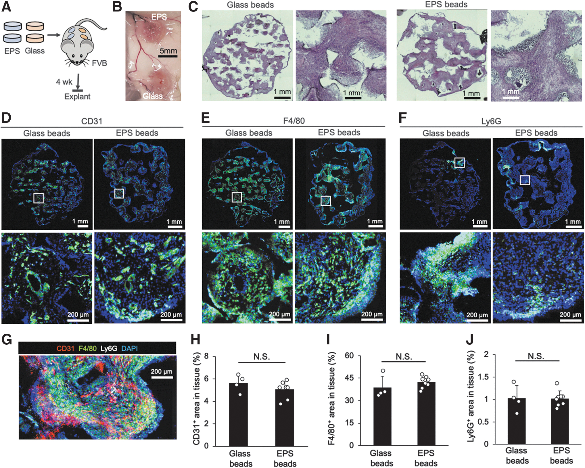
Comparison of tissue microenvironment formed in ICC hydrogel scaffolds made from glass and EPS beads after subdermal implantation.
We further characterized the vascular network and attraction of macrophages and neutrophils by immunohistostaining for CD31, F4/80, and Ly6G, respectively. Tissues developed in EPS-templated ICC hydrogel scaffolds contained an extensive vascular network similar to glass-templated ICC scaffolds (Fig. 6D). Nondegradable synthetic hydrogels direct macrophage response throughout the entire scaffolds (Fig. 6E). Distinct from the widespread vasculature and macrophages, neutrophils were highly localized in two to three sites of each tissue section (Fig. 6F). Overlaid CD31, F4/80, and Ly6G, and nuclei staining substantiate the spatial distribution of vascular, stromal, and immune cells in an ICC pore cavity (Fig. 6G). Quantitative analysis of CD31-, F4/80-, and Ly6G-positive areas confirmed comparable tissue development between EPS- and glass-templated ICC hydrogel scaffolds (Fig. 6H, I). Overall, our results indicate that EPS-templated ICC hydrogel scaffolds retain comparable ability to induce characteristic vascularized and hematopoietic cell attracting lymphoid tissue-mimicking microenvironments to glass-templated ICC hydrogel scaffolds.
Discussion
ICC hydrogel scaffolds have been emerged as a new class of biomaterial and several fabrication methods have been developed. While these methods are conceptually simple, it requires multiple steps and laborious handling of delicate materials. Oftentimes, initial template beads are expensive and dissolving the beads requires highly hazardous chemicals. Under these motivations, we have developed a simple and cost-effective method to fabricate bioactive ICC hydrogel scaffolds using EPS beads. EPS beads consist of more than 95% void space and exhibit intermediate mechanical and structural properties between solid PS and air bubbles, two representative template materials. EPS beads leverage advantages of each material for ICC hydrogel scaffold synthesis: easier to handle than air bubbles with higher mechanical stability and faster to dissolve than solid PS beads due to significant void fraction. In addition, direct inclusion of type I collagen in the precursor solution has greatly simplified the manufacturing procedure for bioactive hydrogel scaffolds. These features together represent great potential to reduce the technical and cost barriers in fabricating bioactive ICC hydrogel scaffolds.
Although we demonstrated the feasibility of EPS-based ICC hydrogel scaffold fabrication, there are several things to be improved further. First, the dimension of EPS beads is not ideal for modeling trabecular bone cavities in hematopoietic bone marrow and other lymphoid tissues. This issue can be resolved by reducing the size of expandable PS resins that are being processed into EPS. Second, the packing of EPS beads is suboptimal as the low density of EPS beads makes it difficult to apply centrifugation and agitation-assisted packing methods. This can be improved by benchmarking the previous methods in building colloidal crystal templates at the air-water interface.46,47 Finally, the coiled morphology of in situ collagen integration is also suboptimal to maximize the function of collagen for stromal cell adhesion. Collagen fibers undergo pH-dependent conformational change; collagen fibers are relaxed in an acidic environment, whereas they are coiled configuration in basic solutions. 48 Alkaline precursor solution likely causes the collagen fibers to undergo a conformational change. By adjusting the pH in the precursor solution, more aligned collagen fibers and better coverage of hydrogel matrix may be achieved.
The biological significance of EPS-based ICC hydrogel scaffolds was demonstrated across multiple aspects of bone marrow tissue models. First, EPS-based ICC scaffolds with collagen fibers supported adhesion of BMSCs and substantiated different secretory profiles of OPG and RANKL, which play an important role in regulating the bone remodeling activity and associated hematopoiesis.49,50 Second, BMSCs on EPS-based ICC scaffolds supported expansion and differentiation of hematopoietic cells as a function of collagen matrix; collagen-embedded ICC hydrogel scaffolds showed a significantly higher number of colonies than collagen-free control. Finally, the in vivo implantation study demonstrated that EPS-based ICC scaffolds formed comparable levels of interscaffold tissue development to conventional glass bead-based ICC hydrogel scaffolds. This functional validation of EPS-based ICC scaffolds in bone marrow tissue modeling gave high confidence for their use in other lymphoid tissue engineering applications.
Besides lymphoid tissue modeling, ICC hydrogel scaffolds have been previously demonstrated to be advantageous in other tissue engineering applications. For example, ICC geometry made with cell-repulsive hydrogel matrix-directed formation of multicellular hepatocyte and stem cell spheroids with narrow size distribution.51,52 Hepatic and embryonic spheroids demonstrated in vivo relevant cellular processes in toxic nanoparticle exposure 37 and differentiation into lineage cells, 53 respectively. ICC hydrogel scaffolds have demonstrated additional in vitro success modeling cartilage, 54 bone, 35 and neuronal 55 tissues, taking advantage of their unique geometry with tailored biomaterials in each target application. ICC hydrogel scaffolds have also demonstrated exceptional capability for rapid vascularization as a function of pore size. 19 This capability has been applied for cardiac tissue engineering and wound healing medical devices. The presented EPS-templated ICC hydrogel scaffold fabrication will leverage these abilities to the broader academic and industrial communities.
ICC hydrogel scaffolds with controlled isotropic spherical pore arrays of hydrogel matrix have emerged as an important class of biomaterials for tissue engineering. Originally, in the context of lymphoid tissue engineering, ICC scaffolds have many promising applications both in vitro and in vivo. An important next step in the advancement of ICC hydrogel scaffolds is improved fabrication techniques that facilitate effective scale-up. Currently, there are no commercial ICC hydrogel scaffolds. This work addressed many of these challenges by using EPS and directly mixing collagen fibers within the synthetic hydrogel matrix. ICC hydrogel scaffolds have already demonstrated many promising applications in lymphoid tissue engineering and beyond. We envision that the presented EPS bead-based scaffold fabrication will greatly facilitate broad and practical applications of ICC hydrogel scaffolds.
Footnotes
Acknowledgments
We thank Dr. Shelly Peyton for access to her confocal microscope. We also thank the UMass-Amherst Animal facility for assistance with mouse experiments, and the Animal Imaging Core, and Light Microscopy Core for access to their IVIS Spectrum CT and multiphoton microscope, respectively.
Disclosure Statement
No competing financial interests exist.
Funding Information
This work was supported by the National Cancer Institute (R00CA163671 and R01CA237171) and National Science Foundation Research Traineeship (1545399).
References
Supplementary Material
Please find the following supplemental material available below.
For Open Access articles published under a Creative Commons License, all supplemental material carries the same license as the article it is associated with.
For non-Open Access articles published, all supplemental material carries a non-exclusive license, and permission requests for re-use of supplemental material or any part of supplemental material shall be sent directly to the copyright owner as specified in the copyright notice associated with the article.