
Abstract
The objective of this study is to investigate the utility of gelatin hydrogel-fragmented fibers (GHFF) as a material to suppress the shrinkage of cell sheets, which often happens upon detaching from a culture plate. The GHFF were fabricated by cutting gelatin hydrogel nonwoven fabrics. MC3T3-E1 cells were simply mixed with different amounts of GHFF, followed by culturing to formulate the cell sheet homogeneously incorporating GHFF. When detached from the culture plate, the cell sheet formulated without GHFF shrunk while the area became about 23% of the original one before detachment. On the contrary, the cell sheet formulated with GHFF hardly shrunk. The lactate/glucose ratio of a metabolic activity was significantly lower and the adenosine triphosphate (ATP) production was higher for the cell sheet formulated with the GHFF than that obtained without the GHFF. An osteogenic activity was high for the cell sheet formulated with the GHFF compared with that obtained without the GHFF. The GHFF addition was a simple and promising method to fabricate active cell sheets without size change.
Impact Statement
This study introduces the utility of gelatin hydrogel-fragmented fibers (GHFF) for cell sheet engineering. Upon detaching from the culture plate, the cell sheet formulated without GHFF shrunk, while the area became about 23% of the original one before detachment. On the contrary, the cell sheet formulated with GHFF hardly shrunk. The GHFF allowed cell sheets to enhance the metabolic and osteogenic activities. The GHFF addition was a simple and promising method to fabricate active cell sheets without size change.
Introduction
Cell sheet engineering has been extensively used for cell-based therapies, drug discovery, regenerative medicine, and tissue engineering.1–6 Generally, cell sheets are prepared in a culture dish. The confluent cells are detached from the surface of culture dish without using enzymatic treatment, and consequently an intact contiguous cell sheet is harvested.
The cell sheets harvested maintain the cell–cell junction, the cell receptors, and deposited ECM important for cell functions, but the shrinkage and reduction of sheets observed in detaching are often of problem. 7 Since cell sheets shrinkage is often seen, it is practically difficult to give a constant size to the sheets harvested from the culture dish. Moreover, it is reported that the shrinkage extent depends on the cell type, for example, oral mucosal epithelial cell and hepatocyte cell sheets showed 508 and 16% 9 decrease of the original size, respectively. For hepatocytes 9 and chondrocytes, 10 the shrinkage causes the change not only in the size of cell sheets, but also in the biological function, due to their cytoskeleton reorganization. 7 Several attempts have been carried out to suppress a decrease in the size of cell sheets, such as the usage of a gelatin manipulator, 11 a cell culture insert,12,13 and a medium mixture. 14 However, it is still necessary to further develop technologies to retain the shape of cell sheet.
When the thickness of cell sheets becomes large, the cells present inside cell sheets often weaken or die due to the lack of oxygen and nutrients supplies. 15 Several strategies are reported to improve the cell viability, such as the combination of vascular endothelial cells and the use of porous membrane.16–18 In addition, it is demonstrated that the incorporation of gelatin hydrogel microspheres (GHM) between cardiovascular cell sheets enables to improve the cell viability and functions. 19
To tackle the problem of cell sheet shrinkage, we try to take advantage of gelatin hydrogel-fragmented fibers (GHFF). In this study, the cell sheet homogeneously incorporating GHFF was prepared to evaluate the size change of cell sheet comparing with the GHFF-free cell sheet. We examine the shrinkage extent, the glucose consumption, the lactate production, the ATP production, and the osteogenic activity of cell sheets formulated with the GHFF.
Materials and Methods
Preparation of gelatin hydrogel-fragmented fibers
GHFF were prepared by simply cutting gelatin hydrogel nonwoven fabrics (GHNF) with a mixer mill. The GHNF were prepared by the solution blow spinning method previously reported. 18 In brief, an aqueous solution of 37.5 wt% gelatin (isoelectric point 5.0; Nitta Gelatin, Inc., Osaka, Japan) was pumped through the nozzle. The spinning was performed at an air pressure of 0.375 MPa and collected in the state of fiber as the GHNF at the collector. Then, the GHNF prepared were air-dried at room temperature. The noncrosslinked and dried GHNF were treated in a vacuum oven at 140°C and 1 × 10⁻⁵ MPa for the dehydrothermal crosslinking of gelatin for 48 h. The crosslinked GHNF were swelled in water and cut with the mixer mill (KMZ-0800; Koizumi, Osaka, Japan) for the processing time of 120 s. The suspension was collected and freeze-dried to obtain GHFF. The pictures of GHFF in the dried and swollen states were taken with a light microscope (BZ-X710; Keyence, Osaka, Japan). The diameter and length of 100 fibers were measured using a computer program (PhotoRuler, Hyogo, Japan), and the mean diameter and length were calculated.
Preparation of MC3T3-E1 cell sheets formulated without or with GHFF
MC3T3-E1 cells (1 × 10⁵) were seeded into each well of a 24-well multiple well plate (24-well plate; Corning, NY) in the minimal essential medium alpha (MEMα; Thermo Scientific, Waltham), supplemented with 10 vol% fetal bovine serum (FBS; Thermo Scientific), 1 vol% penicillin and streptomycin (PS; Nacalai Tesque, Kyoto, Japan), and 100 mM of ascorbic acid phosphate (AA; Fujifilm-Wako, Osaka, Japan). The AA was used to promote the cell sheet formation. 20 After the GHFF dried was swollen and suspended at 1 mg/mL by the medium, the GHFF suspension was added into the cell-preseeded plate. Then, the supernatant of medium in the culture plate was carefully removed to adjust at 0.5 mL. The culture plate was shaken with Vortex (Vortex-Genie 2; Scientific Industries) to disperse GHFF uniformly on the culture plate. The cell culture was performed for 5 days at 37°C in a humidified incubator at 5% CO2–95% air atmosphere. The medium containing AA was exchanged every 2 days. After the culture, the distribution of GHFF in the cell sheets on the well before detachment was observed with a light microscope (BZ-X710; Keyence, Osaka, Japan). For the coverage ratio of GHFF to each well, images were analyzed using the ImageJ software (National Institutes of Health, MD). The confluent cells were detached to obtain a monolayer sheet from each well plate by flushing several times with the culture medium using a 1000-μL micropipette, transferred to a 1.5-mL-microtube, and observed with a digital camera.
Sheet shrinkage assay
MC3T3-E1 cell sheets formulated without or with various amounts of GHFF were prepared in wells of 24-well plates. Similarly, the confluent cells were detached from each well by flushing. After spreading the cell sheet by aspirating the medium, the area of cell sheets was measured using the ImageJ software to determine the shrinkage from the full size. 14 The full size of cell sheet was defined as the area of one well in 24-well plate (1.9 cm2). Experiments were performed independently three times unless mentioned otherwise.
The thickness of cell sheets was measured described previously. 9 The cell sheets were prepared in a temperature-responsive culture plate (UpCell; CellSeed, Tokyo, Japan). To obtain an extended sheet form (Ex-form), before the culture temperature decreased, a support membrane (CellShifter; CellSeed) was covered over the cell sheet. Culture plates were placed at room temperature for 30 min, and the cell sheet with the support membrane was obtained without shrinkage. To obtain a shrinking sheet form (Sh-form), the culture plate was placed at room temperature. The thickness of Ex- and Sh-forms was assessed by measuring randomly selected 20 points per one cell sheet. Both types of cell sheet forms were processed for histological analysis.
Histological analysis
Cell sheets were fixed with 4 wt% paraformaldehyde aqueous solution. Specimens were embedded in an optimal cutting temperature compound (Sakura Finetek Japan, Tokyo, Japan) and frozen. The frozen samples were sectioned using a cryotome (CM3050S; Leica Microsystems, Wetzlar, Germany), and then stained with hematoxylin and eosin. Section specimens prepared were examined under microscope (BZ-X710; Keyence, Tokyo, Japan).
Actin staining
Cell sheets detached from the culture plate were fixed with 4 wt% paraformaldehyde aqueous solution and stained with Alexa Fluor 488 phalloidin (Thermo Scientific) to observe the F-actin and DAPI (Thermo Scientific) of a nuclear staining dye according to the manufacturer's instructions. In addition, the sections prepared described above were also stained with Alexa Fluor 488 phalloidin and DAPI. The whole and section specimens of cell sheets prepared were examined under confocal laser scanning microscope (FV1000; Olympus, Tokyo, Japan).
DNA assay
The cell number of cell sheets formulated without or with GHFF (0.4 mg) was determined by fluorometric quantification of cell DNA. In brief, cell sheets were washed with phosphate-buffered saline (PBS) and lysed in a buffer solution (pH 7.4) containing sodium dodecyl sulfate (0.8 mg/mL), and 30 mM saline–sodium citrate (SSC) at 37°C for 1 h with occasional sonication. The cell lysate (100 μL) was mixed with a dye solution (100 μL) (composition: 30 mM SSC and Hoechst 33258 dye [1 μg/mL]), and the fluorescence intensity of the mixed solution was measured with a fluorescence spectrometer (Spectra Max i3; Molecular Devices, CA). A calibration curve between DNA and cell number was prepared by using cell suspensions with different cell densities. The DNA assay was performed three times independently.
Measurement of glucose consumption, lactate production, and lactate/glucose ratio of cell sheets formulated without or with GHFF
Cell sheets harvested from the culture plate were transferred onto a new 24-well plate. After spreading the cell sheet without any folds by aspirating the medium, the plate was incubated for 60 min at 37°C to allow the cell sheets to adhere to the surface of plate. Cell sheets were recultured for 24 h in the 24-well plate with 2 mL of medium. The amount of glucose consumed by a cell sheet was determined by measuring the change in glucose concentration of the culture medium using Glucose C2 kit (Fujifilm-Wako). The amount of lactate produced by a cell sheet was determined with a Glycolysis Cell-Based Assay Kit (Cayman Chemical). The lactate/glucose ratio was calculated as a measurement of aerobic condition. 21 To calculate the lactate/glucose ratio, the amount of glucose was determined by dividing the weight (mg) by the molecular weight (180 g/mole). Experiments were performed independently six times.
Measurement of adenosine triphosphate production of cell sheets formulated without or with GHFF
Similarly, cell sheets were recultured for 24 h in the 24-well plate with 2 mL of medium. The amount of ATP production by the cell sheet was determined by the ATP Kit for Aggregate (Toyo-b-net, Tokyo, Japan) according to the manufacturer's instructions. Experiments were performed independently three times.
Osteogenesis culture
The confluent cells formulated without or with GHFF were detached as a monolayer sheet from the 24-well plate. After spreading the cell sheet by aspirating the medium, the plate was incubated for 60 min at 37°C to allow the cell sheet to adhere to the surface of plate. The harvested cell sheets were recultured on a new culture plate in MEMα, supplemented with 10 vol% FBS, 1 vol% PS, 10 mM β-glycerophosphate (Nacarai Tesque, Kyoto, Japan), 50 μg/mL AA (Fujifilm-Wako), 10 nM dexamethasone (Nacarai Tesque), and 100 ng/mL Bone morphogenetic protein-2 (BMP-2) and cultured at 37°C in a humidified incubator at 5% CO2–95% air atmosphere. Cell sheets were recultured for 3, 5, and 7 days with 0.5 mL of the medium. The medium was exchanged every 2 days.
Alizarin red S staining
In alizarin red S (ARS; Nacarai Tesque) staining, cell sheets cultured in 24-well plate were washed with PBS twice and fixed 4 wt% paraformaldehyde aqueous solution at 4°C for 20 min. The cell sheets were then washed twice with PBS, and 500 μL of 1% ARS (pH = 6.4) was added to each well. Each cell sheet was incubated at room temperature for 5 min, washed with PBS, and then imaged using a light microscope (BX-X710; Keyence, Osaka, Japan).
Calcium contents and alkaline phosphatase activity assay
Cell sheets were washed with PBS twice, treated with 0.08% sodium lauryl sulfate (SDS) solution, and sonicated in 1 mL of SDS solution. After sonication, the cell lysate was centrifuged and the supernatant was collected for further measurement. Calcium concentration of supernatant was measured using a Calcium E-Test Wako kit (Fujifilm-Wako). Alkaline phosphatase (ALP) activity in supernatant was measured using a LabAssay ALP Kit (Fujifilm-Wako). The results were normalized relative to corresponding the cell number evaluated with DNA assay.
Statistical analysis
All the data were statistically analyzed and expressed as mean ± standard error of the mean. The data were analyzed by Student's t-test to determine the statistically significant difference, while the significance was accepted at p < 0.05.
Results
Characterization of GHFF
Figure 1 shows the microscopic pictures of GHFF in the dried and swollen conditions. The GHFF maintained a nonwoven fabric of fiber shape even in the swollen condition, although both the diameter and length of fibers became large. The diameters of GHFF in the dried and swollen conditions were 25 ± 5.6 and 54 ± 9.3 mm, respectively. The lengths of GHFF in the dried and swollen conditions were 207 ± 98 and 255 ± 111 mm. The GHFF were homogeneous in the diameter of fibers, whereas a distribution in fibers length was observed.
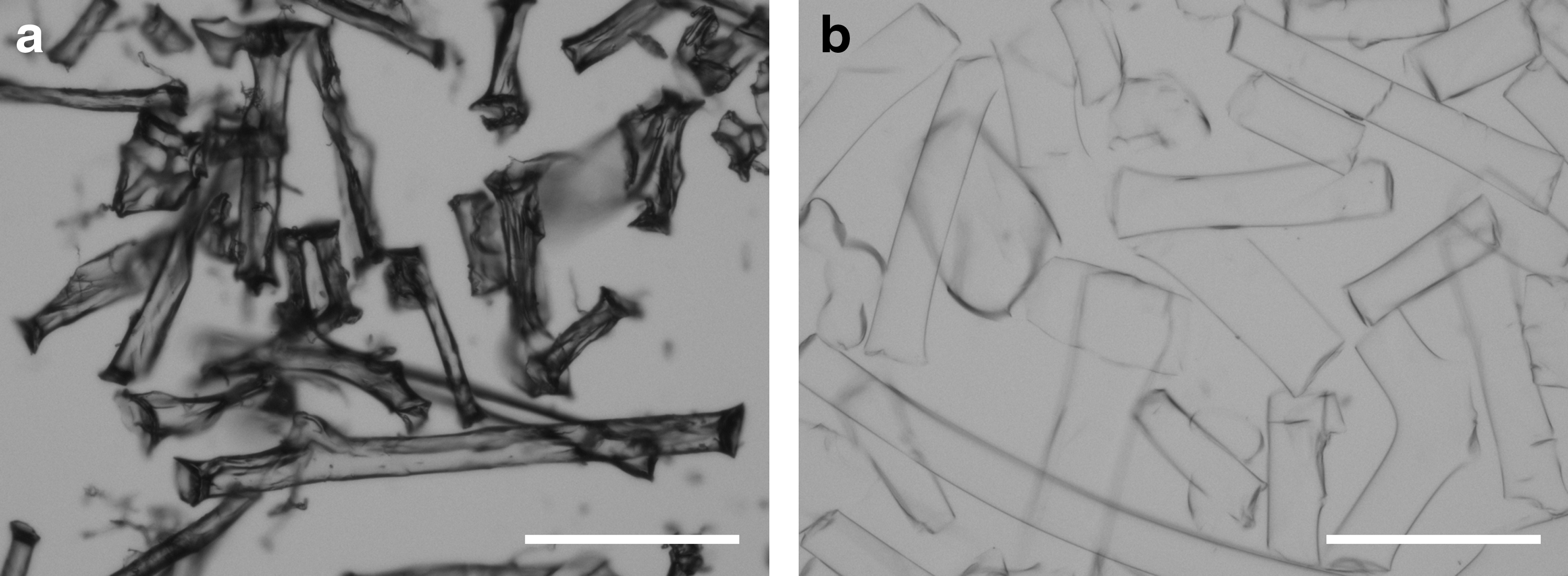
Light microscopic pictures of GHFF in the dried
Cell sheets shrinkage
Figure 2A shows the pictures of GHFF distribution in cell sheets on well of 24-well plate. GHFF were homogeneously incorporated in cell sheets. As the amount of GHFF became large, the coverage ratio of GHFF to cell sheet increased (Fig. 2B). Figure 3A shows the picture of shrunk cell sheets formulated without or with GHFF in PBS. The cell sheet formulated with GHFF kept the original size even in PBS and the shape was maintained. Figure 3B shows the pictures of shrunk cell sheet formulated without or with the addition of various GHFF amounts on the culture plate. Figure 3C shows the area change of cell sheets formulated with or without GHFF. When the amount of GHFF added was 0.2 mg or more, the area of cell sheet formulated with GHFF was significantly larger than that without GHFF. As the amount of GHFF became large, the area of the cell sheet increased.
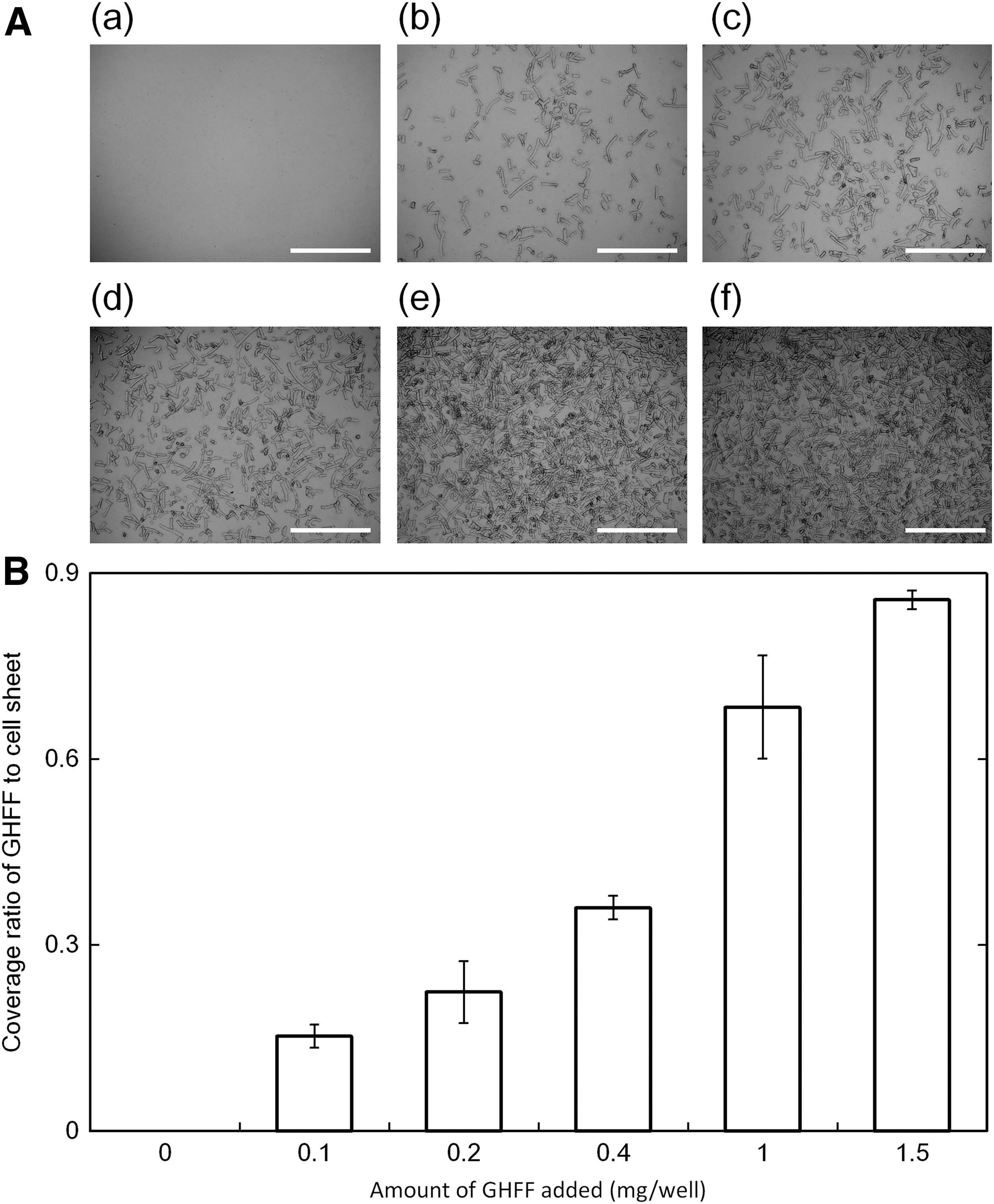

Figure 4A shows the light microscopic pictures of cross section of cell sheets formulated without or with GHFF (0.4 mg). Figure 4B shows the thickness of cell sheet formulated without or with GHFF. For the Ex-form, the thickness of cell sheets was same, irrespective of the GHFF presence. On the contrary, for the Sh-form, the thickness of cell sheets increased. The thickness of cell sheets formulated with GHFF was significantly larger than that without GHFF.

) Sh-form. Ex-form, extended sheet form; Sh-form, shrinking sheet form. Color images are available online.
Figure 5 shows the confocal macroscopic pictures of top view and cross section of Sh-form cell sheets formulated without or with GHFF. The density of F-actin labeled with phalloidin in the cell sheet formulated with GHFF was lower than that without GHFF.
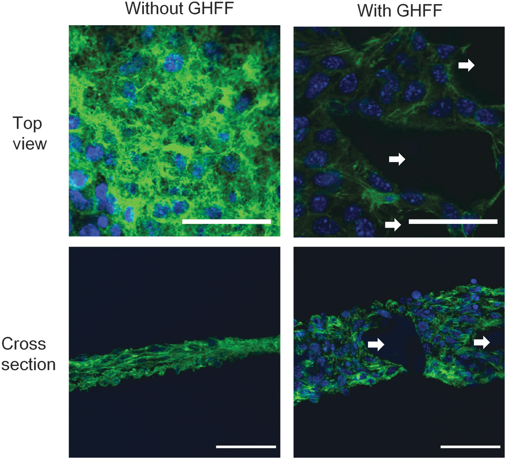
Confocal microscopic pictures of cell sheet formulated without or with GHFF detached from dish. Cell sheets were stained by Alexa Fluor 488 phalloidin to observe the F-actin (green) and DAPI for nucleuses (blue). The white arrow indicates the GHFF. The scale bar indicates 50 μm. Color images are available online.
Cell number of cell sheet formulated without or with GHFF
Figure 6 shows the cell number of cell sheets without or with GHFF (0.4 mg). There was no significant difference in the cell number between the cell sheets formulated without and with GHFF.
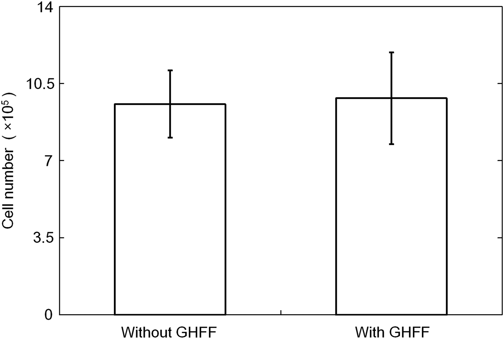
Cell number of cell sheet formulated without or with GHFF (0.4 mg).
Cell activity of cell sheets formulated without or with GHFF
Figure 7 shows the glucose consumption, the lactate/glucose ratio, and ATP production of cell sheet without or with the GHFF 24 h after incubation in the reculture. A significantly higher glucose consumption, lower lactate/glucose ratio, and higher ATP production were observed for cell sheets formulated with GHFF than those without GHFF.
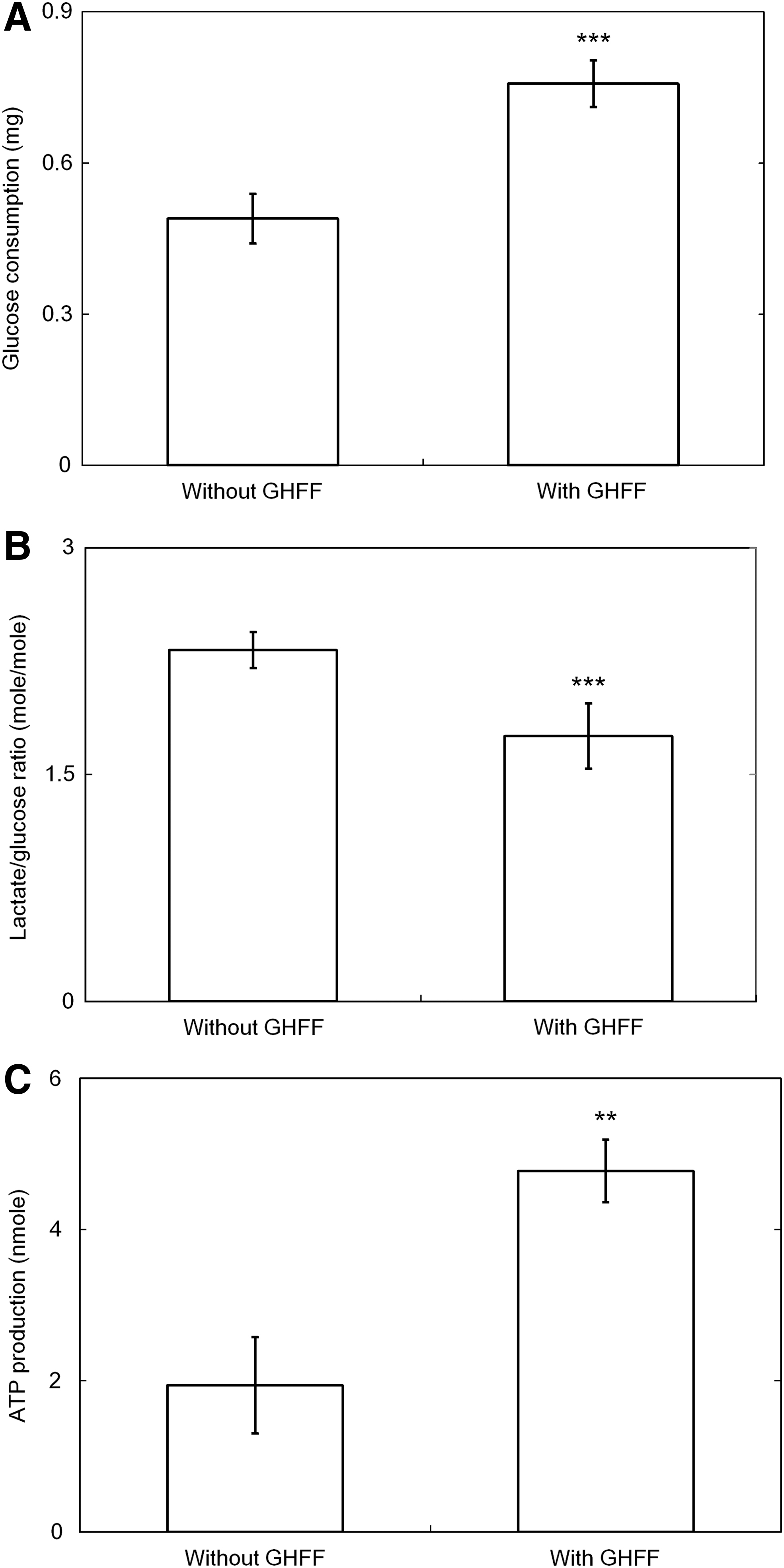
Cell activity of cell sheet formulated without or with GHFF (0.4 mg) 24 h after reculture:
Osteogenic activity of cell sheets formulated without or with GHFF
Figure 8A shows the light macroscopic pictures of ARS staining of cell sheets without and with GHFF (0.4 mg) 7 days after reculture. The mineralization extent of cell sheet with GHFF was larger than that without GHFF. Figures 8B and C show the calcium contents and the ALP production of cell sheet without or with the GHFF. A significantly higher calcium contents and ALP production were observed for cell sheets with GHFF than those without GHFF.
Osteogenic activity of cell sheets formulated without or with GHFF (0.4 mg): ) with GHFF. *p < 0.05, **p < 0.01; significant difference between the two groups. ALP, alkaline phosphatase. Color images are available online.
Discussion
GHFF were fabricated by simply and mechanically cutting GHNF swollen with a mixer mill. Even when swollen in PBS, the GHFF showed a mechanical strength strong enough to maintain the shape by vigorously flushing with micropipette. The present study clearly demonstrates that the incorporation of GHFF effectively suppressed the shrinkage of cell sheets. Regarding the morphology of cell sheets after the detachment process, both the area and thickness of cell sheets formulated with GHFF were larger than those without GHFF (Figs. 3 and 4). Several approaches have been reported to suppress the shrinkage of cell sheets.11–14 However, little has been tried to add a material to the cell sheet for this purpose.
The GHFF were homogeneously incorporated in the cell sheet layer (Fig. 2A). Several fragmented fibers have been reported for cell culture.22–24 Cell aggregates incorporating poly(lactic acid) (PLA)-coated polydopamines are prepared to improve the cell adhesion ability. 22 PLA-coated biomineral has been reported to enhance the osteogenic differentiation of adipose-derived stem cells. 23 Gelatin hydrogel is a biodegradable biomaterial of good cytocompatibility as a cell culture substrate. As the water content of gelatin hydrogels is >95%, 25 the culture medium and body fluids containing oxygen and nutrients can be easily penetrated through the water phase in gelatin hydrogels.26–28 In addition, gelatin has a cell adhesion ability. The cell activity of cell aggregates is improved by the incorporation of the GHM. 29 The ratio of lactic acid production to glucose consumption is a general measure of aerobic metabolism. 25 The lower the ratio the higher the aerobic metabolism of cells. The glucose consumption of cell sheets was increased by the GHFF incorporation (Fig. 7A). In addition, a decrease in the lactate/glucose ratio and an increase in the ATP production suggest that the cell condition was more aerobic (Fig. 7B and C). It is reported that the diffusion limits of nutrients and oxygen are reduced by creating a structure of blood vessels or inserting GHM between layered cell sheets.19,30 Following the implantation, blood vessels were formed between the layers when endothelial cells are inserted between cardiac cell sheet layers. Incorporation of GHM between the cell sheet layers also allowed to form blood vessels. In the GHFF-used method, it is likely that GHFF function as a reductant of oxygen diffusion limits, resulting in an improved cell activity of cell sheets incorporating GHFF.
The cell sheets formulated with GHFF was thicker than that without GHFF, although the thickness of both the sheets before detachment was similar (Fig. 4B). There was no significant difference in the cell number evaluated by the DNA assay between the cell sheets formulated with and without GHFF (Fig. 6). Since for the cell sheets with GHFF, the area and thickness increased, the cell density of cell sheet formulated with GHFF was lower than that without GHFF considering the volume of GHFF added. It is reported that when harvested, cell sheets without a supporting material usually shrink and superpose by themselves during the detachment processes due to their cytoskeleton reorganization. 7 Stress fibers function as an important component of cytoskeleton, which acts as a structural link between cells and their ECM. 31 In this study, the GHFF incorporation did not change the size of cell sheets. This is because there is a limitation of reorganization of cytoskeleton as shown by the stretched shape of stress fibers in the cell (Fig. 5). Without GHFF, cell sheets were spontaneously detached, leading to the shrinkage of cell sheets due to stress fiber contraction (Fig. 5). The change in stress fiber formation would not only cause the shrinkage of cell sheets but also influence the cell function. Changing the cell shape through cytoskeletal reorganization stimulates the pathway of extracellular and intracellular signaling, leading to the regulation of cellular functions, such as the cell proliferation, migration, and differentiation. 32 Wongin et al. reported that the cells in the cell sheet without shrinkage show abundant stress fibers. However, the cells in the cell sheet shrunken suppress the stress fiber formation. 10 The formation of stress fibers are associated with cell differentiation, and also exhibited an increased osteoblastic differentiation. 31 In this study, we used MC3T3-E1 cells of a mouse osteoblastic cell line, which are known to differentiate and form calcified nodules in vitro. 33 The incorporation of GHFF into cell sheets may affect the osteogenic behavior of MC3T3-E1 cells. Osteogenic activity was higher for the cell sheet formulated with the GHFF than that without the GHFF (Fig. 8). It is likely that the shrinkage state of cell sheets affects the osteogenic differentiation. On the contrary, it is reported that the high oxygen tension of culture condition stimulates osteogenic differentiation. 34 In the case of GHFF incorporation, it is highly conceivable that the cell condition is more aerobic (Fig. 7B, C). Therefore, the aerobic effect should be considered.
The area of cell sheets formulated with GHFF detached from the culture plate was much larger than that without GHFF. In addition, it is easy for the cell sheet incorporating GHFF to allow spreading in PBS (Fig. 3A). Cell sheets are often implanted for regenerative medicine, such as heart, liver, and cartilage regeneration. 35 From this viewpoint, no size change of cell sheets and the easy spreading nature in PBS are suitable for the transplantation. The present GHFF formulation of a fiber structure is promising to retain the shape of cell sheets with cells activities remaining. The influence of GHFF fiber diameter and length on the viability and biological functions of cells in cell sheet should be evaluated aiming at the preparation of more active cell sheets.
Conclusion
The GHFF were fabricated by simply cutting GHNF. MC3T3-E1 cells were simply mixed with GHFF, followed by culturing to formulate the cell sheet homogeneously incorporating GHFF. When detached from the culture plate, the cell sheet formulated without GHFF shrunk comparing with the original one before detachment, while the cell sheet formulated with GHFF hardly shrunk. The lactate/glucose ratio of metabolic activity was significantly lower, and the ATP production and the osteogenic activity were higher for the cell sheets formulated with GHFF than those obtained without GHFF. It is concluded that the GHFF addition is a promising method to fabricate the sheets of activeness without size change.
Footnotes
Disclosure Statement
No competing financial interests exist.
Funding Information
No funding was received for this article.