
Abstract
The use of decellularized tissues or organs as cell culture scaffolds has proven to be a promising approach for tissue engineering and regenerative medicine, as these decellularized tissues can provide the instructive niche for cell differentiation and functions. Cartilage is a largely avascular tissue with limited regenerative capacity. Lesions caused by arthritis can lead to severe cartilage degeneration. Previous studies have indicated that decellularized cartilage can be used as scaffolds that support the chondrogenic differentiation of adult stem cells. However, these decellularization protocols all require the use of denaturing agents, such as high salt and detergents, that lead to the artifactual disruption of the chemical and physical integrity of the tissue microenvironment. Here, we established a new decellularization method for cartilage, through a combined effect of freezing-thawing, sectioning, and sonication in water. This protocol achieved the complete removal of cells within minutes, instead of hours or days required by existing procedures, and does not use any detergent. The resulting decellularized cartilage preserved the native ultrastructure and biochemical contents, including glycosaminoglycans, which is typically depleted by traditional decellularization methods. Human mesenchymal stem cells could readily adhere onto the decellularized cartilage. Together, this work unveils a simple new method for decellularizing cartilage, which will be useful in studying how tissue microenvironment supports chondrocyte growth and functions.
Impact statement
In this study, we develop a simple, fast cartilage decellularization method that does not require any detergent, so that the decellularized cartilage chemistry is preserved. Traditional detergent-based decellularization removes the tissue biochemical contents (i.e., glycosaminoglycans). In this new water decellularization protocol, the biochemical contents of cartilage can be preserved. This allows the study of biochemistry and physical content in extracellular matrix as a whole, and this protocol would definitely be useful for studying the effect of tissue microenvironment in supporting chondrocyte growth and functions.
Introduction
Arthritis is a disabling condition caused by the inflammation of joints. Two major types of arthritis are osteoarthritis, triggered by trauma or infection that results in degenerative joints, and rheumatoid arthritis, an autoimmune disease that leads to cartilage damage and bone erosions. Arthritis is associated with other health conditions such as obesity and fractures, but the actual cause and detailed mechanism of this disease have not been fully elucidated. Inflammation due to trauma and infection is instrumental in the initiation and progression of arthritis.1,2 Proinflammatory cytokines (e.g., tumor necrosis factor alpha [TNFα] and interleukin 1 beta [IL-1b]) increase the production of matrix-degrading enzymes that damage cartilage tissue and impair repair.3,4
It is hard to recover from destruction by arthritis and traumatic injuries due to the limited repair and regeneration capacity of the cartilage tissue. 5 Current treatments such as autologous chondrocyte implantation, microfracture, mosaicplasty, and allogeneic transplant can only achieve a partial regeneration of cartilage tissues. The available and appropriate sources of allogeneic and autologous cartilage tissues are also limited. Therefore, there have been increasing interests in engineering cartilage tissue replacement in vitro. 6
One aim of tissue engineering is to restore functions of defect tissues by functional replacements consist of cells delivered to appropriate scaffolds. Previous clinical studies have demonstrated the feasibility of this approach in cartilage tissue engineering. Hollander et al. engineered a cartilage replacement with esterified hyaluronic acid and chondrocytes that were implanted into patients with osteoarthritic knees. 7 Early clinical studies have shown that cartilage grafts composed of autologous chondrocytes in a three-dimensional (3D) polymer scaffold significantly improved the cartilage lesions. 8
More recently, “smart” biomaterials have been designed to construct cartilage replacement that can enhance the extracellular matrix (ECM) deposition in vitro (reviewed by Eslahi et al. 9 ). Despite these promising developments, the scaffolds used in these studies are materials with very different biochemical and architectural properties from the native cartilage tissue.
ECM provides a support network for cell growth as well as an instructive milieu to guide cell behaviors. Recently, tissue decellularization has been used to generate scaffolds that preserve the biochemical, mechanical, and topographical properties of native tissue microenvironment. All decellularization techniques are based on the disruption of cell membrane by physical and chemical agents and the leaching out of the cellular contents.10,11 The harshness of the decellularization agents and the time required to complete decellularization depend on tissue type and size. Exogenous cells inserted into a decellularized tissue will be exposed to the intrinsic cell niche of the tissue and respond accordingly.
Numerous studies have demonstrated the ability of decellularized tissues to guide stem cells toward tissue-specific lineages, even in lieu of exogenous growth factors.12–14 Many attempts have been made to explore the potential of using decellularized cartilage in tissue engineering (reviewed in Xia et al. 15 ).
As cartilage is avascular, its inner structure is relatively inaccessible to the decellularizing reagents. To achieve the decellularization of this tissue, all existing protocols involve the use of enzymes such as chondroitinase and nucleases, 16 and detergents such as sodium dodecylsulfate (SDS) at concentrations up to 2.5%. 17 These harsh conditions are known to destroy the native ECM composition or alter the native microenvironmental architecture. Thus, a milder protocol is desirable. Here, we report a novel, detergent-free method for cartilage decellularization.
Materials and Methods
Cell culture
Human TERT-immortalized mesenchymal stem cells (htMSCs) expressing green fluorescent protein (GFP) (htMSC) were a gift from Dr. Dario Campana (St. Jude Children's Research Hospital, Memphis). htMSC was cultured in high glucose Dulbecco's modified Eagle's medium (DMEM) (31600-026; Invitrogen) supplemented with heat inactivated 10% (v/v) fetal bovine serum (FBL02-500 ML; Caisson), 1% (v/v) Antibiotic-Antimycotic (15240; Invitrogen), and 1% (v/v) Glutamax (35050061; Invitrogen) in a humidified incubator supplied with 5% CO2. The medium was changed every 3 days, and cells were subcultured at 80% confluence.
Decellularization of cartilage
Fresh articular cartilage tissue and growth plate were isolated from the distal femur of a 2-week-old pig, and costal cartilage was isolated from ribs of 2-month-old pig obtained from a local wet market. The cartilage tissues were stored at −80°C before use. For cryosectioning, cartilage tissues were mounted onto the tissue holder from a cryostat (JUNG CM 1500; Leica Instruments) using polyvinyl alcohol and polyethylene glycol (0201 08926; OCT, Jung). The mounted cartilage tissues were sectioned into desirable thickness and harvested into an Eppendorf tube. For histological experiments, cartilage sections were thawed onto glass slides (Menzel-Glaser Superfrost Ultra Plus) and stored at −20°C for further use.
Ten cartilage sections used for decellularization were immersed into deionized water (water decellularization) or 1% sodium dodecyl sulfate (862010; Sigma-Aldrich) in a 50 mL Falcon tube and sonicated by water bath sonicator (Crest Ultrasonic) for 5 min. The decellularized sections were then rinsed with deionized water, freeze-dried onto the glass slides, and stored at −20°C.
The preparation of sections for cell culture was conducted at room temperature. The sections were rinsed with phosphate-buffered saline (PBS) and fixed with 4% (w/v) paraformaldehyde (PFA) (30525-89-4; Sigma-Aldrich) in PBS for 15 min. After fixation, the sections were immersed in 100 mM glycine (50046; Sigma-Aldrich) in PBS to quench the free aldehyde groups. The glycine quenching was performed twice with fresh glycine solution for 15 min each. The sections were further washed three times with PBS for 5 min each time and then could be used for cell culture.
Histological staining
Native cartilage sections and decellularized cartilage sections were rinsed by deionized water (5 min) and stained in Gill's hematoxylin No. 3 (26030–30; Electron Microscopy Sciences) for 8 min and rinsed in running tap water for 5 min. The sections were then stained with different histological dyes as follows: For eosin staining, they were immersed in 1% (v/v) acid ethanol for 30 s, followed by 0.2% (v/v) ammonia water (1336-21-8; Merck) for 1 min. After rinsing in running tap water for 5 min, the slides were washed with 95% (v/v) alcohol and stained with eosin solution (14851; Electron Microscopy Sciences) for 1 min. The stained sections were then included in 70%, 96%, and 100% alcohol (10 min each) and twice in xylene (10 min each). For glycosaminoglycans (GAGs) staining, the sections were stained with 0.05% (w/v) fast green water solution (2353-45-9; Sigma-Aldrich) for 5 min and rinsed with 1% (v/v) acetic acid (64-19-7; Merck) for 10 s.
The sections were stained with 0.1% safranin O water solution (TMS-009; Merck) for 5 min, followed by alcohol and xylene dehydration as described above. For collagen staining, the sections were stained with sirius red solution (ab246832; abcam) for 1 h and rinsed twice with 0.5% (v/v) acetic acid solution (10 s each). The sections were dehydrated with a series of alcohol with increasing concentrations and twice incubated in xylene, followed by alcohol and xylene dehydration as described above. In all cases, the sections were embedded in a resin mounting medium (SP15-100; Permount, Fisher Chemical) and imaged by an inverted microscope (DMI3000; Leica).
In some experiments, to assess the effectiveness of cell removal, the native and decellularized cartilage sections were immersed in an aqueous solution of Hoechst 33342 (1:20 000, 14533; Sigma) for 10 min before imaging on a confocal microscope (Leica SPE). The Hoechst 33342 staining intensity was quantified by using NIH ImageJ (version 1.42q). Four randomly chosen microscopic fields were measured for each sample.
Scanning electron microscopy
Native or decellularized cartilage sections were rinsed with PBS and fixed with 4% PFA as described above. The sections were then dehydrated through a series of ethanol with increasing concentrations (30%, 50%, 70%, 80%, 90%, 95%, and 100%) (5 min each time), and further dehydrated with 100% ethanol and 100% acetone in ratios of 3:1, 1:1, and 1:3 (10 min each time). Finally, the sections were further dried with submersing in hexamethyldisilasane (HMDS, H4875; Sigma-Aldrich) three times (5 min each) and dried against a filter paper. Carbon coating was then performed in a vacuum chamber by evaporation. The resulting sections were examined by an environmental scanning electron microscope (FEI/Philips XL30 Esem-FEG).
Statistical analysis
All experiments were conducted in triplicates and presented in the form of “mean ± standard deviation” unless otherwise indicated. Statistical analysis was performed by one-way ANOVA test. p-Value <0.05 was arbitrarily considered statistically significant.
Results
Detergent-free decellularization of cartilage
This new protocol for decellularizing cartilage is based on the release of chondrocytes from the lacuna by the force of sonication. Preceding the sonication step, the chondrocytes were lysed by freezing-thawing, and the cartilage tissue was cryosectioned to expose the lacunae. To optimize section thickness for the most effective decellularization, porcine articular cartilage was cut into 5, 10, 20, 30, and 50 μm sections (Fig. 1).
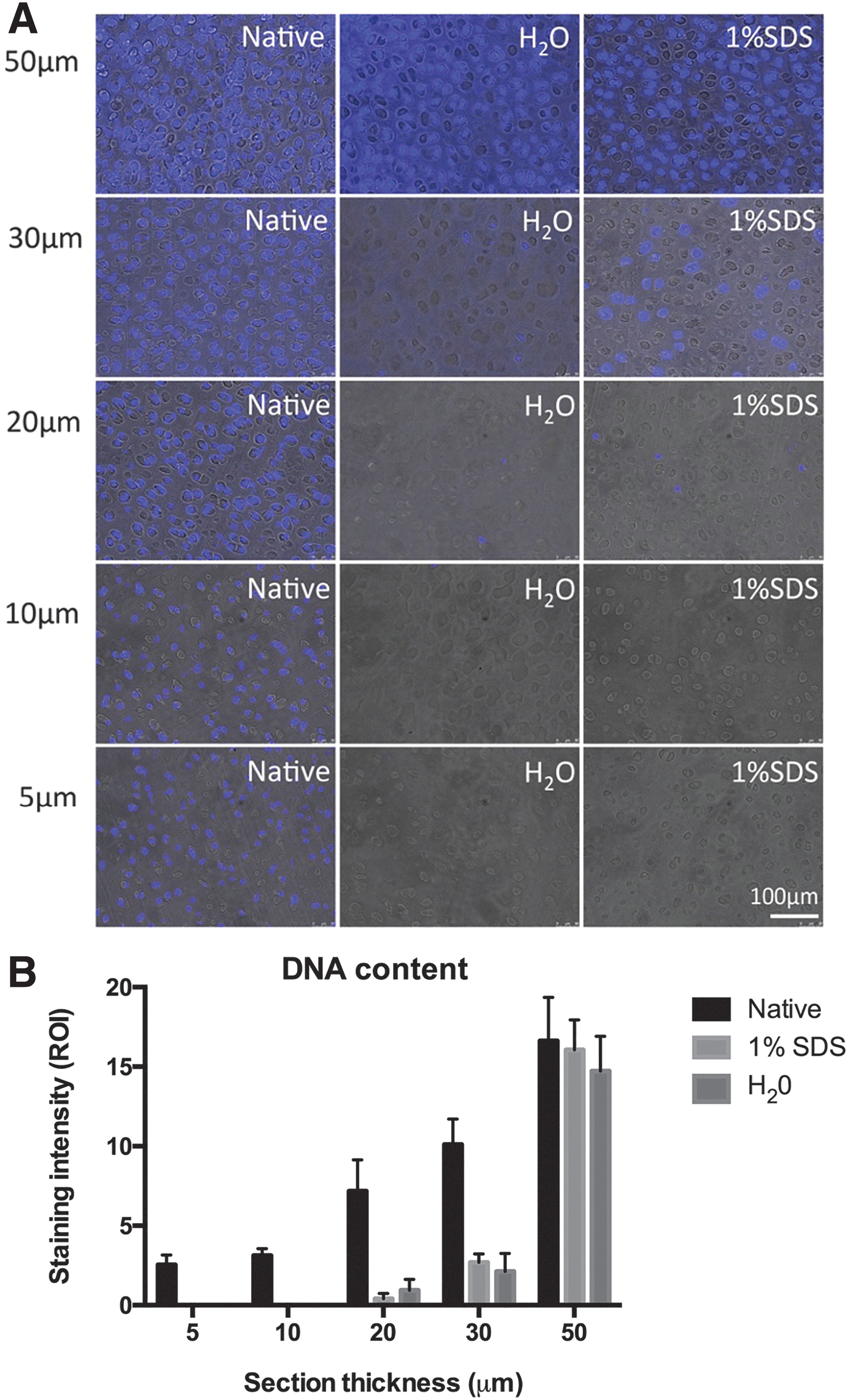
We observed that sonicating 5–20 μm thick cartilage sections in water for 5 min was sufficient to achieve >90% removal of nuclei, as judged by Hoechst DNA staining. For sections thicker than 30 μm, however, cell removal was not complete. Water and SDS (1%) achieved comparable decellularization efficiency (Fig. 1). Hence, the combination of physical disruptions by freezing-thawing, sectioning, and sonication allowed the complete decellularization of cartilage in the absence of any denaturing chemical. This protocol also works for other types of cartilage tissues, including porcine costal cartilage and growth plate (Fig. 3A).
Detergent-free decellularization preserves the topographical and biochemical properties of cartilage
The decellularized cartilage was characterized by scanning electron microscopy (SEM) analysis. Figure 2Ba shows that chondrocytes were observed in some lacunae of native cartilage. In both water (Fig. 2Bb) and SDS (Fig. 2Bc) decellularized cartilage, empty lacunae were observed. This result confirms the complete removal of cells, not just the elimination of DNA, as the lacunae were devoid of any discernible cell debris. Higher magnifications (Fig. 2Ba2, b1 and c2) show that the topography of the SDS-decellularized sections was smoother than those of native tissue and water-decellularized cartilage sections. Collagen fibrils were present in water-decellularized cartilage sections (Fig. 2Bb2), similar to those observed in the native cartilage, indicating that the detergent-free decellularization procedure was more efficient in preserving the ultrastructure of the cartilage tissue microenvironment.
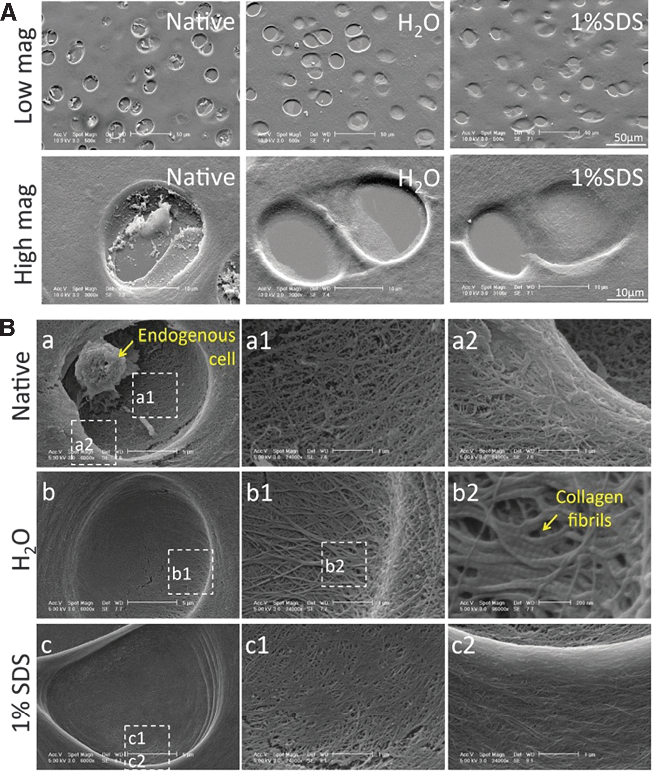
Representative SEM images of decellularized cartilage.
To examine the preservation of biological components, proteoglycan and total collagen in native, water-, and SDS-decellularized cartilage were assessed by histological staining. As shown in Figure 3B, there was no significant difference in the amount of collagen between water- and SDS-decellularized cartilage. However, while SDS has essentially removed all proteoglycan from the cartilage, water decellularization largely preserved the proteoglycan content. The results suggested a better preservation of matrix proteins in water-decellularized cartilage.
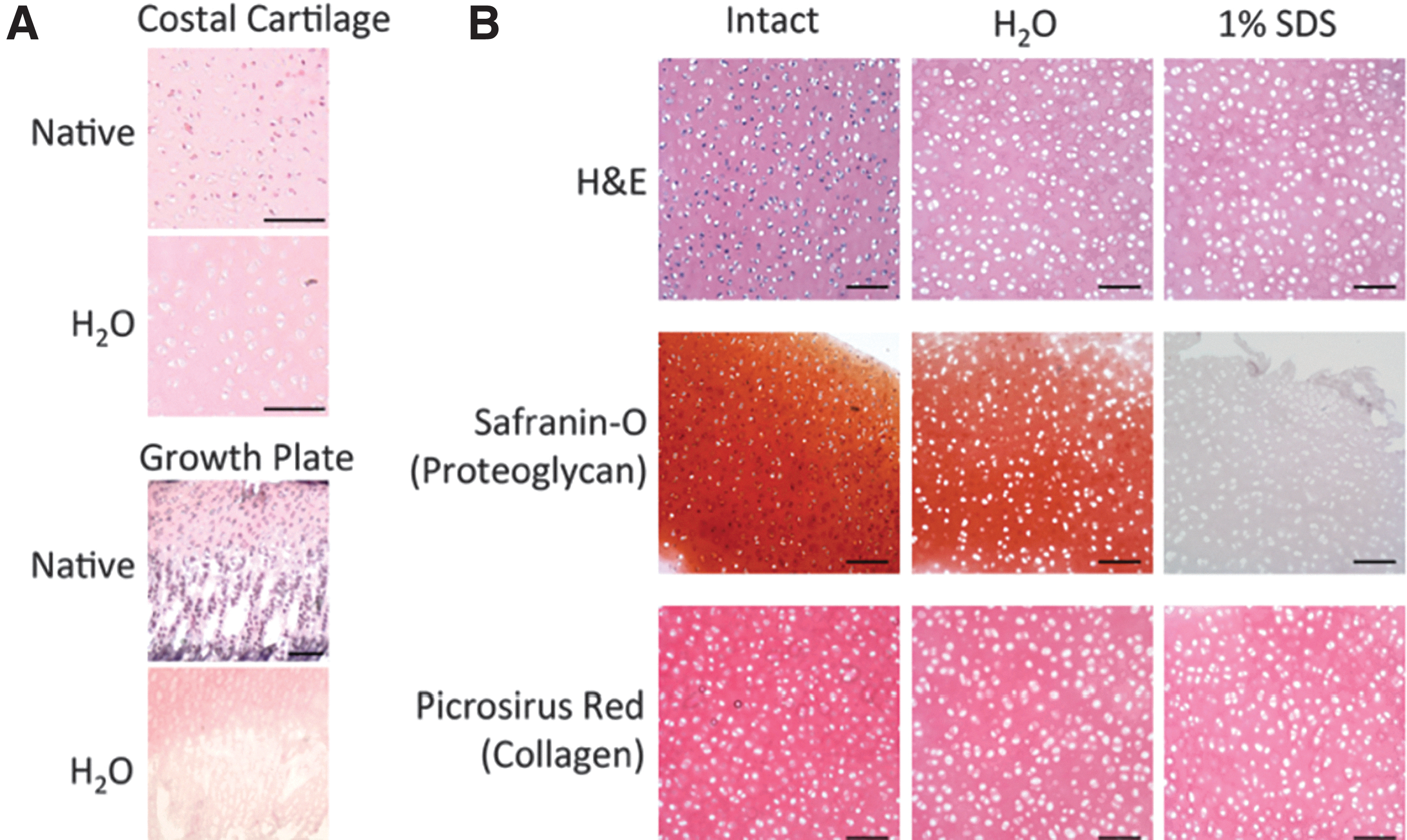
H&E, proteoglycan, and collagen staining of decellularized cartilage sections.
Decellularized cartilage as cell culture substrate
Further investigations on the use of acellular cartilage sections as a cell culture substrate were conducted with human MSC stably expressing GFP. Figure 4A shows that MSC could readily adhere onto both native cartilage sections and cartilage decellularized in either water or SDS, with no significant difference in attachment efficiency compared with glass coverslip. Cells did not show apparent preference for the lacunae, possibly because human MSCs are much larger than the size of a lacuna. SEM analysis (Fig. 4B) shows that MSC cultured on glass coverslip adopted a flatter morphology than cells cultured on native and decellularized cartilage. The data highlight the importance of tissue microenvironment on cell behavior such as adhesion, migration, and morphology.
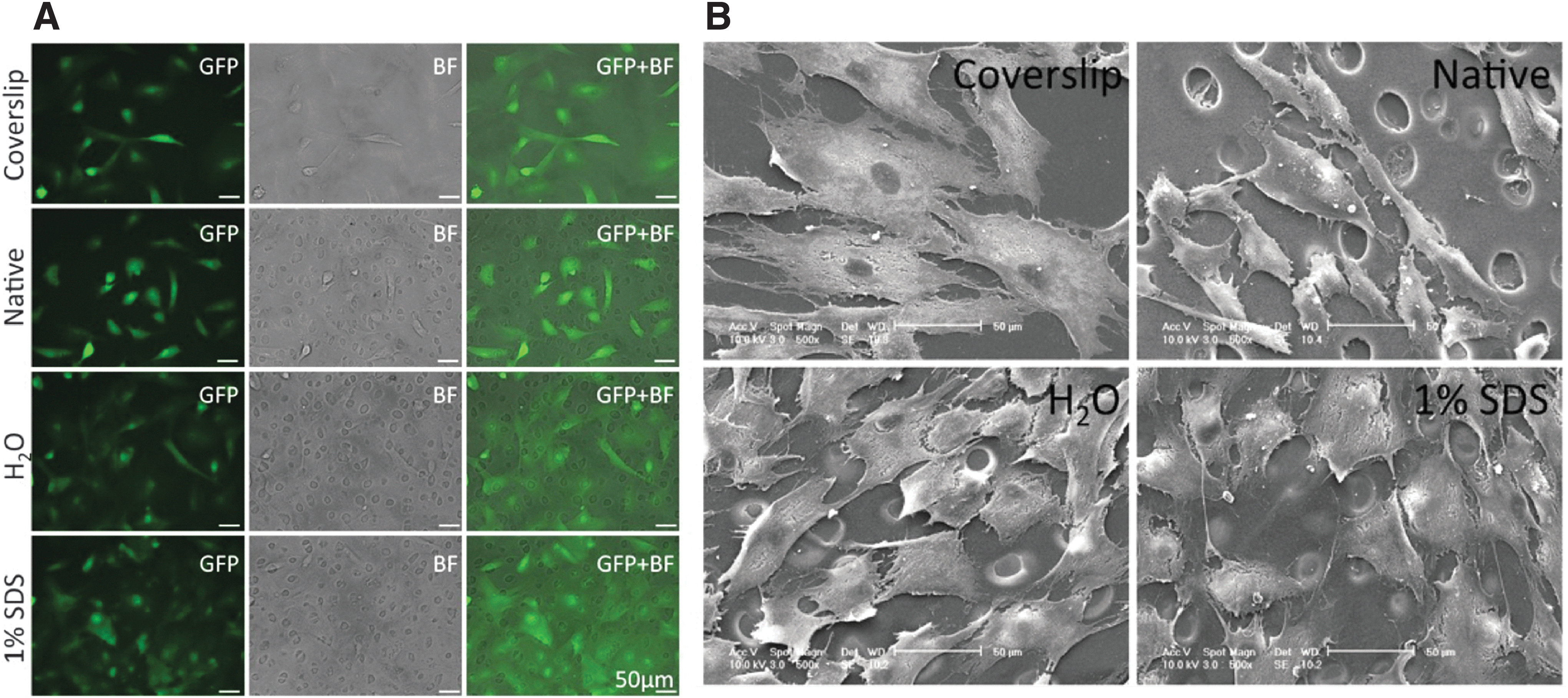
Decellularized cartilage as culture substrate for MSC.
Discussion
Decellularization of cartilage has previously been achieved by protocols that involve the use of detergents and enzymes. 15 While these conditions are necessary to remove chondrocytes that are enveloped in the lacunal cavities of the cartilage, exposure to detergents leads to the leaching of macromolecules, such as GAGs, from the tissue. Previous studies have demonstrated that GAGs such as chondroitin sulfate and aggrecan are important for chondrogenic differentiation.18–20 Decellularized cartilage without GAGs therefore represents disrupted and nonphysiological tissue microenvironment. To circumvent the use of detergent, some investigators cultured cells on cryogrounded cartilage matrix, 21 with the loss of the mechanical and topographical information of the tissue.
In this study, we report a new approach in which cartilage is decellularized by the combined effect of freezing-thawing, sectioning, and sonication in water. Our decellularized protocol eliminated the use of any detergent, resulting in the preservation of GAG content and the detailed architectures of the tissue.
Water-decellularized cartilage can support cell adhesion, growth, and proliferation. As far as we know, this is the first demonstration of detergent-free decellularization of cartilage tissue. The resulting scaffold will be a promising biomaterial for future applications in cartilage tissue engineering. One limitation of this protocol is that effective decellularization was only possible for cartilage sections thinner than 30 μm. Previous studies have demonstrated that sheets of cells can be stacked up into 3D structures.22–24 To date, most attempts of cell sheet assembly involve the culture of cell monolayers on peelable materials such as temperature-responsive polymer poly(N-isopropylacrylamide, 25 allowing the harvest of intact sheets of cells enveloped by ECM.
However, the resulting stacked-up structures still lack the architectural properties of the native tissues. It may be possible to utilize these water-decellularized cartilage sections as the scaffold for cell sheets. Upon recellularization with MSC-derived chondrocytes, these sections will then be stacked into reconstructed tissues, thus providing the necessary histological information for the engineered cartilage.
Conclusion
We have developed a simple, fast, and most importantly, nondetergent-based cartilage decellularization method that could efficiently remove cells while preserving the GAG content. We envisage that decellularized cartilage is an ideal substrate to study chondrocyte–matrix interactions, and that decellularized cartilage would be an ideal platform for arthritis research.
Footnotes
Disclosure Statement
No competing financial interests exist.
Funding Information
S.W.T. was supported by a PhD fellowship (ID: 000618) funded by Hong Kong University Grants Committee (UGC).