
Abstract
Recently, many studies on the three-dimensional (3D) fabrication of cells have been performed. Under these circumstances, it is indispensable to develop the imaging technologies and methodologies for noninvasive visualization of 3D cells fabricated. The objective of this study is to develop the labeling method of human induced pluripotent stem (iPS) cells-derived 3D cartilage tissue with gelatin nanospheres coincorporating three kinds of quantum dots (QD) and iron oxide nanoparticles (IONP) (GNSQD+IONP). In this study, two labeling methods were performed. One is that a cartilage tissue was labeled directly by incubating with octaarginine (R8)-treated GNSQD+IONP (direct labeling method). The other one is a “dissociation and labeling method.” First, the cartilage tissue was dissociated to cells in a single dispersed state. Then, the cells were incubated with R8-GNSQD+IONP in a monolayer culture. Finally, the cells labeled were fabricated to 3D pellets or cell sheets. By the direct labeling method, only cells residing in the surrounding site of cartilage tissue were labeled. On the contrary, the 3D cartilage pellets and the cell sheets were homogenously labeled and maintained fluorescently visualized over 4 weeks. In addition, the cartilage properties were histologically detected even after the process of dissociation and labeling. Homogenous labeling and visualization of human iPS cells-derived 3D cartilage tissue was achieved by the dissociation and labeling method with GNSQD+IONP.
Impact statement
The homogenous labeling and visualization of human iPS cells-derived three-dimensional (3D) cartilage tissue was achieved over 4 weeks by the dissociation and labeling method with gelatin nanospheres coincorporating quantum dots (QD) and iron oxide nanoparticles (IONP) (GNSQD+IONP). The cartilage properties of cells treated were maintained. It is concluded that the dissociation and labeling method with GNSQD+IONP is a promising to visualize the human iPS cells-derived 3D cartilage tissue.
Introduction
With the recent progress of molecular and cell biology and tissue engineering technologies, a lot of studies on the three-dimensional (3D) fabrication of cells have been reported.1,2 It is well recognized that most cells in the body exist in the 3D state to enhance their survival and functions.3,4 Based on the idea, the 3D cell fabrication has been widely applied to the basic cell research,5,6 drug discovery,7,8 and cell transplantation therapy.9,10 Under these circumstances, it is indispensable to develop the imaging technologies and methodologies for noninvasive visualization of 3D cells fabricated.
Gelatin is a biodegradable material and has been widely used for food, pharmaceutical, and medical purposes. The biosafety and biocompatibility have been proved through their long-term practical applications. Gelatin is readily crosslinked to form various shapes of hydrogels, such as sheet, 11 sponge, 12 microspheres, 13 and nanospheres. 14 The gelatin nanospheres are effective carrier of plasmid DNA, 15 small interfering RNA, 16 and imaging probes17–20 for the internalization into the cells. We have previously demonstrated that the feasibility of gelatin nanospheres incorporating quantum dots (QD) and iron oxide nanoparticles (IONP) with octaarginine (R8) of a cell penetrating peptide. 21 The R8-treated gelatin nanospheres incorporating QD with an intense fluorescence and narrow fluorescent spectrum 22 and IONP of a negative contrast agent for magnetic resonance (MR) imaging 23 were readily internalized into human chondrocytes and visualized by both the optical and MR imaging modalities. QD is one of useful imaging probes for the cellular labeling, and a lot of nanocarriers of QD, including the use of R8, have been reported.24–28 However, most of the studies focused on the use of single QD. In this study, we design a multimodal and multicolor imaging probe by utilizing three kinds of QD with different fluorescent wavelengths and IONP. The multimodality to visualize more than two imaging modalities can compensate the disadvantages of each imaging modality, leading to an enhancement of visualization.29,30 In addition, the multicolor imaging is potentially useful to visualize different intracellular targets as well as the enhancement of imaging reliability.
To visualize the 3D cells fabricated, it is technically necessary to develop the labeling method as well as imaging probes. The objective of this study is to develop the labeling method of human induced pluripotent stem (iPS) cells-derived 3D cartilage tissue with the gelatin nanospheres coincorporating three kinds of QD and IONP (GNSQD+IONP). To label the human iPS cells-derived 3D cartilage tissue, two labeling approaches are tried. One is that the cartilage tissue was labeled directly by incubating with R8-GNSQD+IONP (direct labeling method). The other one is a “dissociation and labeling method.” First, the cartilage tissue was dissociated to cells in a single dispersed state. Then, the cells were incubated with R8-GNSQD+IONP in a monolayer culture. Finally, the cells labeled were fabricated to 3D pellets or cell sheets. The labeling efficiency was compared in terms of the fluorescent visualization. We perform the immunohistological evaluation of cartilage tissue after the labeling with GNSQD+IONP.
Materials and Methods
Materials
Gelatin with an isoelectric point of 5.0 and the weight-averaged molecular weight of 100,000, prepared by an alkaline treatment of bovine bone collagen, was kindly supplied from Nitta Gelatin, Inc. (Osaka, Japan). Qdot 525, 605, and 705 ITK Carboxyl Quantum Dots (QD525, 605, and 705, 8 μM in water) were purchased from Invitrogen Co. (Tokyo, Japan). Alkali-treated dextran-coated magnetic IONP (5 mg Fe/mL in water) was purchased from Meito Sangyo Co., Ltd. (Nagoya, Japan). Isooctane, polyoxyethylene sorbitan monooleate (Tween 80), glutaraldehyde (GA, 25 wt% in water), glycine, and concentrated hydrochloric acid (HCl) were purchased from Nacalai Tesque, Inc. (Kyoto, Japan). Sorbitan monooleate (Span 80) was purchased from Tokyo Chemical Industry Co., Ltd. (Tokyo, Japan). R8 was purchased from Sigma-Aldrich, Inc. (St. Louis, MO). The reagents were used without further purification.
Preparation of gelatin nanospheres coincorporating quantum dots and IONP (GNSQD+IONP)
According to the preparation procedure previously reported,18,21 gelatin nanospheres coincorporating three kinds of QD and IONP (GNSQD+IONP) were prepared by the conventional emulsion method. In brief, QD525 (200 nM), QD605 (80 nM), QD705 (160 nM), and IONP (25 μg Fe/mL) were mixed with 2 mL of gelatin aqueous solution (5 mg/mL). The mixed solution was added into 40 mL of isooctane containing Tween 80 (480 mg) and Span 80 (480 mg), and then sonicated for 3 min at room temperature to obtain gelatin/isooctane emulsion. Next, 0.5 wt% GA aqueous solution (200 μL) was mixed with isooctane (40 mL) containing Tween 80 (480 mg) and Span 80 (480 mg), followed by sonication to prepare the GA/isooctane emulsion. Then, the gelatin/isooctane and GA/isooctane emulsions were mixed and sonicated for 3 min on ice, and the mixture was agitated for 3 h at 4°C to allow gelatin to crosslink in emulsion with GA. After that, the glycine/isooctane emulsion prepared similarly was added to block the aldehyde groups unreacted. The final reactant was centrifuged at 8500 rpm for 60 min at 4°C to collect gelatin nanospheres. The gelatin nanospheres were washed by acetone, and then double distilled water (DDW) by the centrifugation of 5000 rpm for 5 min at 4°C and 20,000 rpm for 30 min at 4°C, respectively. Finally, the gelatin nanospheres coincorporating three kinds of QD and IONP (GNSQD+IONP) were dispersed in 1 mL of DDW.
Characterization of GNSQD+IONP
The apparent size and polydispersity index of GNSQD+IONP resuspended in 10 mM phosphate-buffered saline solution (PBS, pH 7.4) were measured by dynamic light scattering (Zetasizer Nano-ZS; Malvern Instruments Ltd., Worcestershire, UK). The zeta potential of GNSQD+IONP resuspended in 10 mM PBS (pH 7.4) was measured by electrophoresis light scattering (Zetasizer Nano-ZS; Malvern Instruments Ltd.). The amount of QD and IONP incorporated in the gelatin nanospheres was measured by an atomic absorption spectrophotometer of Cd and Fe (AA-6800; Shimadzu Corp., Kyoto, Japan) after the degradation by the concentrated HCl. The fluorescent intensity of GNSQD+IONP was measured by a microplate reader (SpectraMax i3x; Molecular Devices Japan Co., Ltd., Tokyo, Japan). The excitation wavelength was 350 nm, and the emission wavelengths were 525, 605, and 705 nm, respectively. The experiments were independently performed three times unless otherwise mentioned.
Cell culture experiments
Cartilage tissue differentiated from human iPS cells (human iPS cells-derived cartilage tissue) was kindly supplied from Professor Tsumaki's laboratory. 31 The cartilage tissue was suspended to maintain in Dulbecco's modified Eagle's medium (DMEM; Sigma-Aldrich, Inc.) supplemented with 1 vol% bovine fetal calf serum (HyClone Laboratories, Inc., UT), 1 vol% insulin, transferrin, selenium, and ethanolamine solution (ITS-X; Thermo Fisher Scientific, Inc., MA), 1 mM sodium pyruvate (Thermo Fisher Scientific, Inc.), 0.1 mM nonessential amino acid (Thermo Fisher Scientific, Inc.), 5 mg/mL L-ascorbic acid phosphate (Wako Pure Chemical Industries, Ltd., Osaka, Japan), 10 ng/mL bone morphogenic protein-2 (PeproTech, Inc., Rocky Hill, NJ), 10 ng/mL transforming growth factor-β1 (PeproTech, Inc.), 10 ng/mL growth differentiation factor-5 (ProSpec, Ness Ziona, Israel), and 1 vol% penicillin/streptomycin (Nacalai Tesque, Inc.) at 37°C in a 5% CO2–95% air atmospheric condition.
Direct labeling of human induced pluripotent stem cells-derived cartilage tissue with GNSQD+IONP
The human iPS cells-derived cartilage tissue was directly labeled with GNSQD+IONP. In brief, GNSQD+IONP were mixed with R8 in OPTI MEM (Gibco Lifetechnologies Co.) for 15 min at room temperature (R8-GNSQD+IONP). The cartilage tissue was transferred to a 15 mL centrifuge tube (Greiner Bio-One, Kremsmuenster, Austria), and incubated with R8-GNSQD+IONP (final concentrations of R8 and averaged QD were 8 μM and 8 nM, respectively) in OPTI MEM for 3 h. After that, the cartilage tissue was washed by PBS, and the fluorescent images were taken by LAS-4000 (Fujifilm Co., Tokyo, Japan), with UV light (365 nm) excitation and 515 nm filter (QD525), 605 nm filter (QD605), and 670 nm filter (QD705). The labeled cartilage tissue was gradually fixed with 4 vol% paraformaldehyde, and 15 and 30 wt% sucrose. After the fixation, the tissue was embedded in Tissue-Tek OCT compound (Sakura Finetek, Inc., Tokyo, Japan), followed by freezing in liquid nitrogen. The cryosection was prepared by Cryostat (Leica CM3030S; Leica Biosystems Ltd., Germany) and observed by confocal laser microscopy FV1000-D IX81 (Olympus Co., Ltd., Tokyo, Japan).
Dissociation and labeling of human iPS cells-derived cartilage tissue with GNSQD+IONP
The human iPS cells-derived cartilage tissue was completely dissociated to cells in the single dispersed state, 32 and the monolayerd cells were labeled with GNSQD+IONP as reported previously. 21 In brief, the cartilage tissue was incubated with 0.25 wt% trypsin-containing 1 mM ethylenediaminetetra acetic acid (EDTA) solution (Nacalai Tesque, Inc.) for 1 h, and then washed by PBS. Collagenase D solution (4 mg/mL) (Roche Diagnostics, Indianapolis, IN) was added, and incubated for 3 h. After the incubation, the cartilage tissue was dissociated by pipetting to obtain the chondrocytes suspension.
The chondrocytes were seeded on each well of six-well multidish culture plate (Corning, Inc., Corning, NY) at a density of 1 × 10⁵ cells/well, and cultured for 24 h. The medium was changed to OPTI MEM, and the R8-GNSQD+IONP (8 μM R8 and 8 nM QD) were added to each well. After the incubation for 3 h, the cells were washed by PBS and observed by the confocal laser microscopy. The percentage of labeled cells was evaluated by the flow cytometry analysis (FACSCanto II; Becton Dickinson, Franklin Lakes, NJ).
Fabrication of 3D cartilage pellets after the dissociation and labeling with GNSQD+IONP
The chondrocytes labeled were detached by the trypsinization, and 3 × 10⁵ cells were resuspended in a 15 mL centrifuge tube (Greiner Bio-One GmbH). After the centrifugation at 1000 rpm for 5 min, the cell pellet was cultured for 1, 2, 3, and 4 weeks to fabricate the 3D cartilage pellet. The fluorescent images were taken and the cryosection prepared as described above was observed by the confocal laser microscopy.
Fabrication of cell sheets after the dissociation and labeling with GNSQD+IONP
The chondrocytes dissociated from the cartilage tissue were seeded on each well of 24 well multidish culture plate (Corning, Inc.) at a density of 1 × 10⁵ cells/well, and cultured for 24 h. The cells were incubated with R8-GNSQD+IONP at the same procedure described above, and cultured for 1 week to fabricate the cell sheet. The cell sheet was detached by the pipetting and collected by Cell Shifter™ (CellSeed, Inc., Tokyo, Japan) according to the manufacture's instruction. The fluorescent images were taken and the cryosection prepared as described above was observed by the confocal laser microscopy similarly.
Histological analysis
To immunohistologically evaluate the 3D pellets and 2D sheets, the original human iPS cells-derived cartilage tissue, the 3D cartilage pellet (4 weeks after the labeling with GNSQD+IONP), and the cell sheet (1 week after the labeling with GNSQD+IONP) were stained with the conventional hematoxylin and eosin (HE), alcian blue, and safranin O/fast green. The expression of collagen type I and II was evaluated by immunohistochemical staining. In brief, the sections were treated with 0.3 vol% hydrogen peroxide in methanol solution and blocked with 1 wt% bovine serum albumin in PBS for 30 min. Then, primary antibodies (1:20, goat anticollagen type I and type II; Southern Biotech, Birmingham, AL) were incubated overnight at 4°C. Following the incubation, the samples were treated with peroxidase-conjugated secondary antibody (Simple Stain™ MAX PO (G); Nichirei Biosciences, Inc., Tokyo, Japan). The immunoreactions were visualized by 3,3′-diaminobenzidine substrate in chromogen solution (Agilent Technologies, Inc., Santa Clara, CA). The samples were counter stained with hematoxylin and observed by a microscope (BZ-X700; Keyence Co., Ltd., Osaka, Japan).
Results
Physicochemical and fluorescent properties of GNSQD+IONP
Table 1 shows the physicochemical properties of gelatin nanospheres coincorporating three kinds of QD and IONP (GNSQD+IONP). The apparent size was around 200 nm, and the zeta potential was of a negative charge. The percentage of averaged QD incorporation was around 60%. On the contrary, the fluorescent efficiency of GNSQD+IONP at each wavelength was lower than the percentage of QD incorporated (Table 2).
Physicochemical Properties of GNSQD+IONP
Values are in average ± standard deviation.
IONP, iron oxide nanoparticles; QD, quantum dots.
Fluorescent Properties of GNSQD+IONP
Values are in average ± standard deviation.
Fluorescent efficiency: Percentage of fluorescent intensity relative to QD aqueous solution (200 nM QD525, 80 nM QD605, and 160 nM QD705).
Direct labeling of human iPS cells-derived cartilage tissue with GNSQD+IONP
The human iPS cells-derived cartilage tissue was tried to label directly with GNSQD+IONP (direct labeling method). Figure 1A shows the fluorescent images of cartilage tissue coincubated with R8-GNSQD+IONP. Fluorescence was hardly observed for any QD. Figure 1B shows the confocal microscopic images of cryosectional cartilage tissue. The fluorescence of cells was detected only at the surrounding site of cartilage tissue.
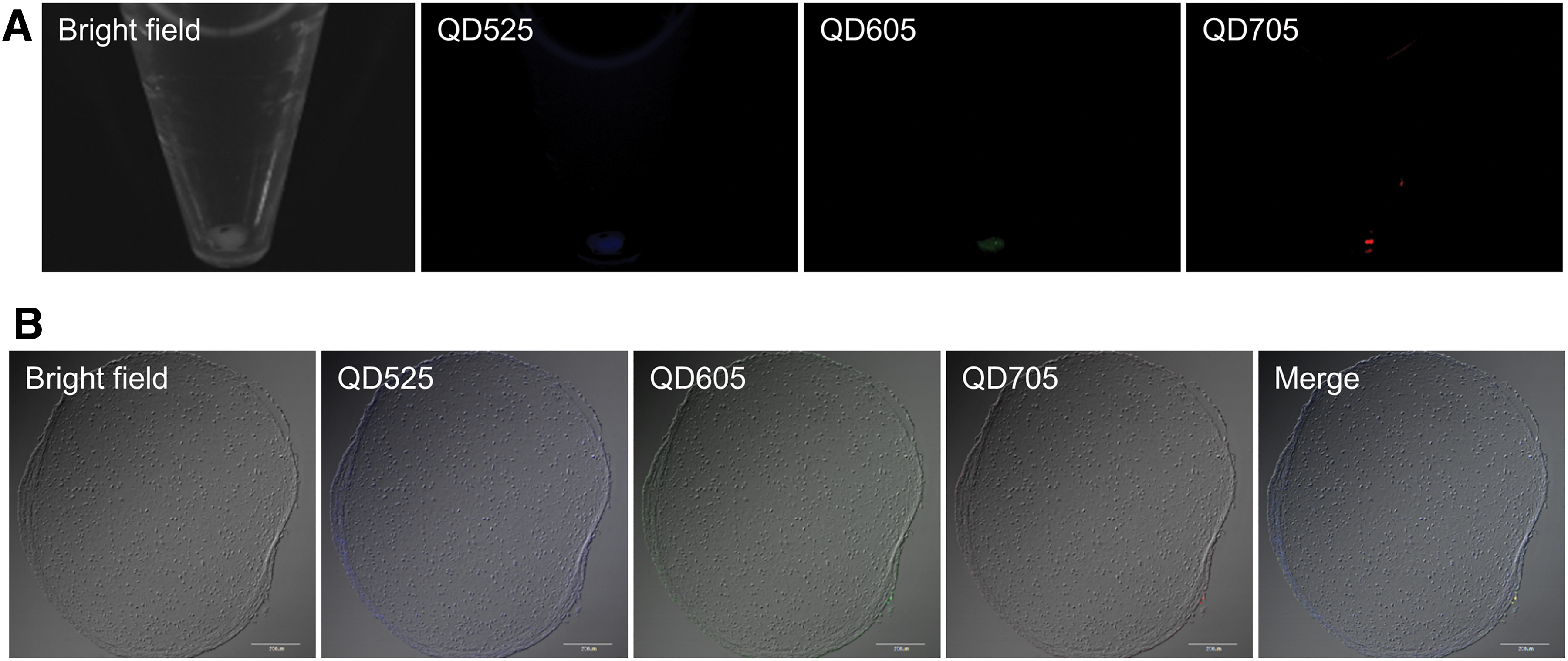
Direct labeling of human iPS cells-derived cartilage tissue with GNSQD+IONP. Fluorescent images of cartilage tissue in 15 mL centrifuge tube incubated with R8-GNSQD+IONP
Dissociation and labeling of human iPS cells-derived cartilage tissue with GNSQD+IONP
The cartilage tissue was dissociated and the cells of a single state were labeled with GNSQD+IONP in monolayer culture (dissociation and labeling method). The GNSQD+IONP were efficiently internalized into the monolayer cells and the fluorescence was observed (Fig. 2A). Figure 2B shows the flow cytometric analysis after the incubation with R8-GNSQD+IONP in the monolayer culture. Almost 100% cells were fluorescently positive for QD605 and QD705, whereas around 40% cells were positive for QD525.
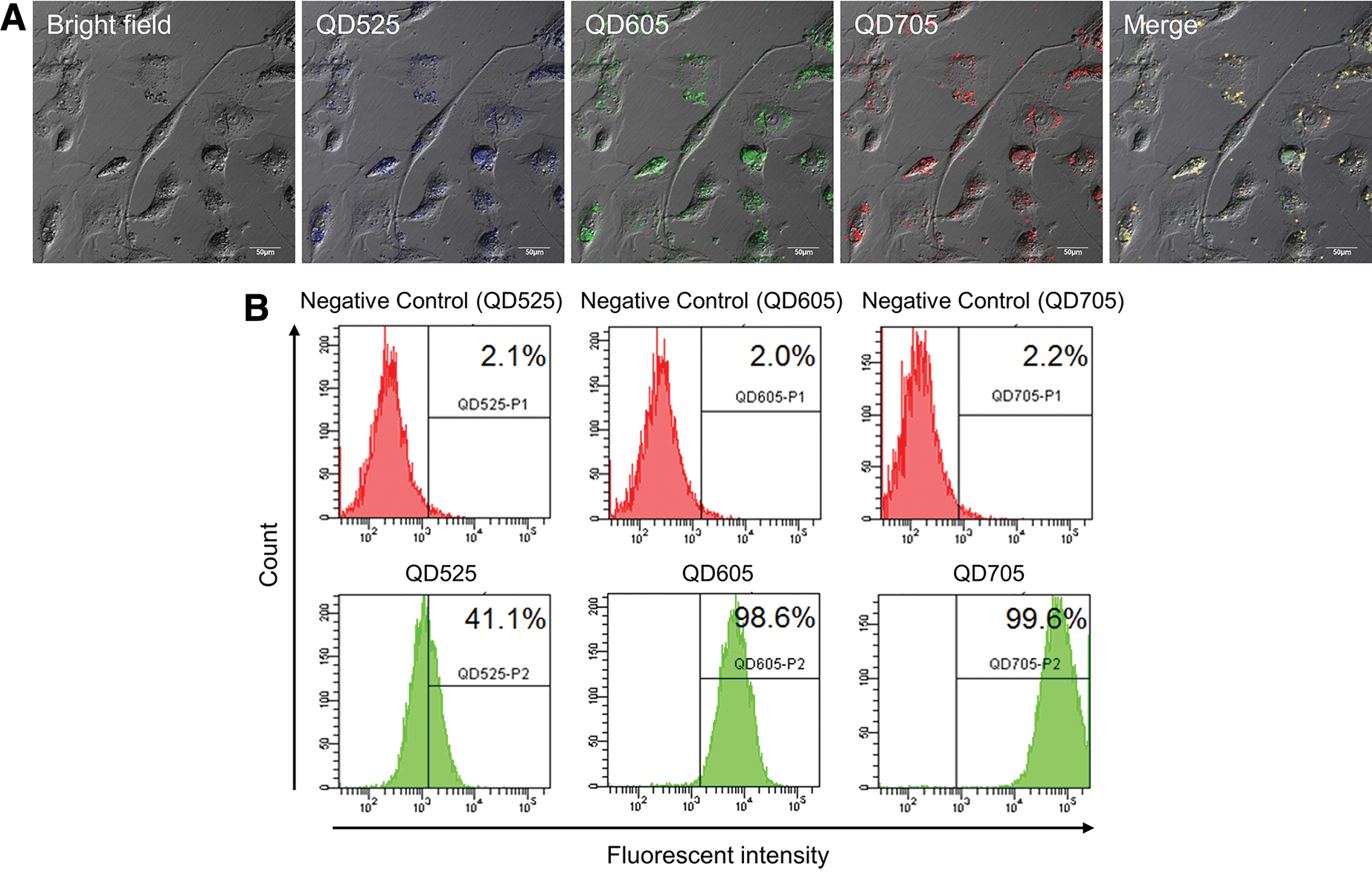
Labeling after dissociation of human iPS cells-derived cartilage tissue with GNSQD+IONP. Confocal microscopic images of monolayer cells incubated with R8-GNSQD+IONP
Fabrication of 3D cartilage pellets and cell sheets
The labeled cells with GNSQD+IONP were cultured in a pellet state to fabricate the 3D cartilage pellets, and the 3D pellets were fluorescently visualized (Fig. 3A). The fluorescence of GNSQD+IONP was homogenously observed in the pellet cryosection (Fig. 3B). Figure 4 shows the fluorescent time course of pellets labeled with GNSQD+IONP. All the fluorescence of pellet (QD525, 605, and 705) could be detected even 4 weeks later. Figure 5 shows the fluorescent observation of cell sheets labeled with GNSQD+IONP. The cells of sheet were fluorescently visualized for all wavelength of QD.
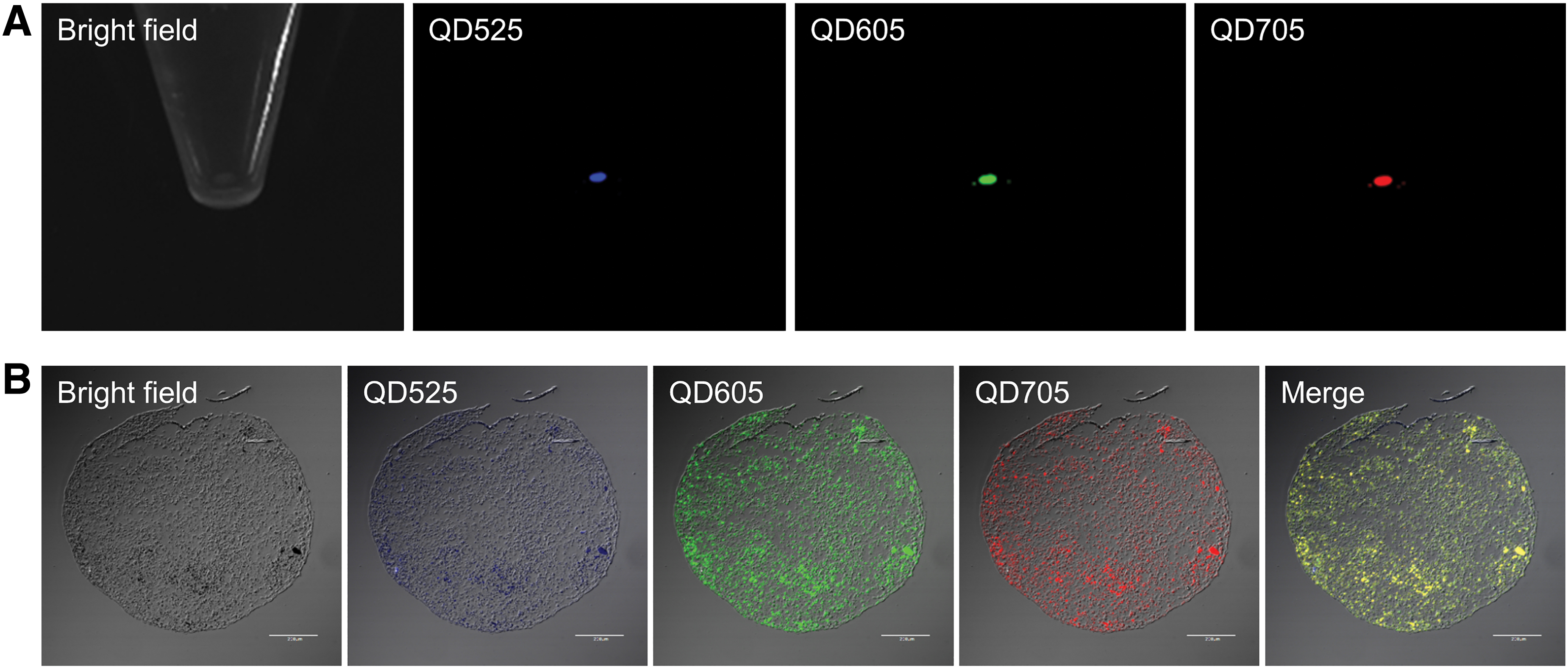
Fabrication of 3D cartilage pellet from cells labeled with GNSQD+IONP. Fluorescent images of cartilage pellet in 15 mL centrifuge tube
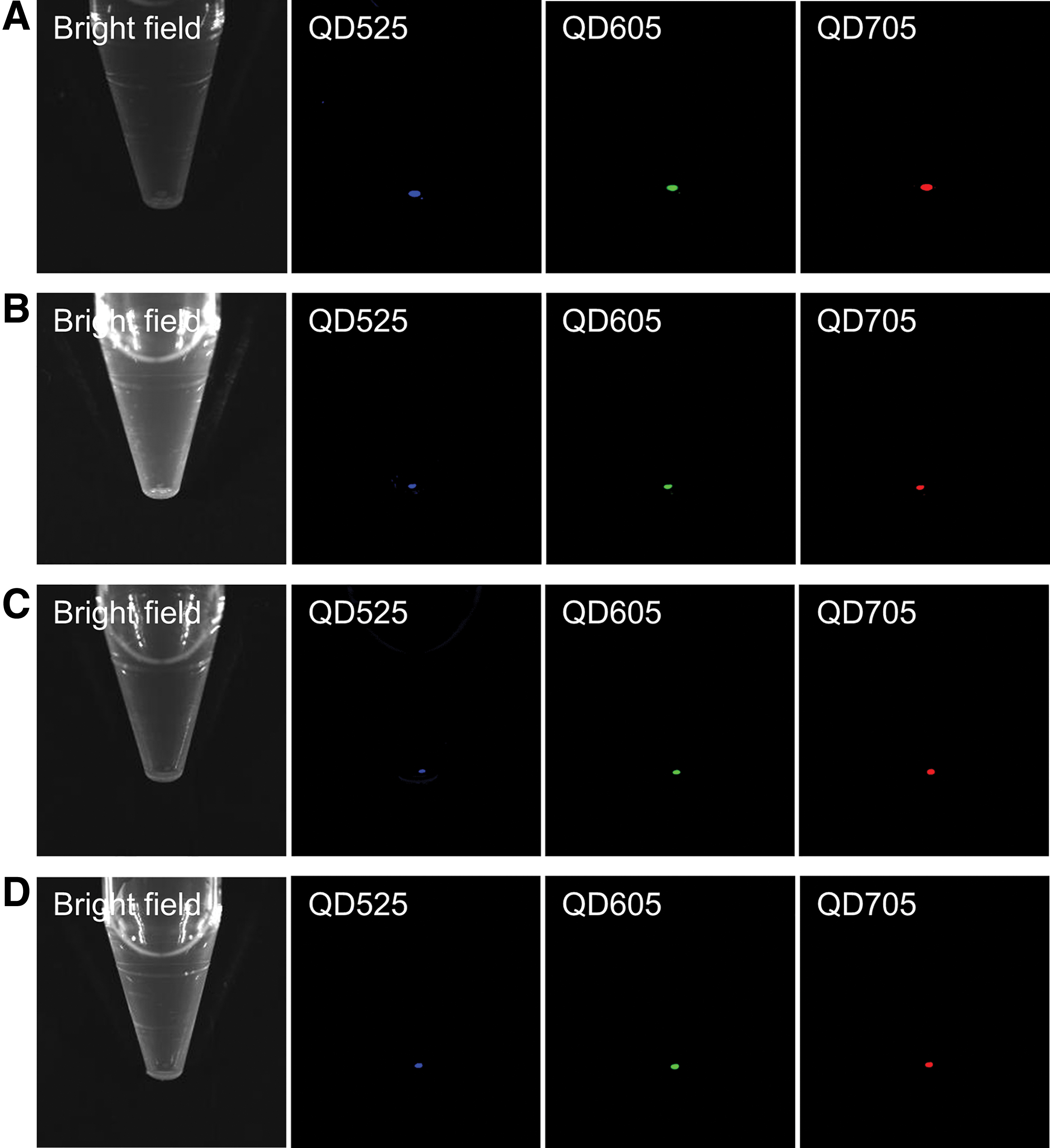
Fluorescent time course of 3D cartilage pellet from cells labeled with GNSQD+IONP. Fluorescent images of cartilage pellet prepared 1

Fluorescent images of cell sheet prepared 1 week after incubation with R8-GNSQD+IONP
Histological analysis
The histological analysis was performed for the original cartilage tissue (Fig. 6A), the cartilage pellet 4 weeks after label with GNSQD+IONP (Fig. 6B), and the cell sheet 1 week after the label with GNSQD+IONP (Fig. 6C). Both the pellet and sheet of cells labeled with GNSQD+IONP were stained with the cartilage specific staining (alcian blue, safranin O/fast green, collagen type I, and II) to a similar extent as the original cartilage tissue.

Histological analysis for the original cartilage tissue
Discussion
Gelatin nanospheres coincorporating three kinds of QD and IONP (GNSQD+IONP) were prepared to visualize cells in the 3D formation. The human iPS cells-derived 3D cartilage tissue was directly incubated with R8-treated GNSQD+IONP (direct labeling method). However, in this case, only cells residing in the surrounding site of cartilage tissue were labeled. On the contrary, the 3D cartilage pellet and the cell sheet prepared by the “dissociation and labeling method” were homogenously and fluorescently labeled. Both the pellet and the sheet of cells labeled with GNSQD+IONP were cartilage specifically stained to the similar extent as the original cartilage tissue.
The apparent size of GNSQD+IONP was around 200 nm, and this small size is advantageous to the cellular internalization. 33 On the contrary, the zeta potential of GNSQD+IONP was negative charge. R8 is one of common cell penetrating peptides to enhance the cellular internalization via an endocytotic pathway. 34 We can say with certainty that the electrostatic interaction between the negative charge of GNSQD+IONP and the positive charge of R8 permitted the surface modification of GNSQD+IONP. This modification may also be advantageous for the cellular internalization due to the easy interaction with the negative charge of cell membrane. 35 Although the percentage of averaged QD incorporation was around 60% (Table 1), the fluorescent efficiency of GNSQD+IONP at each wavelength was lower than the percentage of QD incorporated (Table 2). The reason why the fluorescent intensity decreased relative to the QD aqueous solution is not clear at present. It is highly conceivable that the fluorescence of QD interacts with IONP, and the interaction may increase in the hydrogel of gelatin nanospheres in the concentrated state.
By the direct labeling method, only cells present in the surrounding site of human iPS cells-derived 3D cartilage tissue were labeled (Fig. 1). This is due to the poor penetration of GNSQD+IONP to the interior of tissue. In general, it is known that the natural cartilage tissue is surrounded by extracellular matrix (ECM) to maintain the mechanical structures. 36 The human iPS cells-derived cartilage tissue used in this study also has rich ECM. It is possible that the ECM presence of cartilage tissue prevents GNSQD+IONP from the penetration into the inside of tissue.
As one trial to label the cartilage tissue homogenously, we tried to perform the “dissociation and labeling method.” The cartilage tissue was enzymatically dissociated to cells of a single state due to the ECM degradation by the trypsin and collagenase treatments. 32 Then, the R8-GNSQD+IONP were efficiently internalized into the monolayer cells dissociated from the cartilage tissue (Fig. 2A). In our previous studies, under the QD concentration of 8 nM, QD neither showed the cytotoxicity nor affected the cell proliferation. 37 In addition, the 8 μM R8-treated gelatin nanospheres did not show the cytotoxicity, either. 21 Therefore, we can say with certainty that the labeling condition used in this study has no cytotoxicity. The fluorescence of all QD was well colocalized in the cells. Although the fluorescence of QD was observed from the cytosol (Fig. 2A), the detail localization is not clear at present. However, it has been reported that the liposomes modified with R8 had an ability for endosomal escape. 34 It is likely that the R8-GNSQD+IONP were internalized into the cells via an endocytotic pathway, followed by their endosomal escape into the cytosol. On the contrary, the flow cytometric assay revealed that almost 100% cells were fluorescently positive for QD605 and QD705, while around 40% cells were positive for QD525 (Fig. 2B). The difference might be due to the weak fluorescence of QD525 (Table 2). The poor incorporation in the gelatin nanospheres and the weak fluorescence of QD525 might lead to the low detection level of flow cytometry. However, we would like to claim here that the 3D cartilage pellet fabricated after the labeling with GNSQD+IONP was fluorescently visualized for all wavelength of QD (Fig. 3A). In addition, the homogenous fluorescence of GNSQD+IONP from the 3D cartilage pellet was observed (Fig. 3B). The homogenous labeling should be lead to the retention of fluorescence even after 4 weeks (Fig. 4). Another advantage of the “dissociation and labeling method” is the versatility to fabricate various structures of cells after the labeling. In this study, in addition to the 3D cartilage pellet, the cell sheet was fabricated after the labeling. The cell sheet was fluorescently visualized for all wavelength of QD (Fig. 5A), while it was homogenously labeled as well (Fig. 5B). Figure 6 shows the histological evaluation for the pellet and the sheet of cells labeled with GNSQD+IONP. Both of 3D cartilage pellets and sheets were cartilage-specifically stained. The alcian blue and safranin O/fast green stainings revealed the glycosaminoglycans in the cartilage ECM. In addition, both the collagen types I and II in the cartilage ECM were stained, while no homogenous staining was observed. Although it is further necessary to quantitatively evaluate the expression of cartilage markers, the results indicate that the cartilage properties were maintained even after the process of dissociation and labeling.
It is well recognized that the homogenous labeling of cells in the 3D fabricate is sometimes difficult due to the poor penetration of imaging probes into the cell fabricates. 38 Therefore, some researches on the construction of 3D structure of cells, such as neurosphere, 39 embryoid body, 40 and spheroid of mesenchymal stem cells (MSC), 41 after the cellular labeling in a monolayer culture, have been reported. The 3D cells fabricated after the labeling could be visualized for several weeks after the transplanted in vivo. However, those often focus on the cellular labeling with a single imaging probe. In this study, we designed a multimodal imaging probe of gelatin nanospheres coincorporating three kinds of QD and IOPN. The multimodal imaging is one of the promising approaches to enhance the reliability of visualization by compensating the disadvantages of each imaging modality.29,30 The multimodal imaging probes, such as liposomes, 42 poly(lactic-co-glycolic acid) nanoparticles, 43 and other polymeric nanoparticles 44 coincorporating QD and IONP, have been reported. In addition to the use of single QD and IONP, we used three kinds of QD to perform the multicolor imaging. One of the advantages for multicolor imaging is to visualize different intracellular targets by one single probe. On the contrary, functionalized QD, which changes the fluorescent signal based on the fluorescence resonance energy transfer responding to the intracellular environment, have been investigated. 45 Combination with the functionalized QD may allow our system to visualize an intracellular environmental change more pleiotropically.
Since the self-repairing and regeneration abilities of cartilages are poor, the cell transplantation therapy is one of the promising approaches for cartilage injuries. 46 Some researches using animal models demonstrated that therapeutic potential of human iPS cells-derived 3D cartilage tissue.31,32 The labeling technology developed in this study may be a powerful tool to evaluate the localization and distribution of cartilage tissues transplanted. In the future, the labeled cartilage tissue with GNSQD+IONP will be transplanted to an animal model, and the optical and MR imaging should be performed to evaluate the in vivo efficacy.
Footnotes
Disclosure Statement
No competing financial interests exist.
Funding Information
This work was financially supported by Japan Agency for Medical Research and Development (AMED) through its Research Center Network for Realization of Regenerative Medicine.