
Abstract
Despite considerable research effort, there is a significant need for safe agents that stimulate bone formation. Treatment of large or complex bone defects remains a challenge. Implantation of small molecule-induced human bone marrow-derived mesenchymal stromal cells (hBMSCs) on an appropriate tricalcium phosphate (TCP) scaffold offers a robust system for noninvasive therapy for spinal fusion. To show the efficacy of this approach, we identified a small molecule curcuminoid that when combined with TCP ceramic in the presence of hBMSCs selectively induced growth of bone cells: after 8- or 25-day incubations, alkaline phosphatase was elevated. Treatment of hBMSCs with curcuminoid 1 and TCP ceramic increased osteogenic target gene expression (i.e., Runx2, BMP2, Osteopontin, and Osteocalcin) over time. In the presence of curcuminoid 1 and TCP ceramic, osteogenesis of hBMSCs, including proliferation, differentiation, and mineralization, was observed. No evidence of chondrogenic or adipogenic potential using this protocol was observed. Transplantation of curcuminoid 1-treated hBMSC/TCP mixtures into the spine of immunodeficient rats showed that it achieved spinal fusion and provided greater stability of the spinal column than untreated hBMSC-TCP implants or TCP alone implants. On the basis of histological analysis, greater bone formation was associated with curcuminoid 1-treated hBMSC implants manifested as contiguous growth plates with extensive hematopoietic territories. Stimulation of hBMSCs by administration of small molecule curcuminoid 1 in the presence of TCP ceramic afforded an effective noninvasive strategy that increased spinal fusion repair and provided greater stability of the spinal column after 8 weeks in immunodeficient rats.
Impact statement
Bone defects only slowly regenerate themselves in humans. Current procedures to restore spinal defects are not always effective. Some have side effects. In this article, a new method to produce bone growth within 8 weeks in rats is presented. In the presence of tricalcium phosphate ceramic, curcuminoid-1 small molecule-stimulated human bone marrow-derived mesenchymal stromal cells showed robust bone cell growth in vitro. Transplantation of this mixture into the spine showed efficient spinal fusion in rats. The approach presented herein provides an efficient biocompatible scaffold for delivery of a potentially clinically useful system that could be applicable in patients.
Introduction
Despite success of bone grafts, treatment of large or complex bone defects remains a challenge. 1 Autograft bone harvest limitations include prolonged recovery time, increased surgical blood loss, and pain. 2 Use of autologous bone may be compromised by underlying bone or metabolic disorders that limit patient eligibility. Allografts may be applicable to individuals with underlying conditions, but allograft comes with safety concerns. 3 Alternatively, osteoinductive bioceramics can be used as graft substitutes to stimulate bone formation. 4 Bioceramics overcome disadvantages of autografts and allografts because ceramics are not derived from human tissue. Human bone marrow-derived mesenchymal stromal cells (hBMSCs) represent another approach. hBMSC-based therapy can provide exogenous cells in bone repair and regeneration.5,6
hBMSCs play a fundamental role in bone biology and osteogenesis. 7 hBMSCs can differentiate into osteoblasts, adipocytes, or chondrocytes. 8 hBMSCs are considered “immune privileged” because hBMSCs repress recipient T cells. Implantation of allogeneic hBMSCs does not lead to host immune rejection 9 nor develop MHC-Class II receptors. 10 hBMSCs can be isolated from allogeneic sources, culture expanded, and transplanted into humans without host immune rejection. hBMSCs produce cytokines and trophic factors that modulate adjacent cells and affect their functional activity, immune response, and differentiation. 11 Accordingly, allogeneic hBMSCs have been investigated clinically. 12
Recombinant bone morphogenetic proteins (BMPs) are used as scaffold modifiers for bone grafts, 13 but safety concerns have emerged. 14 Small molecules that mimic functions of endogenous growth factors to stimulate stem cell self-renewal or differentiation15,16 have been developed that stimulate differentiation of osteoblasts from hBMSCs17–20 or potentiate effects of BMP2 on hBMSCs. 21 However, it is yet to be shown if a small molecule approach can directly stimulate bone growth in vivo. New osteogenic agents are desired, and safe methods for integration with currently practiced bone repair interventions are needed.
Herein, we show that small molecules and an osteoinductive tricalcium phosphate (TCP) ceramic can be used together to induce hBMSC osteogenic lineage commitment in a cell-based therapy. Safe, small-molecule osteogenic curcuminoids afforded hBMSC osteogenesis.22,23 Curcumin was reported to increase rat MSC differentiation into osteoblasts in vitro when added to osteogenic medium. 24 To date, minor curcuminoids (e.g., bisdemethoxycurcumin, BDC) and synthetic curcumin derivatives have not been examined in osteogenesis.
Herein, water-soluble prodrugs of BDC (1) and synthetic curcuminoid (2) showed potent osteogenesis. Compound 1 and 2 incubated with hBMSCs, and osteoinductive TCP showed potent effects on hBMSC osteogenic differentiation. Compound 1-induced hBMSC-TCP implants afforded significant osteoinductivity using an intramuscular implant model in immunodeficient rats and showed that small-molecule osteogenic agent induced hBMSCs in the presence of a ceramic cell carrier are of great utility as a cell-based therapy for bone growth and repair.
Materials and Methods
Compounds
Curcumins, including bisdemethoxy curcumin di-L-valinyl ester dihydrochloride salt, 1, were prepared25–29 and purity was >95%.28,29 TCP ceramic (0.5–1.0 mm granule composition) was determined by XRD to be 95/5 TCP/hydroxyapatite.4,30 TCP was provided by Dr. Tim Moseley (NuVasive). TCP was sterilized by γ-irradiation before use. TCP scaffolds were sheets of TCP:collagen (88%:12%), see the Supplementary Data.
Cell culture
hBMSCs were provided by Dr. Pam Robey of the NIH Center for Regenerative Medicine (Bethesda, MD). hBMSCs were cultured in Dulbecco's modified Eagle medium (DMEM) containing 20% fetal bovine serum (FBS) and 1 × GlutaMAX. 31
Alkaline phosphatase activity
hBMSCs were cultured at a density of 1800 hBMS cells/cm2 for 8 days in 12-well plates with test compounds + TCP (5 mg/mL). Cells were lysed and alkaline phosphatase (ALP) activity determined. 32
Quantification of gene expression by qPCR
hBMSCs were cultured the same as that in the ALP studies. Total RNA was extracted using TRIzol (Life Technologies). Quantitative polymer chain reaction (qPCR) (Supplementary Table S1) was run as previously described. 32
Cell viability
hBMSCs were seeded at a density of 500 cells/well in 96-well plates and grown for 24 h before adding test compounds + TCP (5 mg/mL). Cell viability was measured using an Alamar Blue assay.
Cell staining
Alizarin Red S staining was modified from a kit (Millipore). Oil Red O (ORO) was stained for the presence of adipocytes (0.5% stock solution in isopropanol and diluted to 0.03% in deionized water). Alcian Blue was stained for the presence of chondrocytes (1% wt/v in 3% acetic acid, pH 2.5). Cell culture incubations were as described above for ALP studies.
Apoptosis
hBMSCs were incubated with 1 or 2 (500 nM) for 25 days. Apoptosis biomarkers Bax and c-Fos mRNA quantification was done by qPCR.
In vivo studies
Twenty-one male 10-week-old immunodeficient rats (200 g, Taconic Biosciences) were used in this study. Rats were purchased and housed at UC San Diego. All experiments were conducted with the approval of the IACUC committees of HBRI and UCSD. After anesthesia, rats were euthanized, and L4-L5 spinal fusion implants were removed and subjected to constant bending forces and visually ranked by blinded trained observers as described in the Supplementary Data.
Three groups of nude rats were used as follows: (A) DMEM on TCP scaffold, (B) untreated hBMSCs placed on TCP scaffold, and (C) 1-treated (500 nM) hBMSCs placed on TCP scaffold. Early passage hBMSCs (≤5 passages) were seeded (density of 17,500 cells/well in six-well plates) and cultured for 24 h before cell induction. For groups B and C, hBMSCs were then cultured for 8 days with TCP granules (5 mg/mL) or in the case of group C, TCP granules (5 mg/mL) + 1 (500 nM) before implantation in immunodeficient rats. For hBMSC induction (i.e., group C), 1 (500 nM) was added on day 0, and media replenished every 48 h with 1 (500 nM) until day 8. After 8 days, cells were harvested and separated from TCP granules by centrifugation. For implantation, cell samples were placed on the precut TCP scaffold before implantation.
On the day of surgery, implants containing (A) 200 μL DMEM and Tisseel (10 μL of Tisseel component A containing 0.16 mg/μL fibrinogen, ASD) placed on TCP scaffold, (B) 0.7 million untreated hBMSCs combined with 200 μL DMEM and Tisseel placed on TCP scaffold, or (C) 1-induced (500 nM) hBMSCs (0.7 million) combined with 200 μL DMEM and Tisseel and placed on TCP scaffold were prepared and kept cold (4°C). Tisseel component B (10 μL containing 0.036 mg/μL thrombin) was added to each implant. TCP scaffold alone or cell scaffold with Fibrin gel was immediately used for implantation.
Rats received implants of: (A) TCP scaffold alone (n = 7 rats, 14 implants), (B) untreated hBMSCs on TCP scaffold (n = 7, 14 implants), and (C) implants of 1-treated hBMSCs on TCP scaffold (n = 7, 14 implants). Each week for 8 weeks, rats were analyzed with imaging. 30 At 8 weeks, rats were evaluated by computed tomography for spinal fusion 31 and, thereafter, euthanized. Rat spines containing implants were subjected to constant bending force analysis and visually ranked by blinded trained observers. μCT images of L4-L5 vertebral disc height measurements were used for determining stability of the lumbar spinal segments under controlled lateral displacement. Distance of left length and right length of L4-L5 disc height was determined by μCT at the neutral position and then under a constant left (±10 mm) and right (−10 mm) lateral displacement. Deviation in disc height was calculated from the averaged differences in disc height from the neutral position that represented the absolute sum of contraction and elongation deviations. Implants were harvested and immediately fixed in 10% neutral buffered formalin as described in the Supplementary Data.
Statistical analysis
Statistical analysis was done with GraphPad Prism showing the mean and standard deviation (SD) or standard error of mean (SEM) of at least triplicate samples for each biological assay. Student t-tests were used for pairwise comparisons. p < 0.05 was considered significant. * p < 0.05, ** p < 0.01, *** p < 0.001. In some cases, other statistical treatments were used.
Results
Stimulation of hBMSC osteogenesis by curcumins
Bisdemethoxycurcumin 3 (BDC), a minor curcuminoid of turmeric (5–10%), and curcumin 4 (Curc), a major curcuminoid (65–80%) and amino acid conjugates 1 and 2 that increased the water solubility and stability of curcumins (Fig. 1A–C), were independently prepared (Supplementary Data). Curcumins (i.e., 500 nM) stimulated hBMSC osteogenesis based on ALP activity. 31 ALP activity was linearly dependent on concentration of curcumins up to 500 nM. A measure of 500 nM was chosen for further studies, and significant differences in hBMSC ALP activity were observed after 8 days (Fig. 1B). Curcumin 4 was similar to curcumin extract in stimulating ALP in hBMSCs. BDC 3 gave a 1.2-fold increase in ALP activity that was significantly (p < 0.01) greater than curcumin 4. hBMSCs treated with CyBDC 6 increased ALP activity by 1.2-fold that was greater than CyCurc 7 (i.e., 1.1-fold, p < 0.1). CyBDC 6 was less potent than BDC 3 in stimulating ALP activity in hBMSCs. Water-soluble curcumin amino acid conjugates were generally more effective at stimulating ALP activity in hBMSCs. BisVal-BDC 1 gave a 1.2-fold increase in ALP activity similar to results obtained using BDC 3. BisVal-CyBDC 2 gave a 1.2-fold increase in ALP activity (p < 0.05). Water-soluble BDC 1 and 2 stimulated greater ALP activity than water-soluble curcumins 5 and 8. The results showed that BDC and derivatives (i.e., 1, 2, 3, and 6) were more potent stimulators of ALP activity than curcumin and derivatives (i.e., 4, 5, 7, and 8). Based on the potency of 1 and 2 and increased water solubility and stability, BisVal-BDC 1 and BisVal-CyBDC 2 (Fig. 1C) were selected for further studies.
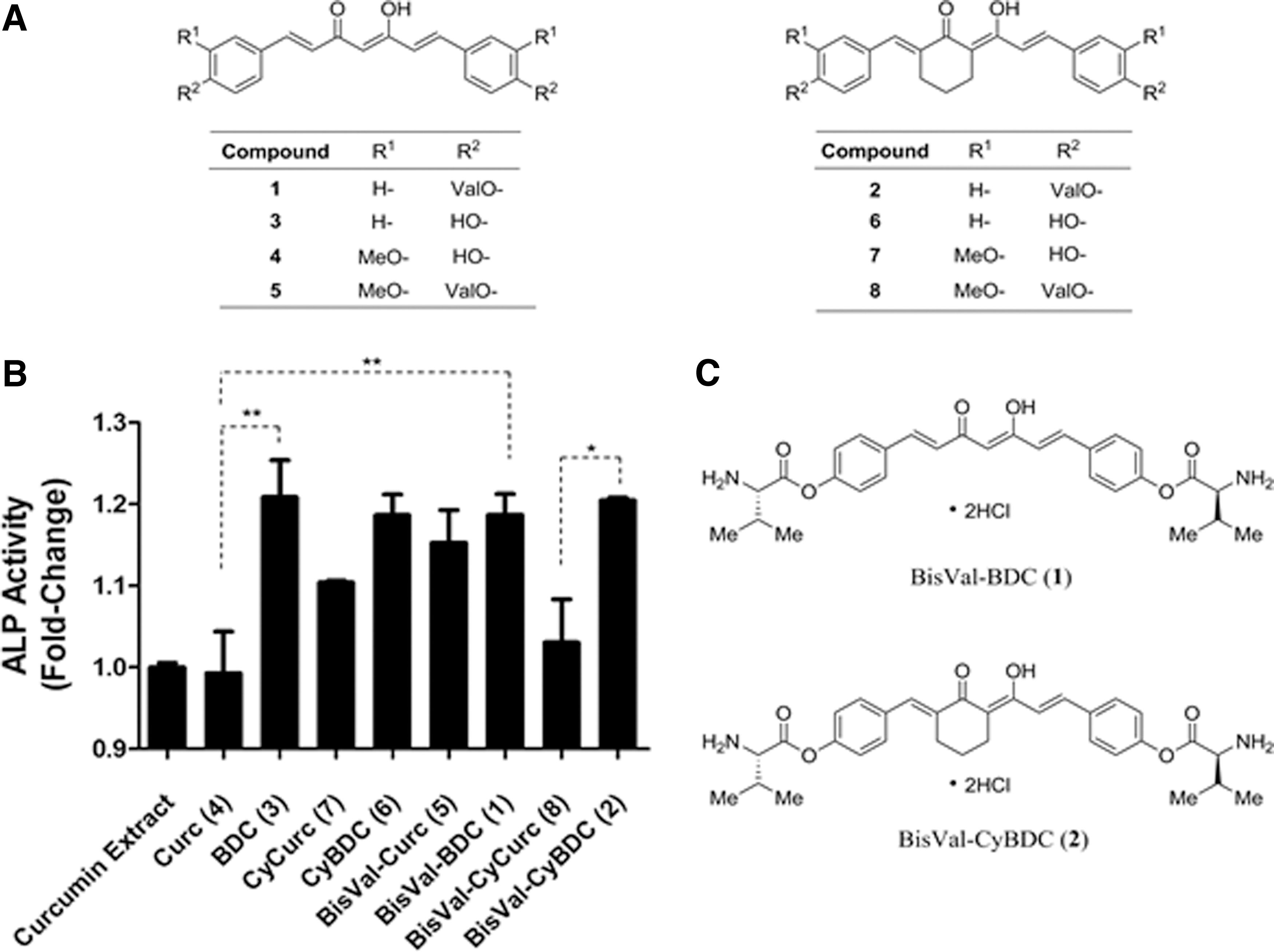
Naturally-occuring and synthetic curcumins tested for ALP activity in hBMSC osteogenic differentiation.
hBMSC differentiation with TCP and 1 or 2
Compounds 1 or 2 stimulated hBMSC osteogenic lineage commitment in the presence of osteoinductive TCP granules (i.e., 500–1000 μm granules). 4 Compounds 1 or 2 (500 nM) administered to hBMSCs incubated with TCP granules (5 mg/mL) for 8 or 25 days showed marked osteogenic differentiation based on ALP activity. For hBMSCs, ALP activity increased linearly from 0 to 5 mg/mL of TCP granules. Accordingly, 5 mg/mL TCP granules were used in osteogenesis experiments.
hBMSCs incubated with 1 (500 nM) for 8 days provided a 1.2-fold increase in ALP activity compared to vehicle-treated cells (Fig. 2A). hBMSCs treated with 1 (500 nM) + TCP granules (5 mg/mL) increased ALP activity by 2.0-fold after 8 days (significantly greater than TCP granules (1.4-fold, p < 0.05) or 1 alone (1.2-fold, p < 0.01)). After 25 days, hBMSCs treated with 1 or TCP granules alone increased ALP activity 1.8-fold and 2.2-fold, respectively. hBMSCs incubated with 1 and TCP granules showed a 3.1-fold increase in ALP activity (significantly greater than 1 (p < 0.001) or TCP granules alone (p < 0.001)). In hBMSCs, addition of 1 showed time-dependent osteogenesis based on ALP activity over 25 days that was enhanced by TCP granules.
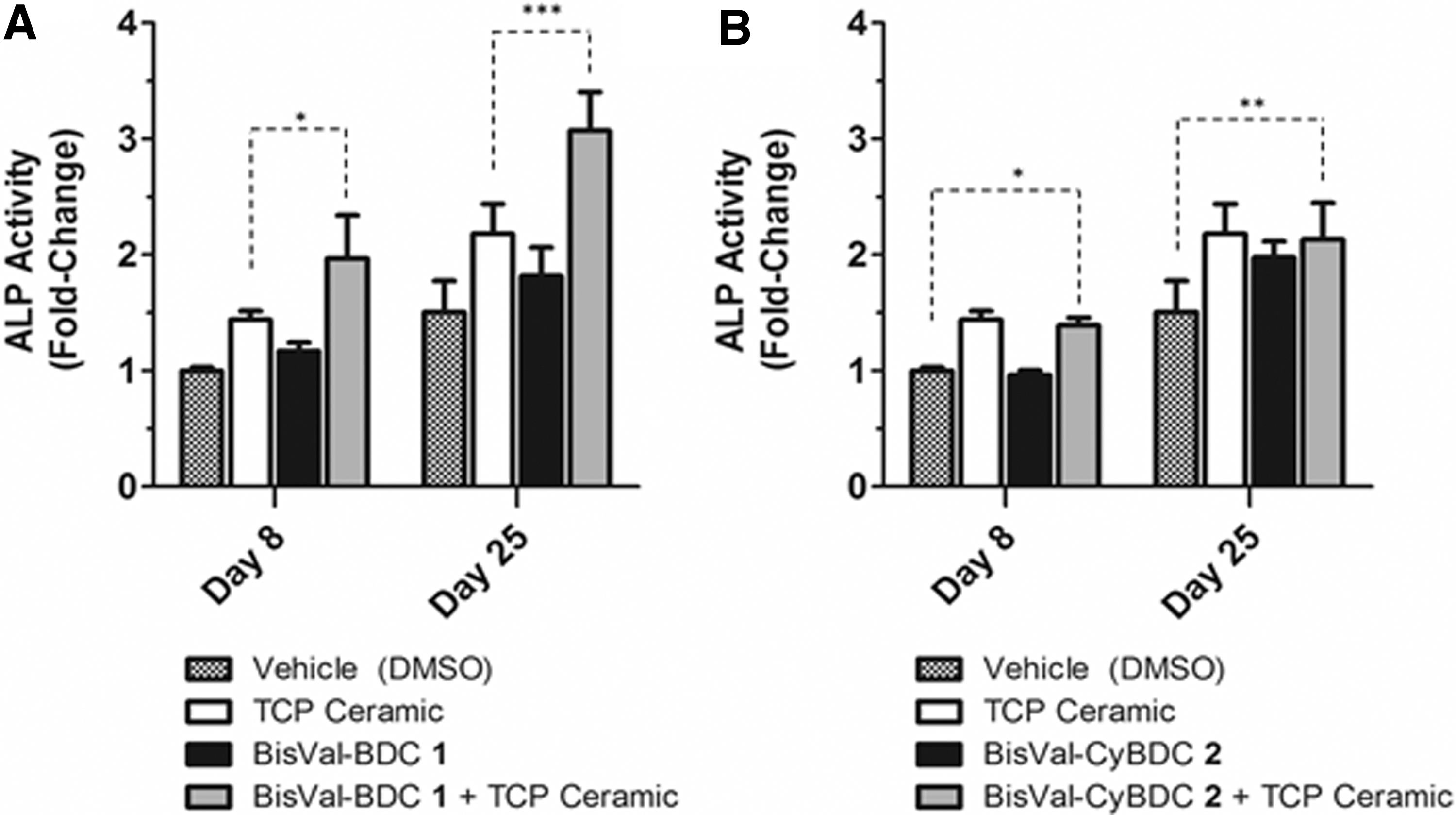
Effect of 1 or 2 on alkaline phosphatase activity in hBMSCs incubated in the presence or absence of TCP granules. hBMSCs were incubated with compounds 1 or 2 (500 nM), TCP granules alone (5 mg/mL), or compounds 1 or 2 (500 nM) in the presence of TCP granules. ALP activity was measured after 8 and 25 days, and results were normalized to vehicle (i.e., DMSO)-treated cells on day 8.
In hBMSCs, 2 + TCP granules stimulated osteogenesis (Fig. 2B). In the presence of TCP granules (5 mg/mL), hBMSCs treated with 2 (500 nM) stimulated ALP activity 1.4-fold after 8 days and 2.1-fold after 25 days. This effect was not statistically different than the effect of 2 or TCP granules alone at either time point. In summary, 1 and 2 both stimulated ALP activity in the absence of TCP granules, but the effects of 1 were greater than 2 in the presence of TCP granules.
Treatment of hBMSCs with 1 (500 nM) for 8 days induced a 1.7-fold, 1.6-fold, and 4.1-fold increase in Runx2, BMP2, and Osteocalcin, respectively (p < 0.001), compared to vehicle-treated cells (Fig. 3 and Supplementary Table S2). Treatment of hBMSCs with TCP granules alone (5 mg/mL) resulted in a 2.3-fold increase in Runx2 (p < 0.01) and a 4.7-fold increase in BMP2 (p < 0.01) compared to vehicle-treated cells. These results showed that either 1 alone or TCP granules alone could stimulate hBMSC osteogenic differentiation. However, greatest induction of osteogenic target genes for hBMSCs occurred in the presence of 1 and TCP granules (2.4-fold, 5.4-fold, 8.7-fold, and 6.3-fold increases in Runx2, BMP2, Osteopontin, and Osteocalcin, respectively (p < 0.001)). Effects of 1 and TCP granules on Osteopontin and Osteocalcin expression significantly exceeded the effect of 1 or TCP granules alone. To extend these studies, additional osteogenic biomarker quantification at 8- and 20-day hBMSC cultivation showed increases although each biomarker had its own time-dependent profile (Supplementary Data). Results showed that administration of 1 and TCP granules to hBMSCs significantly induced osteogenic target gene expression. To confirm the PCR results, Western blot studies were done for cells treated for 8 days. Western blot analysis of cell extracts from 8-day cultivated cells treated with 1 (500 nM) + TCP (5 mg/mL) showed a 31-fold increase in immunoreactive ALP. As a control, a separate Western blot for cells treated with VD3 (500 nM) showed a 13-fold increase in immunoreactive ALP.
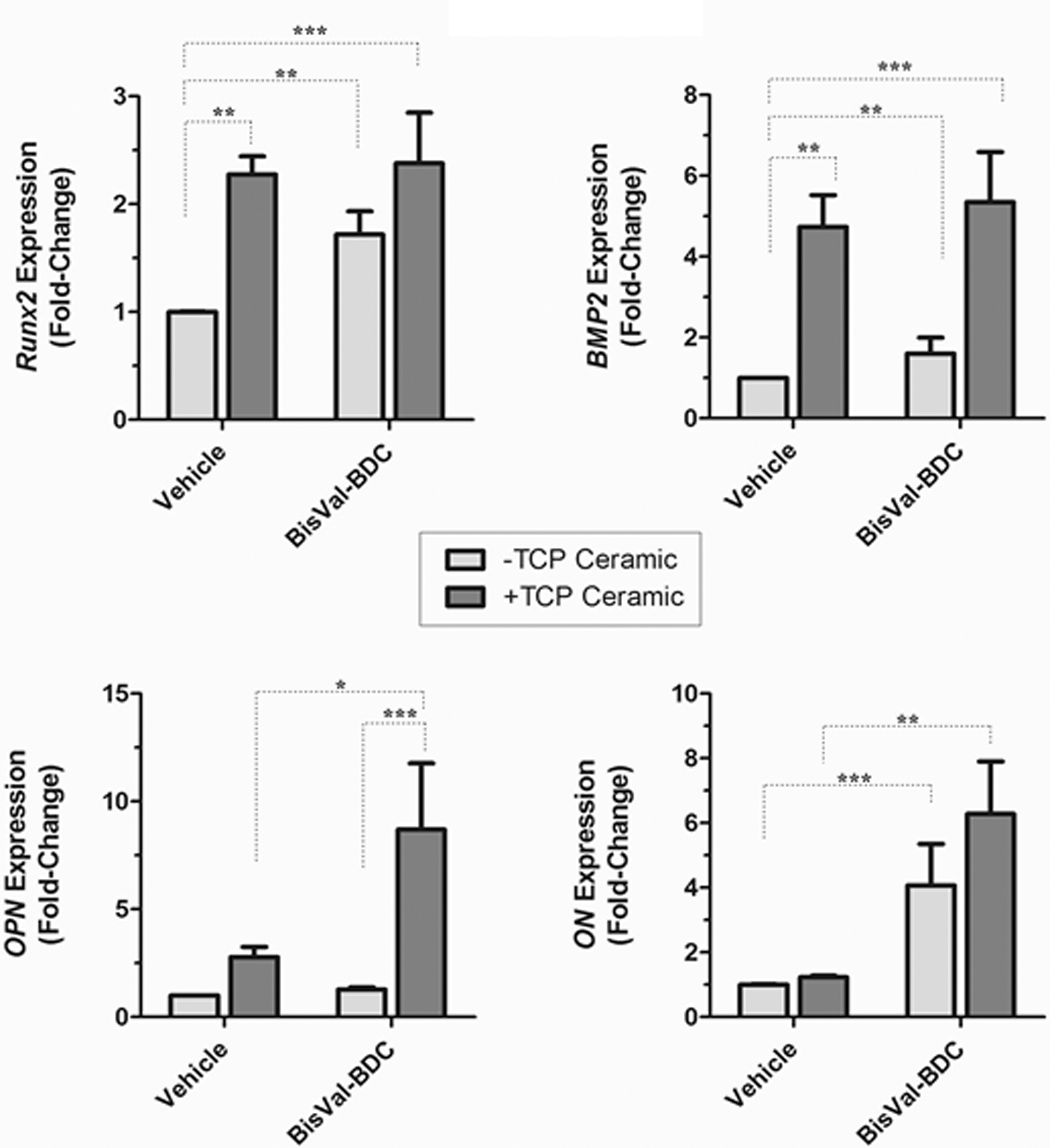
Effect of 1 or 2 on osteogenic gene expression in hBMSCs incubated in the presence or absence of TCP granules. hBMSCs were incubated with compounds 1 or 2 (500 nM) and TCP granules (5 mg/mL). Runx2, BMP2, OPN, and ON gene expression was determined by qPCR after 8 days. Relative gene expression levels were normalized to vehicle (i.e., DMSO)-treated cells and expressed as fold-change in expression levels. Data were mean with error bars for SEM (n = 4). *p < 0.05, **p < 0.01, ***p < 0.001.
Effect of 1 or 2 on Ca2+ deposition in hBMSCs
Accumulation of calcium is a biomarker of functional osteoblasts. Compared to vehicle-treated hBMSCs (Fig. 4A), 1 or 2 (500 nM) significantly increased Alizarin Red S calcium staining after 25 days of treatment (Fig. 4B, C). hBMSCs incubated with 1 or 2 + TCP granules (5 mg/mL) increased calcium deposition (Fig. 4D–F). Staining density quantification showed 1 and 2 increased calcium deposition in hBMSCs (2.0-fold), although analysis of staining in the presence of TCP granules was confounded by intrinsic calcium released from the ceramic (Fig. 4G, H). Results showed 1 or 2 to be equipotent for induction of calcium deposition in hBMSCs after 25 days in culture. In parallel, Oil Red O or Alcian Blue staining of hBMSCs treated with 1 or 2 ± TCP granules at day 25 showed no apparent hBMSC differentiation to adipocytes or chondrocytes, respectively (Supplementary Figs. S1 and S2).
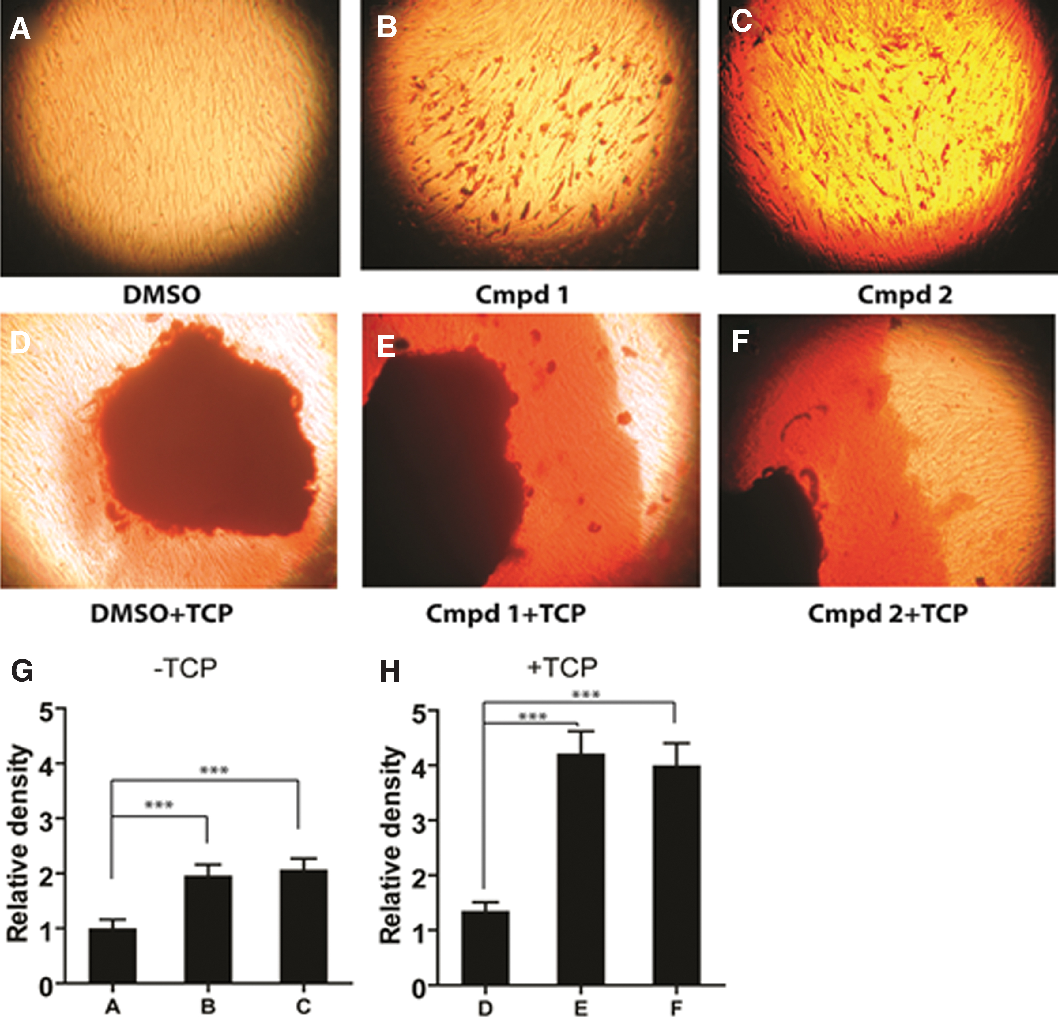
Effect of 1 or 2 on Ca2+ Mineral Deposition in hBMSCs in the presence or absence of TCP granules. hBMSCs stained with Alizarin Red S after incubation with 1 or 2 (500 nM) in the presence or absence of TCP granules (5 mg/mL) for 25 days. Staining intensity in the images was quantified by densitometry and normalized to vehicle (i.e., DMSO)-treated cells.
Effect of 1 or 2 on hBMSC viability
Cytotoxic effects of 1 or 2 (500 and 5000 nM) ± TCP granules were tested on hBMSCs in vitro. Gene expression for Bax and c-Fos that are upregulated in apoptosis showed that Bax and c-Fos were not detectable in any of the treatment groups as determined by qPCR. Cell viability of hBMSCs treated with 1 or 2 (500 nM and 5000 nM) ± TCP granules (5 mg/mL) measured by an Alamar Blue assay on day 25 showed no detectable decrease in cell viability compared to vehicle-treated hBMSCs. The results showed that in vitro differentiation of hBMSCs with curcumins ± TCP granules had no detrimental effects on the viability of hBMSCs.
Mechanistic studies of 1
Previously, we reported that small molecules modulate Wnt signaling and stimulate expression of Runx2 and BMP2 and promote osteogenesis. 32 The effect of curcumin 1 on expression of Runx2 or BMP2 and other target genes of Wnt-mediated osteogenesis that showed administration of 1 alone to hBMSCs showed a 101-fold enhancement of Runx2 (Fig. 5A). Coadministration of 1 and TCP granules showed an 8.2-fold increase in Runx2 expression (Fig. 5A). Administration of 1 or TCP granules did not have as strong an effect on expression of BMP2, but 1 increased BMP2 expression 7.5 fold in the presence of TCP granules (Fig. 5B). One stimulated Runx2 expression in hBMSCs, while in the presence of TCP granules, 1 caused a synergistic increase in expression of BMP2.

Effect of compound 1 on induction of hBMSC Wnt gene expression in the presence or absence of TCP granules: Mechanistic studies. hBMSCs were incubated with compound 1 (500 nM) in the presence or absence of TCP granules (5 mg/mL), and expression of Wnt inducible target genes was determined after 8 days in culture by quantifying mRNA with qPCR. Gene expression was normalized to vehicle (i.e., DMSO)-treated cells.
Expression of Wnt-related genes Axin2, Wnt3a, Wnt5a, and Sox9 was examined as biomarkers of Wnt-mediated hBMSC osteogenic differentiation. Axin2 is a negative regulator of Wnt signaling. One (500 nM) or TCP granules (5 mg/mL) inhibited Axin2 expression by 76% (Fig. 5C). Compound 1 or TCP granules alone did not significantly induce Wnt3a expression in hBMSCs, but 1 and TCP granules increased Wnt3a expression 62.3-fold (Fig. 5D). Thus, 1 plus TCP granules promoted Wnt3a and simultaneously inhibited expression of Axin2, a Wnt repressor gene. Administration of 1 to hBMSCs in the presence of TCP granules caused a 6.3-fold increase in Wnt5a expression (Fig. 5E). Wnt5a expression is an antiadipogenic biomarker in hBMSCs. 33 In hBMSCs, 1 plus TCP granules caused a 40% decrease in Sox9 expression (Fig. 5F). Sox9 expression is a chondrogenic biomarker. 34 Thus, 1 inhibited adipogenesis and chondrogenesis by inhibiting master regulatory genes in their respectful lineages.
Characterization of hBMSC-TCP implants
Based on the pharmaceutical properties and promising pharmacological properties of 1 plus TCP, we examined their ability to grow bone cells in vivo. A TCP-collagen scaffold that comprised 88% (wt/wt) TCP granules and 12% (wt/wt) bovine collagen I was used as a cell carrier scaffold for testing cell implants in an L4-L5 spinal fusion model in rats. Implants were designed to overlay on the dorsal aspect of a rat's vertebrae and extend 10 mm in total length to ensure extensive surface contacts with L4 and L5 transverse processes (Fig. 6A). The TCP scaffold had a 4-mm long projection designed to fill the void space between L4 and L5 transverse processes where fusion mass was desired. Average mass of the shaped TCP collagen scaffold was 145 ± 25 mg and absorbed 150 μL of an aqueous-cell solution. A radiograph of a rat that received bilateral 1-stimulated hBMSC implants of shaped TCP-collagen scaffold after 6 weeks is shown in Figure 6B. Greater than 95% of cells were adsorbed on a TCP-collagen scaffold after direct application of an hBMSC suspension in DMEM as determined by cell counting. Adsorbed hBMSCs remained viable on the TCP scaffold over the time frame of surgical implantation (i.e., <6 h) as shown by Live/Dead staining of implants (Fig. 6C). Results confirmed the presence of viable cells in implants used in spinal fusion experiments.
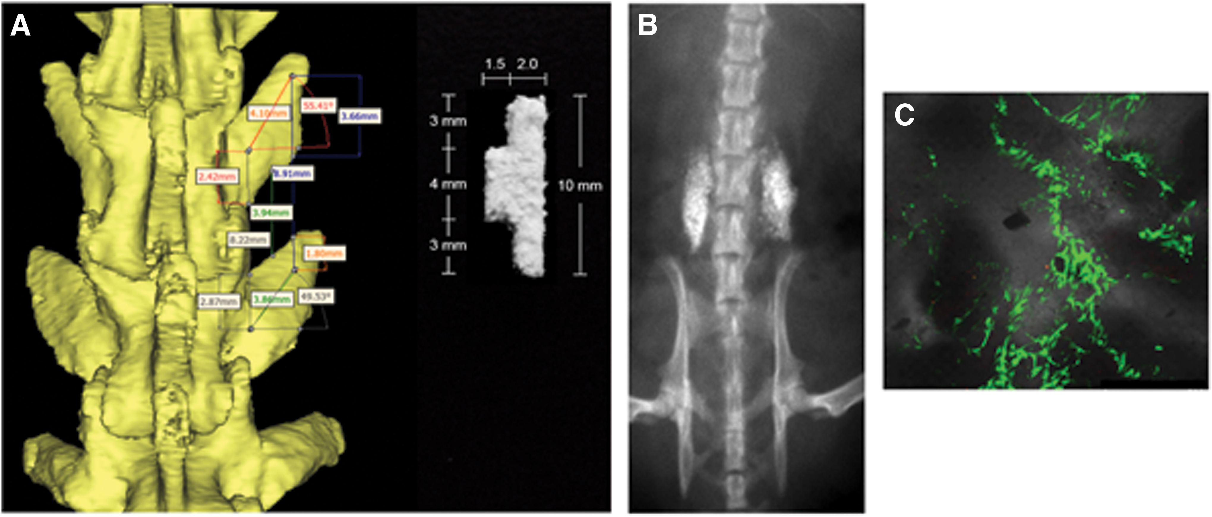
TCP-collagen scaffold used for posterolateral spinal fusion.
Excess hBMSCs that were not used in the in vivo studies (i.e., cells treated with 1 (500 nM) + TCP (5 mg/mL) or TCP alone) were used for another experiment. After 6 days of cultivation in vitro, excess hBMSCs that were not used in the in vivo fusion studies (Figs. 6 and 7) were cultured for an additional 2 days in maintenance media (α-MEM, 20% FBS, and 1 × GlutaMAX) in the absence of any additional compound treatment. At the end of this 2-day period, cells were isolated and prepared for ALP functional activity as described herein. Compared to TCP alone treated cells, cells previously treated with 1 + TCP showed a 1.2 ± 0.1-fold increase (p = < 0.05).

Effect of implants on L4-L5 spinal fusion in spinal segments.
Effect of 1-treated hBMSC-TCP scaffold implants in an immunodeficient rat model
Compound 1-treated hBMSCs combined with TCP scaffold afforded implants that were compared to untreated hBMSC-TCP scaffold or TCP scaffold alone implants in a rat model of posterolateral spinal fusion. Previously, we determined that an 8-day in vitro incubation period gave significant osteogenic lineage commitment. Thus, 8-day cultivated cells were harvested, or media alone was adsorbed onto TCP-collagen scaffolds. Three groups of immunodeficient rats (n = 7/group) received implants: (i) 1-stimulated hBMSC-TCP scaffold implants containing 0.7 M cells, (ii) untreated hBMSC-TCP scaffold implants containing 0.7 M cells, and (iii) TCP scaffold alone (i.e., no cells).
Following surgical implantation at L4 and L5 transverse processes, biweekly radiographs were taken. Weekly X-Ray time courses of spinal implants of animals analyzed with an exponential growth equation showed that 1-stimulated hBMSC-TCP scaffold implants, untreated hBMSC-TCP scaffold implants, and TCP scaffold alone (i.e., no cells) had a doubling time of 0.54 ± 0.03, 0.5 ± 0.02, and 0.53 ± 0.03 weeks, respectively. After 8 weeks, the growth was not statistically significantly different among groups (Fishers information parameter (p ≥ 0.05)). After 8 weeks rats were euthanized and analyzed further.
Intact rat lumbar spine segments (i.e., S4-L5) were removed from euthanized rats and analyzed for spinal fusion. Visual analysis was done by blinded trained observers. Motion of implants was evaluated under constant bending force, and implants were ranked for fusion (i.e., secure fusion mass between transverse processes), partial fusion (i.e., fusion mass bridging transverse processes maintained, but some motion observed), or nonfusion (i.e., implants were unsecure or did not bridge the transverse processes). Cell implants were ranked as either fused or partially fused by 8 weeks (Fig. 7A). No statistically significant difference was observed between 1-treated hBMSC-TCP implants (65% fused, 35% partially fused) and untreated hBMSC-TCP implants (75% fused, 25% partially fused). However, both cell implants gave greater fusion than TCP alone implants (42% fusion, 42% partial fusion, and 12% nonfusion). Visual bending evaluation showed for 1-treated hBMSC-TCP scaffold implants, untreated hBMSC-TCP scaffold implants, and TCP scaffold alone implants (i.e., 0.36 ± 0.1, 0.25 ± 0.1, and 0.71 ± 0.2, respectively) was not statistically significantly different (unpaired means comparison, p ≥ 0.05).
Accordingly, a more quantitative analysis of L4-L5 lateral bending of spine segments was done using three dimensional μCT disc height measurements as a quantitative spinal stability measure under constant bending forces. 35 Lumbar spinal segments (i.e., S1 to L1) were displaced in left lateral-bending (+10.0 mm) and right lateral-bending (−10.0 mm) positions from the neutral position for μCT disc height measurements using a custom radiopaque bending apparatus. Lateral displacement resulted in an average 15° bend in lumbar spines. Disc height deviation from the neutral position was determined by taking the sum of contraction and elongation of disc heights on left and right sides of the disc under lateral bending forces (Fig. 7B). Independent measurements from left and right bending were averaged to give disc height deviation from neutral position for each spinal segment. Using this quantifiable metric, 1-treated hBMSC-TCP scaffold implants showed the greatest stabilization of rat spinal segments at L4-L5 under lateral bending forces with an average total displacement from the neutral position of 22 μm (Fig. 7C).
By comparison, untreated hBMSC-TCP scaffold implants showed greater deviation of disc heights under lateral bending forces with an average total displacement of 34 μm. TCP scaffold alone implants gave an average of 44 μm total disc height deviation that represented a two-fold greater deviation (i.e., p < 0.05) in disc height deviation compared to 1-treated hBMSC-TCP scaffold implants. Results showed that 1-treated hBMSC-TCP scaffold implants achieved spinal fusion and provided greater stability of the spinal column than untreated hBMSC-TCP scaffold implants or TCP alone implants. 36
Explants were analyzed for extent of bone formation by histology (Fig. 8). Nondecalcified histological sections (Goldner's Trichrome) were quantified by densitometry analysis to compare surface area of bone in sections of fusion mass. Compound 1-treated hBMSC TCP scaffold implants resulted in new bone tissue in 1% of total histological surface area of tissue sections examined, while much of the other tissue was a dense collagenous tissue that bone growth plates appeared to have emerged. Compound 1-treated hBMSC-TCP scaffold implants had 2.0-fold greater bone surface area in fusion mass than untreated hBMSC-TCP scaffold implants (0.5% surface area, p < 0.1). Compound 1-treated hBMSC-TCP scaffold implants afforded 5.0-fold greater bone surface area in the fusion mass compared to TCP scaffold alone implants (0.2% surface area, p < 0.05). This analysis specifically excluded regions of contiguous bone mass with the transverse processes that largely underestimated total new bone formation in implants (i.e., de novo bone tissue could not be definitively resolved against the transverse processes despite administration of fluorescent bone labels and was excluded from this analysis).
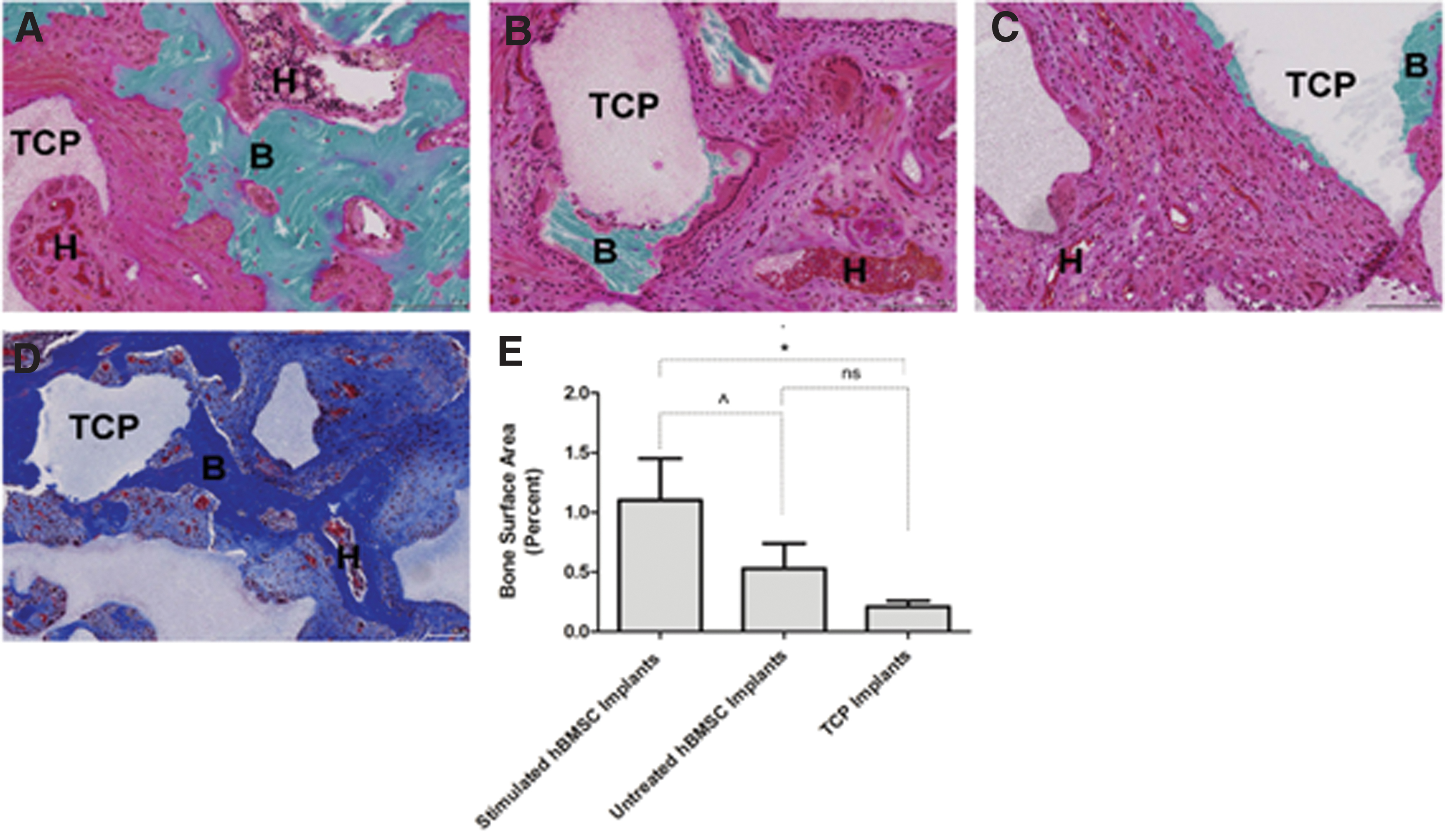
Histology of L4-L5 fusion mass from implants
Greater bone formation associated with 1-treated hBMSC TCP scaffold implants manifested as contiguous growth plates that emerged from an amorphous tissue surrounding TCP scaffold. Residual TCP scaffold was observed to have incorporated osteoblasts on their periphery, but the scaffold themselves were not noticeably remodeled after 8 weeks. In addition to bone tissue, extensive hematopoietic territories in histological sections were observed that supported good biological integration of implants in all treatment groups, including the noncell group (i.e., TCP scaffold alone). No significant differences were found among treatment groups for hematopoietic territory surface area by densitometry analysis of histological sections. Overall, the histological results showed that spinal segments receiving 1-treated hBMSC-TCP scaffold implants provided more mature fusion mass and twice as much bone after 8 weeks compared to untreated hBMSC-TCP scaffold implants.
Discussion
Curcuminoids can be used safely37,38 to promote osteogenesis in hBMSCs. Curcumin and minor component BDC (i.e., 3, Fig. 1) 25 have been used safely for oncology, inflammation, and immune modulation. 36 BDC is more potent than curcumin in upregulation of certain genes, including vitamin D3.28,37
Comparison of BDC prodrug (i.e., 1) to BDC (3) showed that water-soluble prodrug 1 was a more potent inducer of hBMSC proliferation. Administration of 1 or 2 to hBMSCs promoted osteogenic differentiation in vitro, but in the presence of osteoinductive TCP granules, cell differentiation was markedly increased. Results with 1 and 2 contrast those of curcumin on rat MSCs that showed induction of ALP but only at elevated concentrations (i.e., 10–20 μM). 24 1 and 2 induced hBMSC osteogenic differentiation 20-fold greater than 4 likely due to increased solubility or stability/availability.38–40 Prodrug 1 is a more potent inducer of hBMSC osteogenic differentiation than curcumin (3).
Previously, in C2C12 cells, we showed that small molecules induced Runx2 and BMP2 expression for synergistic osteogenic differentiation. 32 This is similar to cooperativity between Wnt and BMP2 signaling for osteogenic differentiation of hBMSCs.41–43 In C2C12 cells, compared to BMP2 (100 ng/mL), compound 1 (500 nM) + TCP granules (5 mg/mL) was equal or superior to induction of gene markers BMP2, ALP2, Col 1, and Runx2 (unpublished). Small molecule compound 1 may thus possess properties that simulate BMP2 but avoid adverse effects of BMP2.
In hBMSCs, 1 upregulates Runx2, Wnt3a, and BMP2 and decreases expression of Axin2 (a pro-adipogenic factor44,45) in hBMSCs (Fig. 5). Compound 1 decreased Sox9 (a chondrogenic factor in hBMSCs46,47). Thus, 1 inhibited both adipogenesis and chondrogenesis during promotion of osteogenesis in hBMSCs. While the molecular target of 1 is unknown, curcumin is an inhibitor of glycogen synthase kinase-3β (GSK-3β).48,49 Wnt activation by 1 extends the scope of curcumins that activate this signaling pathway. Compound 1 increased TLR expression during osteogenic differentiation. hBMSCs show a wide range of TLR expression. 50 TLR4 stimulation is correlated with osteogenesis in hBMSCs, and TLR3 stimulates adipogenesis. 51 Compound 1-treated hBMSCs increased cell migration and TLR4 expression and may help explain osteogenic differentiation and migration of hBMSCs to TCP like the postulated roles of TLRs in chemotaxis. 52
hBMSCs treated with 1 synergized osteogenesis of TCP. 53 Osteoinductive properties of TCP used herein correlated with microstructure of particle surface. 4 Protein binding might be an important factor in mediating osteoinduction whereby growth factors and morphogens adsorb onto the surface of TCP to promote osteogenesis54,55 and provide a microenvironment for bone growth. 56 It is notable that compound 1 adheres to TCP granules very efficiently (nearly 100%), and this may contribute to the synergistic potency of the combination. Calcium release from TCP may also contribute to this microenvironment.57,58 Stimulation of hBMSCs with 1 that modulates osteogenic signaling affords marked effects on cell fate and provides a novel approach to inducing cell differentiation. In contrast, TCP alone showed little effect on expression of Wnt target genes examined in this study.
hBMSCs have been cultured in the presence of osteogenic media 31 and combined with a carrier substance for transplantation.59–62 Administration of dexamethasone to hBMSCs in osteogenic media before implantation resulted in improved bone formation. 63 Similarly, hBMSCs cultured with calcium phosphate microspheres and transplanted afforded improved bone formation in a mouse calvarial defect model. 64 Osteogenic media has not always provided improved bone growth following transplantation of cells, 65 and paradoxically, long-term effects of glucocorticoids in vivo led to promotion of bone loss. 66 Purmorphamine applied to hydroxyapatite discs increased bone formation after implantation. 67 Compound 1-treated hBMSCs grown in the presence of TCP granules can serve as a standalone replacement of osteogenic media. The advantage of this ex vivo cell model approach is that cells can then be placed onto a similar TCP scaffold microenvironment used in in vitro induction and implanted in vivo. Addition of 1 in the presence of TCP granules to osteogenic media (unpublished) showed greater stimulation of osteogenic markers. Taken together, results show that 1 with osteoinductive TCP granules was highly osteoinductive to hBMSCs.
Eight-day ex vivo induction of hBMSCs by 1 and TCP granules and then implantation on a TCP scaffold afforded osteoid-like tissue formation following implantation in vivo. hBMSCs induced with 1, implanted in rats, and analyzed 8 weeks later showed dense osteoid-like tissue present in induced cell implants that were generally lacking in implants from untreated cells. Progression to mature bone may require a longer time course.
In conclusion, 1 modulated osteogenesis of hBMSCs, including proliferation, differentiation, and mineralization. These effects were greatly enhanced when 1 was administered to cells cultured in the presence of TCP granules. Administration of small-molecule 1 to hBMSCs in the presence of TCP to induce osteogenesis avoids challenges of systemic administration of small molecules or a biologic. This procedure may also be beneficial in a situation where stem cells originate from patients with risk factors or older age. Thus, findings reported herein represent a new strategy to prepare cell therapies for bone repair and regeneration.
Footnotes
Acknowledgments
The authors thank Dr. Pam Robey of the National Institute of Health for hBMSC cells. The authors also thank Dr. Tim Moseley of NuVasive, Inc., (San Diego, CA) for the TCP granules and scaffold.
Disclosure Statement
No competing financial interests exist.
Funding Information
No external funding was received. Funding was from the Human BioMolecular Research Institute.
References
Supplementary Material
Please find the following supplemental material available below.
For Open Access articles published under a Creative Commons License, all supplemental material carries the same license as the article it is associated with.
For non-Open Access articles published, all supplemental material carries a non-exclusive license, and permission requests for re-use of supplemental material or any part of supplemental material shall be sent directly to the copyright owner as specified in the copyright notice associated with the article.