
Abstract
Porous materials containing cells—prepared via cell seeding on scaffolds or gelation of cell-containing solutions—have been widely studied to investigate tissue regeneration and three-dimensional cultures. However, these methods cannot introduce cells into porous materials that have low water absorption or scaffolds that require cytotoxic solvents or processes for their production. In this study, first, three different impregnation treatments conditions (vacuum, pressure, and vacuum pressure impregnation: VPI) were applied to cell suspensions to evaluate the effect of each treatment on cells. Following all three treatments, fibroblasts adhered to the cell culture dish and proliferated in the same manner as untreated cells, which confirmed that the three impregnation treatments did not affect cell function. Second, cells were introduced into a poly-
Impact statement
Poor cell infiltration into the porous scaffold material is a major issue for tissue regeneration. In this article, a new method for introducing cells into porous materials using impregnation techniques is presented. This method makes it possible to introduce various cells into the porous material and has potential applications in tissue regeneration and three-dimensional culture using cell-containing porous materials.
Introduction
Tissue regeneration using cell transplantation has been reported for tissues, including cartilage, bone, and soft tissue.1–3 In particular, cell sheet techniques have been applied to the treatment of heart infarcts and cornea diseases.4,5 Using these techniques, tissue regeneration, by small-scale cell transplantation, or cell sheet transplantation up to several layers, is becoming possible. However, large-scale cell transplantation and using five or more cell sheets for transplantation has been found to be insufficient for tissue regeneration because of a shortage of blood and nutrient supply. 6 Furthermore, because cells have low mechanical strength, it is difficult to maintain a structure at sites requiring strength using only cells.
Hence, scaffolds have been used for the regeneration of tissues that are not suitable for cell transplantation. These scaffolds have a role in supporting cells and are used as alternatives to the extracellular matrix. Porous materials, which are one type of scaffold, are materials with a large number of continuous holes, and cells are thought to infiltrate into the voids. 7 Bone tissue and adipose tissue regeneration using porous materials have been reported based on host cell infiltration into the material in vivo, and seed cell infiltration into the material, respectively.7,8
However, this method has limitations; for example, cell infiltration into the porous material can lead to oxygen and nutritional deficiencies inside the porous material, 9 and cells may not infiltrate into the porous material depending on the pore size and nature of the material. In addition, cell infiltration cannot be expected in transplantation into tissues whose volume comprises few cells and dense extracellular matrix, such as cartilage tissue.10,11
The seeding of cells into porous materials in vitro has been studied to address these limitations. 12 The cell-containing material is prepared by allowing the seeded cells to migrate from the surface of the porous material through the pores. Because this happens through cell migration or diffusion, it takes time for the cells to infiltrate the material and the cells may not infiltrate depending on the pore size and the type of porous material. 13 In addition, because the inside of the material is hypoxic and malnourished, it is difficult for cells to migrate into a large porous material. 14 Furthermore, cell seeding is not suitable for cells with low migration ability such as bone marrow stem cells or for the introduction of cells into hydrophobic porous materials. 15
Poly-
The gelation of cell dispersions is an alternative method to cell seeding for rapidly producing cell-containing materials. 19 Cell-containing materials containing collagen and agarose are also used for three-dimensional cell culture. 20 The gelation of mixtures containing cells is only possible for materials that can induce gelation under conditions, in which the cells can survive, which results in low mechanical strength. Because each method has disadvantages, a method for introducing cells into various porous materials in a short time remains desirable.
Various composite materials have been produced using vacuum treatment, pressure treatment, and vacuum pressure impregnation (VPI); a procedure that combines the two treatment methods.21,22 The VPI method is used in the fields of metal and food processing for scratching the surface of materials and for introducing a solution into the interior of foods. When the porous material and the solution are brought into contact with each other under vacuum, the solution permeates into the interior of the material and then pressurization enables the efficient introduction of even highly viscous solutions.
Our research group has reported the use of VPI impregnation for the introduction of a solution into a biological material and for the preparation of a composite porous material. It was found that in the introduction of saline into lyophilized decellularized blood vessels and the cornea, the solution was introduced in a shorter time by VPI than by immersion. 23 It was also shown that PLA-collagen composite material can be prepared by introducing collagen solution into PLA porous material by VPI. 24
In this study, we investigated the applicability of VPI—a combined vacuum and pressure treatment found to be an improvement over the previous method—as a method of introducing cells into a porous material. PLA was used as the porous material and cells were introduced into the PLA by VPI impregnation, as well as vacuum and pressure treatments, and the number of cells after culture and distribution of cells in the material were evaluated.
Methods
In the first experiment, the effects of the three impregnation treatments on cell function (adhesion, proliferation) were evaluated using cell suspensions only. For the vacuum treatment, the cell suspension was introduced into a vacuum sealed vial through a syringe and left for 2 min. For the pressure treatment, the cell suspension was sealed in a polyethylene bag and hydrostatic pressure was applied for 5 min. For the VPI treatment, the vacuum-treated cell suspension was sealed in a polyethylene bag, and hydrostatic pressure was applied for 5 min. The three treated cell suspensions and an untreated control suspension were seeded in a cell culture dish, cultured, and cell morphology observation and cell number measurement (days 1, 3, and 5) were performed.
In the second experiment, the cell suspension was introduced into PLA scaffolds using the three impregnation treatments (Fig. 1). Vacuum treatment was performed using frozen vials containing PLA scaffolds that were sealed using a freeze dryer. The cell suspension was injected into the vacuum sealed vial containing a PLA scaffold using a syringe, and left undisturbed for 2 min. In the pressure treatment, the PLA scaffold and the cell suspension were sealed in a polyethylene bag, and then hydrostatic pressure was applied for 5 min. In addition, VPI was performed by pressure treatment after vacuum treatment. The PLA scaffolds impregnated with cells using the three treatments, and a PLA scaffold immersed in a cell suspension as a control, were cultured for a given period of time, and then the cell introduction and cell proliferation were evaluated.

Schematic diagrams illustrating the three impregnation treatments. Color images are available online.
Experiment
Experimental design
Materials
PLA powder was purchased from Akina, Inc. (West Lafayette, IN). 1,4-Dioxane and ethanol were purchased from Wako. L929 cells were selected for the experiment and Dulbecco's modified Eagle's medium (with 10% fetal bovine serum and 1% penicillin/streptomycin) was used for cell culture. A Cell Counting Kit-8 (Dojindo, Kumamoto, Japan) was used to quantify live cells. A Live/Dead Cell Staining Kit II (PromoCell, Heidelberg, Germany) was used to distinguish between live and dead cells. In addition, the amount of DNA in the PLA scaffold was measured using a Quant-iT™ PicoGreen® dsDNA Assay Kit (Invitrogen, Carlsbad, CA).
Evaluation of the effect of the impregnation methods on cells
A mouse fibroblast (L929) suspension (5.0 × 10⁵ cells/mL) was used. In the vacuum treatment, this suspension (3 mL) was added to a vacuum sealed vial (11 Pa) and left at room temperature for 2 min. In the pressure treatment, a polyethylene bag containing the cell suspension (3 mL) was subjected to a hydrostatic pressure of 1.45 MPa for 5 min. In the VPI treatment, the cell suspension was evacuated for 2 min, then the cell suspension was sealed in a polyethylene bag and pressure treated for 5 min. The three impregnation method treated cell suspensions were diluted with medium (1.0 × 10⁴ cells/mL) and seeded on a 24-well plate. In addition, an untreated cell suspension was seeded as a control.
The cell morphology was observed under a microscope on days 1, 3, and 5 of the culture. Cell Counting Kit-8 was added to each well, and the number of live cells was evaluated by measuring the absorbance (450 nm). On the first day of seeding, live cells were stained with calcein-AM and dead cells were stained with Ethidium Homodimer III, and samples were observed with a fluorescence microscope.
PLA scaffold preparation
A dioxane solution containing 10% PLA was placed in a stainless-steel container and sealed in a polyethylene bag. The PLA solution was frozen by immersing the polyethylene bag in ethanol at −80°C overnight. The frozen PLA was immersed in ethanol at −80°C to remove the dioxane, and then the PLA was immersed in 70% ethanol to sterilize. Finally, the PLA scaffold (cylindrical shape; diameter 8 mm, height 10 mm) was obtained by air-drying on a clean bench. 25
Cell introduction into the PLA scaffold and cell culture in the PLA scaffold
Cell suspension (3 mL, 5.0 × 10⁵ cells/mL) was injected into vials containing the PLA scaffold and kept at room temperature for 2 min (vacuum-treated PLA: V-PLA), or the PLA scaffold and cell suspension (3 mL) were sealed in polyethylene bags and a hydrostatic pressure of 1.45 MPa was applied for 5 min (pressure-treated PLA: P-PLA), or hydrostatic pressure was applied to the PLA scaffold into which the cell suspension had been introduced by vacuum treatment (VPI-treated PLA: VPI-PLA). In addition, as a control for the three impregnation treatments, the PLA scaffold was immersed in the cell suspension for cell seeding (immersion-treated PLA: I-PLA).
The introduction rate of the cell suspension was calculated from the weight of the PLA scaffold before introducing the cell suspension (Wp) and the weight of PLA after introducing the cell suspension (Wi) using the following formula.
In addition, cell-free medium was introduced into the PLA scaffold using the three impregnation treatments and the immersion treatment, and the introduction rate of the medium was calculated.
Observation of cells in the PLA scaffold by scanning electron microscope (SEM)
Cell-free PLA scaffold and the PLA scaffolds, into which the cell suspension was introduced, were fixed with 4% paraformaldehyde, dehydrated stepwise, and immersed in t-butyl alcohol. The PLA scaffold soaked in t-butyl alcohol was freeze-fractured into two pieces at the center, dried under reduced pressure, and observed using a scanning electron microscope. The observation ranges were from the surface of the PLA scaffold to 500 μm deep at the end of the cylinder and 5 mm from the surface of the PLA scaffold in the center.
DNA quantification to evaluate cell proliferation within the PLA scaffolds
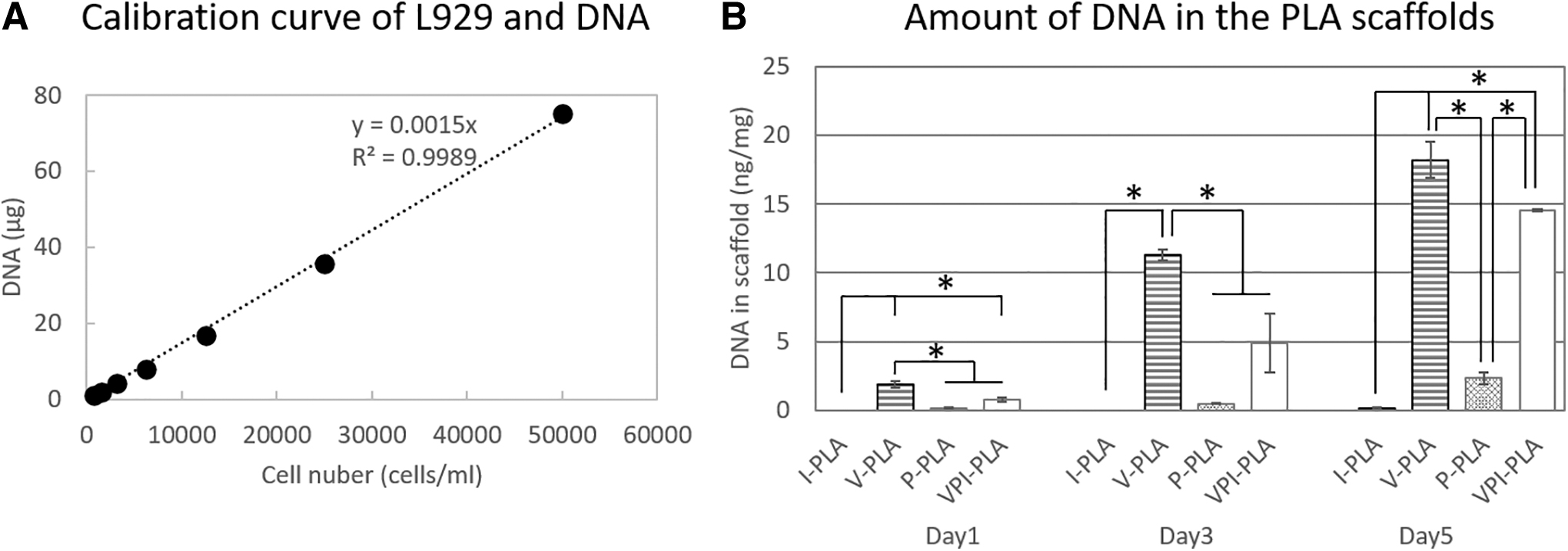
After introducing the cells into the PLA scaffold, each PLA scaffold was immersed in medium and cultured. After culturing for a certain period of time, the PLA scaffold was solubilized, subjected to phenol/chloroform extraction and ethanol precipitation, and the DNA in the PLA scaffold was purified. The purified DNA was quantified using PicoGreen, and the amount of DNA in each PLA scaffold was calculated. In addition, L929 cells (5.0 × 10⁵, 2.5 × 10⁵, 1.25 × 10⁵, 6.25 × 10⁴, 3.12 × 10⁴, 1.56 × 10⁴, and 7.81 × 10³ cells/mL) were seeded on a 96-well plate, DNA was extracted, and after purification, the DNA was quantified with PicoGreen to prepare a calibration curve.
Statistical analysis
The statistical significance of differences was evaluated by analysis of variance, with probability values for the in vitro assays calculated by Student's t-test. A value of p < 0.05 was considered statistically significant. Data are presented as mean ± standard deviation (*p < 0.01).
Experimental Results
Evaluation of the effect of the impregnation methods on cells
The impregnation treatments were applied to the cell suspensions, the number of adhered cells was measured, and the influence of the impregnation treatment on the cells was evaluated (Fig. 2). The numbers of cells in the impregnated groups were comparable to the number of untreated cells at day 1. No significant difference in the number of adhered cells was observed between the untreated and treatment groups at 3 and 5 days after cell seeding.
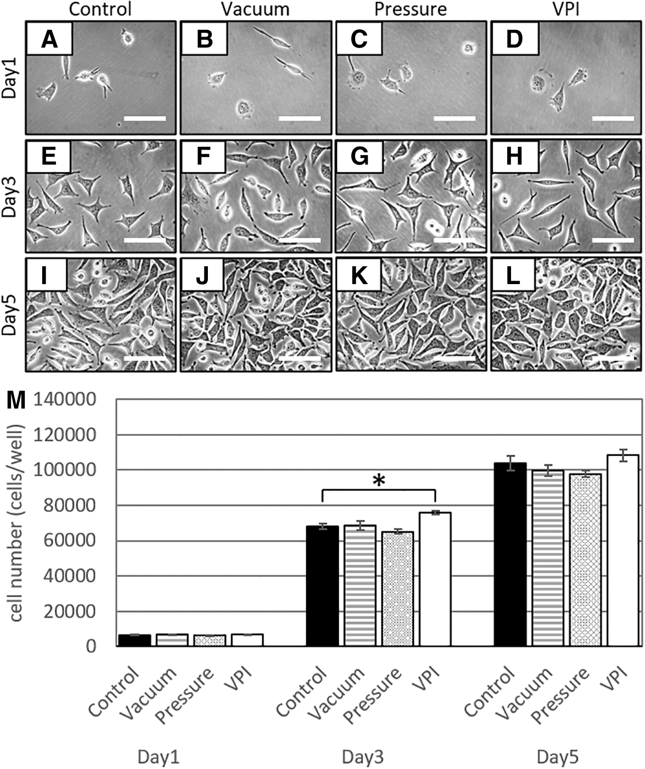
Images of L929 cells after different impregnation treatments.
The cell morphology, dead cells, and living cells were observed using Live and Dead staining 1 day after the impregnation treatment (Fig. 3). The cells in all treatment groups adhered to the dish and were spindle-shaped. Most of the cells in the treated groups were live cells and few dead cells were observed, similar to the observation for the untreated group. The cell viabilities were as follows; untreated 99% ± 2%, vacuum treatment 99% ± 2%, pressure treatment 98% ± 2%, and vacuum pressure treatment 98% ± 2%.
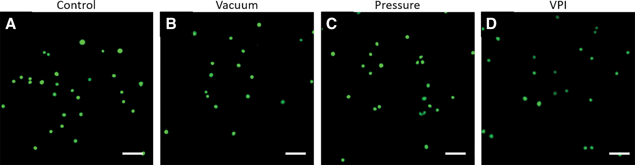
Images of Live and Dead stained cells after different impregnation treatments.
Comparison of the introduction rates of the cell suspensions into the PLA scaffolds
The immersed, pressurized, and evacuated PLA scaffolds floated on the medium. Conversely, the VPI-PLA scaffold material sank into the medium (Fig. 4A). The PLA scaffold immersed in the cell suspension had almost no cell suspension introduced and the surface and cross section remained white. In the P-PLA scaffold, some cell suspension was observed near the surface; however, the center of the scaffold was white. V-PLA and VPI-PLA had cell suspension introduced throughout the material (Fig. 4B).
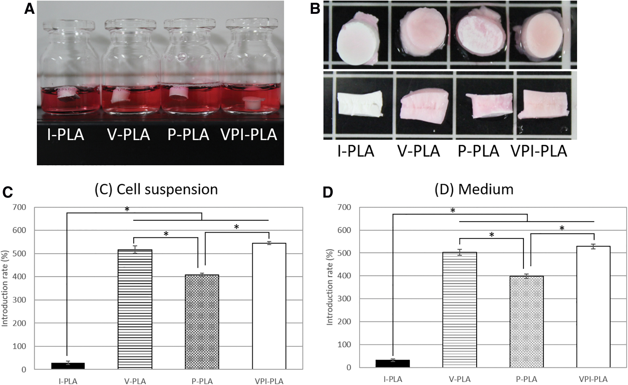
The cell suspensions and cell-free medium were introduced into the PLA scaffold, and the introduction rate for each treatment was calculated from the weights before and after the introduction (Fig. 4C, D). The PLA scaffold that had been immersed in the cell suspension and the PLA scaffold into which the suspension was introduced by pressure treatment floated in the cell suspension, and the cell suspension introduction rates were 29.2% and 407.5%, respectively. The vacuum-treated and VPI-PLA scaffolds were submerged in the cell suspension, and the cell-suspension introduction rates were 517.5% and 544.6%, respectively. Similarly, for the PLA scaffolds treated with cell-free medium, the introduction rate resulting from immersion treatment was the lowest, followed by the introduction rate because of pressure treatment, and the introduction rates because of vacuum treatment and VPI treatment were almost the same.
PLA scaffold structure and introduction of cells into the PLA scaffolds by the impregnation treatments
The PLA scaffold was prepared as a cell introduction material using a freeze extraction method, and the structure of the PLA scaffold was observed by SEM (Fig. 5A, B). The SEM image of the PLA scaffold showed many pores of ∼25 μm in the PLA material.
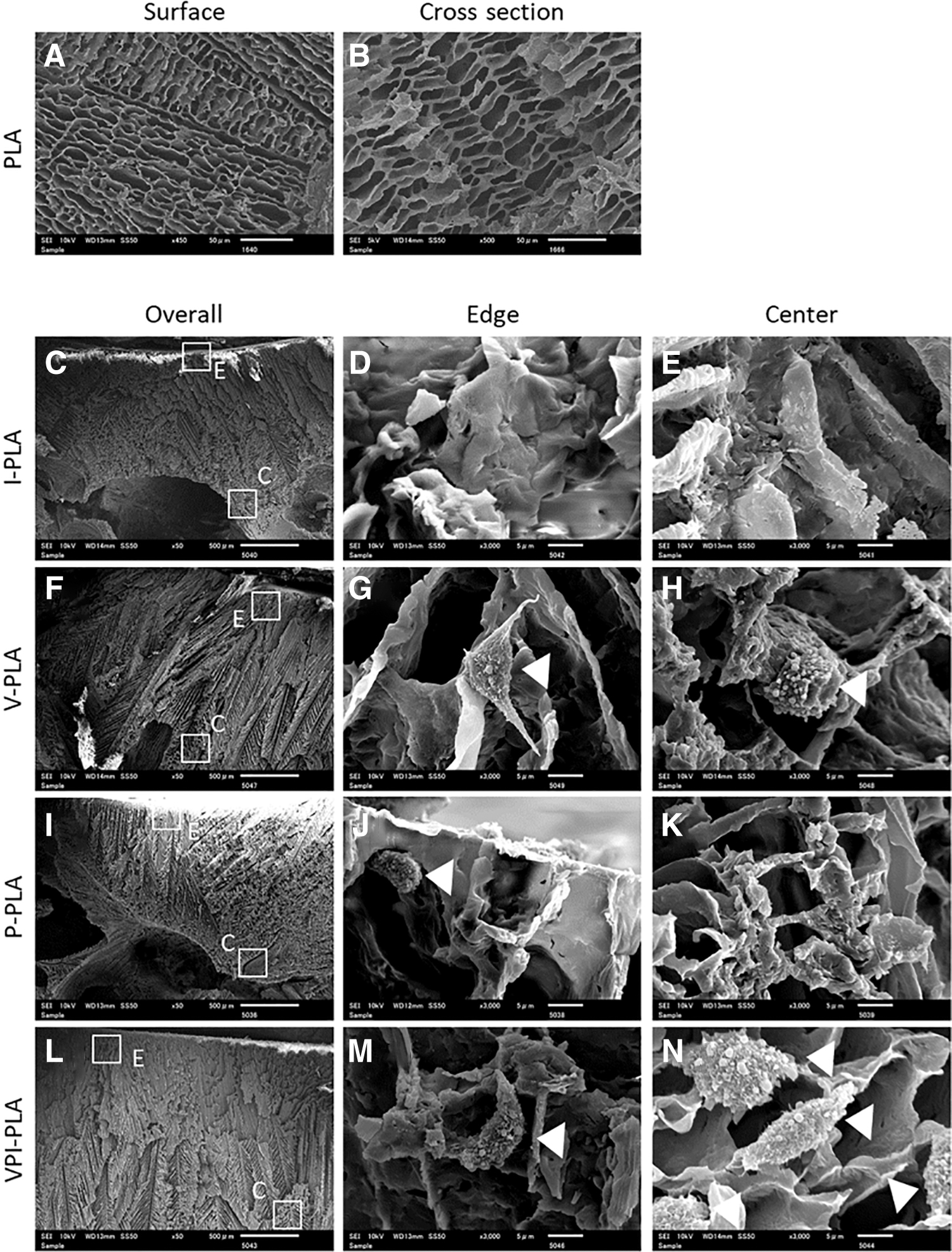
SEM images of cell-free PLA scaffold and PLA scaffolds 5 days after cell impregnation.
The cells within the PLA scaffold were observed using SEM. In the SEM observations immediately after cell introduction, no cells were observed in any of the PLA scaffolds (data not shown). On day 5, no cells were observed on the surface or inside of I-PLA. Furthermore, cells were found on the surface of P-PLA, but no cells were found inside P-PLA. Conversely, in V-PLA and VPI-PLA, cells were observed both on the surface and inside the scaffolds (Fig. 5).
The amount of cells within the PLA scaffolds was evaluated by DNA quantification (Fig. 6). Because there is a positive correlation between the amount of DNA and the number of L929 cells on the calibration curve, the number of cells could be evaluated from the amount of DNA in the PLA scaffold. One day after transfection, the I-PLA scaffold had a DNA content of 0.01 ng/mg, the V-PLA scaffold had a DNA content of 1.89 ng/mg, the P-PLA scaffold had a DNA content of 0.17 ng/mg, and the DNA content of the VP-PLA scaffold was 0.81 ng/mg. The amount of DNA at 3 and 5 days after cell introduction was 0.03 and 0.17 ng/mg for the I-PLA scaffold, 11.3 and 18.22 ng/mg for the V-PLA scaffold, 0.51 and 2.34 ng/mg for P-PLA, and 4.91 and 14.54 ng/mg for the VP-PLA scaffold, respectively.
Discussion
Cell-containing porous materials are mainly produced by seeding cells on a scaffold or gelation of a cell-containing solution. Cell seeding is not dependent on cell diffusion or migration and is difficult to apply when using scaffolds with low water absorption or slow migration rates. Gelation of cell-containing solutions requires noncytotoxic solutions and gel processes, which limits the materials that can be used. The PLA scaffold used in this study has low water absorption, and the manufacturing process involves a cytotoxic solution or a cytotoxic process, which makes it difficult to introduce cells by conventional methods.
In this study, the introduction of cells into porous materials using impregnation techniques (vacuum, pressure, and VPI) was examined. To evaluate the effect of cell introduction by the different impregnation techniques on the cell function, a cell suspension was subjected to vacuum, pressure, and VPI treatments, and the number of adhered cells was evaluated. The number of adhered cells after each treatment was similar to that of untreated cells, and the cell morphology was also similar to that of untreated cells. These results indicated that the impregnation treatments did not affect cell adhesion and spreading. In addition, the number of adhered cells after treatment for 3 and 5 days did not differ to that of untreated cells, and the number of cells was increased in all groups, confirming that impregnation did not affect cell proliferation.
Live/Dead staining on the first day of seeding showed almost no dead cells for all impregnation treatments, and it was revealed that the three impregnation treatments did not affect the survival of fibroblasts. Previous studies have shown that pretreatment of mammalian cells with a solution containing glycerol suppressed the decline in survival and proliferative potential even after exposure to vacuum conditions for 200 s. 26 Based on this study, we speculate that a short evacuation does not affect cell function when a solution is present around the cell. Because the vacuum treatment was performed by injecting the cell suspension into a vacuum vial, the vacuum state was short, which suggests that the dehydration of the cells did not occur because the cells were dispersed in the medium. In the pressure treatment, a hydrostatic pressure of 1.45 MPa was applied to the cell suspension enclosed in a polyethylene bag. Cells are present in the living body under pressure, and a pressure of ∼2.8 MPa is used for the culture of chondrocytes. 27 In addition, Crenshaw et al. measured intracellular calcium in hydrostatic fibroblasts and concluded that hydrostatic pressure up to 40 MPa did not affect fibroblasts. 28 It was therefore believed that the pressure treatment had little influence on the cell function of fibroblasts used in this study. It is considered that the VPI treatment, which is a combination of vacuum treatment and pressure treatment, did not affect the function of the fibroblasts because the treatment times were short (under 10 min).
Cell suspensions were introduced into the PLA scaffolds using impregnation treatments. After the introduction of the cell suspensions, the PLA scaffolds that had been subjected to the immersion, pressure, or vacuum treatments floated in the medium, but the PLA scaffolds that had been subjected to VPI treatment sank. I-PLA had the lowest cell suspension introduction rate, followed by the P-PLA scaffold, which also had a low cell suspension introduction rate, whereas the cell suspension introduction rates of the PLA scaffolds treated with vacuum and VPI were equally high. The PLA scaffold has low water absorbency, and it is believed that air inside the scaffold hindered the permeation of the cell suspensions upon immersion or pressure treatments.
In the vacuum treatment and the VPI treatment, the PLA scaffold and the cell suspension were in contact in the vacuum state, and it is suggested that the cell suspension penetrated the entire PLA scaffold unimpeded by air. In addition, V-PLA floated on the medium and VPI-PLA sank in the medium. The vacuum process allowed the medium to penetrate most of the voids in the PLA scaffold, but a small amount of air remained and may have caused the scaffold to not be submerged in the medium. Conversely, in VPI-PLA, residual air was compressed by the pressurization treatment after vacuum treatment, and is thought to have been easily discharged from the PLA scaffold to the outside; therefore, the scaffold sank in the medium.
Tran et al. reported that hydrophilic Ag nanoparticles could be introduced into hydrophobic polycaprolactone using impregnation techniques. 29 In addition, our previous research showed that collagen solution could be introduced into a hydrophobic PLA scaffold using the impregnation technique. 24 Based on these studies, the impregnation technique was believed to be suitable for introducing water-soluble substances into hydrophobic materials. The present study extended this principle to introducing a medium containing cells into a highly hydrophobic PLA scaffold.
The amount and distribution of cells within the PLA scaffolds were evaluated by DNA quantification and SEM observations. One day after introduction, the I-PLA and P-PLA scaffolds had less DNA than the V-PLA and VP-PLA scaffolds. It is believed that the PLA scaffolds that were immersed and pressurized had only small amounts of DNA because it was difficult for the cell suspension to penetrate into the material; therefore, not as many cells were introduced.
The amount of DNA in the V-PLA scaffold was higher than that in the VPI-PLA scaffold. Because the introduction rates of the cell suspensions were the same for the vacuum treatment and VPI treatment, it is believed that only the vacuum treatment caused the cell suspension to penetrate into the PLA scaffold. Therefore, the observed reduction in the number of cells introduced in the VPI treatment was thought to be due to cells in the medium being precipitated during the pressure procedure, leading to a solution with unevenly dispersed cells being introduced into the PLA scaffold.
Cell seeding on PLA scaffolds was pretreated by immersing the PLA scaffolds in medium. Although PLA is highly hydrophobic, the immersion treatment allows the medium and serum to penetrate the PLA material, providing cells with an opportunity to adhere to the surface of the material. However, in the pretreatment, the medium and serum gradually permeate from the surface of the PLA scaffold, meaning cells can adhere to the surface of the PLA scaffold, but it is difficult for cells to infiltrate into the scaffold. 30 In the three impregnation treatments used in this study (particularly the vacuum and VPI treatments), cells, medium, and serum permeated throughout the entire PLA scaffold in a short time, so that the cells permeated and adhered not only to the surface of the material but also to the inside.
In the DNA quantification, 3 and 5 days after the introduction of the cell suspension, it was observed that the amount of DNA in the PLA scaffold subjected to the vacuum treatment and the VPI treatment increased with time. The above findings indicated that cells introduced into the PLA scaffold could attach to the material and proliferate. Because the vacuum treatment and the vacuum-pressurization treatment were performed under conditions that did not affect cell function, including the impregnation treatment of the cell suspension, it is believed that the cells functioned normally in the material.
Murphy et al. seeded preosteoblastic cells in collagen–glycosaminoglycan scaffolds with different pore sizes and evaluated the effect of pore size on scaffold material cell function. 31 It has been suggested that the smaller the pore size, the better the initial adhesion of cells in the pore size range 85–325 μm, and it has been reported that the cell adhesion changes depending on the pore size. In addition, because the area where cells can adhere to each other decreases as the pore size increases, it is presumed that it is more conducive to cell proliferation if the pore size is not too large.
The pore size of the PLA scaffold used in this study was ∼25 μm, which is smaller than in previous studies. Because the pore size was small, cells were not introduced by immersion, but it is thought that fibroblasts were able to proliferate in the PLA scaffold because the cells penetrated into the scaffold as a result of the three impregnation treatments and then the area where they could adhere was large. It is known that the pore size of a porous material affects various cell functions.31,32 In this study, only PLA scaffolds with a pore size of 25 μm and fibroblasts were used. In future work, functional analysis of porous materials with various pore sizes and material types, using various cell types, will be conducted to determine the properties of a range of materials. We hope that the relationship between cell functions will be elucidated.
Conclusion
This work shows that vacuum treatment, pressure treatment, and VPI treatment do not affect the adhesion, proliferation, and survival of fibroblasts. It was also shown that vacuum treatment and VPI treatment can introduce cells throughout an entire scaffold composed of highly hydrophobic PLA, and that the cells introduced into the PLA scaffold can proliferate. This study demonstrates the possibility of introducing cells into porous materials without affecting cell function; analyzing cell functions by three-dimensional culture; and regenerating tissues with cell-containing porous materials for cartilage, bone, and soft tissues.
Footnotes
Acknowledgment
We thank Edanz Group for editing a draft of this article.
Disclosure Statement
No competing financial interests exist.
Funding Information
This work is supported by the Futaba Research Grant Program of the Futaba Foundation and JSPS KAKENHI Grant Number 20K20198.