
Abstract
Caffeine is therapeutically effective for treating apnea, cellulite formation, and pain management. It also exhibits neuroprotective and antioxidant activities in different models of Parkinson's disease and Alzheimer's disease. However, caffeine administration in a minimally invasive and sustainable manner through the transdermal route is challenging owing to its hydrophilic nature. Therefore, this study demonstrated a transdermal delivery approach for caffeine by utilizing hydrogel microneedle (MN) as a permeation enhancer. The influence of formulation parameters such as molecular weight (MW) of PMVE/MA (polymethyl vinyl ether/maleic anhydride) copolymer and sodium bicarbonate (NaHCO3) concentration on the swelling kinetics and mechanical integrity of the hydrogel MNs was investigated. In addition, the effect of different MN application methods and needle densities of hydrogel MN on the skin insertion efficiency and penetration depth was also evaluated. The swelling degree at equilibrium percentage (% Seq) recorded for hydrogels fabricated with Gantrez S-97 (MW = 1,500,000 Da) was significantly higher than formulation with Gantrez AN-139 (MW = 1,080,000 Da). Increasing the concentration of NaHCO3 also significantly increased the % Seq. Moreover, a 100% penetration was recorded for both the applicator and combination of applicator and thumb pressure compared with only 11% for thumb pressure alone. The average diameter of micropores created by the applicator method was 62.94 μm, which was significantly lower than the combination of both applicator and thumb pressure MN application (100.53 μm). Based on histological imaging, the penetration depth of hydrogel MN increased as the MN density per array decreased. The hydrogel MN with the optimized formulation and skin insertion parameters was tested for caffeine delivery in an in vitro Franz diffusion cell setup. Approximately 2.9 mg of caffeine was delivered within 24 h, and the drug release profile was best fitted to the Korsmeyer–Peppas model, displaying Super Case II kinetics. In conclusion, a combination of thumb and impact application methods and reduced needle density improved the skin penetration efficiency of hydrogel MNs. The results also show that hydrogel MNs fabricated from 3% w/w NaHCO3 and high MW of copolymer exhibit optimum physical and swelling properties for enhanced transdermal delivery.
Impact statement
Caffeine has been proven to be effective as a pharmaceutical and cosmetic agent. However, transdermal delivery of caffeine is challenging owing to its hydrophilic nature. In this study, a hydrogel microneedle (MN) system was developed for a painless and sustainable transdermal delivery of caffeine. The optimized hydrogel MN formulation parameters and skin insertion methods were incorporated in an in vitro experiment. In in vitro Franz diffusion model, the hydrogel MN was able to deliver ∼2.9 mg caffeine, which was significantly higher than microneedle untreated groups. The approaches investigated here could also be applied for the transdermal delivery of other hydrophilic drugs.
Introduction
Caffeine (1,3,7-trimethylxanthine) is a weak base that belongs to the methylxanthine alkaloid group and has nitrogen atoms in a two-ring structure. Besides being the most commonly consumed psychoactive agent in the form of beverages, caffeine is also beneficial as a therapeutic and cosmetic agent.1,2 Pharmaceutically, caffeine is used to treat apnea in premature infants and is coadministered with other analgesic drugs in pain management. 3 Moreover, recent findings show that caffeine exhibits remarkable anti-apoptotic, anti-inflammatory, and antioxidant activities in in vitro and in vivo models of neurodegenerative diseases such as Parkinson's disease and Alzheimer's disease. 4 Caffeine is also an active component in topical anticellulite products. It inhibits the phosphodiesterase enzyme activity to stimulate lipolysis and prevents the excessive build-up of fat in adipose cells. 5
However, owing to its hydrophilic nature (log p = −0.072), the permeation of caffeine through the lipophilic skin layers in subcutaneous tissue caffeine is challenging. 6 In addition, the outermost skin layer, the stratum corneum (SC), comprises lamellar lipid and corneocytes that hinder the transdermal penetration of hydrophilic drugs such as caffeine into the body. Therefore, permeation enhancers such as ultrasound, iontophoresis, and microneedles (MNs) have been investigated to facilitate and improve the transdermal delivery of caffeine.6,7
MNs are made of arrays of micron-sized projections manufactured with various source materials, with 25–2000 μm in length, diverse tip shapes, and interneedle spacings. Unlike in parenteral injection modes where the hypodermic needles are used, the goal of the MN approach is to penetrate the rigid epidermal SC to gain access to the highly vascularized dermal layer. 8 MN technology is widely investigated as a drug delivery vehicle for treating various illnesses such as cancer, Alzheimer's disease, arthritis, migraine, diabetes, and immunological diseases. 9 MNs are generally categorized into hollow, solid, coated dissolving, and hydrogel forming. 8 Among these, hydrogel MNs are gaining more attention recently as these MNs offer benefits such as ease of production, cost-efficient, and suitability for big-scale manufacturing. 10
Hydrogel MNs are fabricated from swellable polymers and integrated with a drug reservoir. When applied, these MNs permeate the skin and gain access to the interstitial fluid underneath. Consequently, these hydrogel MNs undergo swelling and provide an interconnected channel for the drug to be transported across the crosslinked hydrogel networks and enter the skin. 11 The polymers used to prepare hydrogel MNs are biocompatible, nontoxic, and biodegradable with the FDA approval. 12 For instance, polymethyl vinyl ether/maleic anhydride (PMVE/MA), polyvinylpyrrolidone (PVP), poly(2-hydroxyethyl methacrylate) (pHEMA), and poly(vinyl alcohol) (PVA) are the commonly used synthetic polymers in the fabrication of hydrogel-forming MNs.10,13
PMVE/MA-based hydrogel MN approach has been studied to improve transdermal delivery of various therapeutics such as small drugs (theophylline, metronidazole, amoxicillin, levodopa, carbidopa, and levofloxacin), proteins (insulin, bevacizumab), and vaccines. This technology was proven to facilitate sustainable transdermal administration of therapeutics in vitro and in vivo.14–17 Based on optical coherence tomography observations, hydrogel MN-treated skin become accustomed to the swelling behavior of the MN. After removing MN, skin barrier function was fully recovered by 24 h, irrespective of the MN application period or swelling degree. Moreover, there was no adverse reaction recorded from volunteers besides minor erythema, which was settled within 48 h after MN removal. 18 Al-Kasasbeh et al. reported that repeated administration of hydrogel MN did not cause any extended skin reaction or disturbances in the skin barrier. Furthermore, no significant changes in the levels of biomarkers of inflammation, allergy, immunity, and infection were measured among the volunteers. 19
Pressing force or impact application are the standard methods for microneedle application. 20 MN application by pressing force is performed manually using the thumb or applicator.20,21 An applicator with a predetermined impact force and velocity is used to apply MNs by impact insertion method. 22 Comparatively, thumb pressure is the commonly adopted application method for hydrogel MNs.16,19 However, it has been shown that compared with the manual application by thumb pressure, an impact applicator or a controlled force applicator ensures reproducible MN insertion independent of the consumer.23,24
Therefore, this study aimed to evaluate the effect of different MN application methods and needle densities on the skin penetration efficiency (PE) and permeation depth of hydrogel MN. Furthermore, the synergistic effect of formulation parameters such as molecular weight (MW) of Gantrez copolymer and sodium bicarbonate (NaHCO3) concentration on the swelling kinetics and mechanical integrity of the hydrogel MNs was also evaluated. As the swelling property and PE of hydrogel MNs are the critical factors in the transdermal delivery of drugs, an optimized hydrogel formulation with desired swelling and mechanical property and the most effective MN application method was investigated for caffeine delivery in an in vitro Franz diffusion cell (FDC) model.
Materials
Gantrez AN-139, a copolymer of methyl vinyl ether and maleic anhydride (MW = 1,080,000 Da) and Gantrez S-97, a copolymer of methyl vinyl ether and maleic acid (PMVE/MA, MW = 1,500,000) were obtained from Ashland. Polyethylene glycol (PEG; MW = 12,000 Da), tripropyleneglycol methyl ether, and methylene blue were purchased from Sigma–Aldrich. Commercial food grade silicone molds were obtained from Malaysia Clay Art. A 10 × phosphate-buffered saline (PBS) was obtained from 1st BASE. NaHCO3 and caffeine were purchased from Nacalai Tesque, Inc. Frozen section compound (FSC 22 Clear) was obtained from Leica Biosystems. Hematoxylin and eosin were purchased from Sakura Finetek.
Methods
Preparation of PMVE/MA hydrogel films
Aqueous blends containing Gantrez AN-139 or Gantrez S-97 (polymer), PEG 12,000 (crosslinking agent), and NaHCO3 (foaming agent) were prepared with deionized water (dH2O) to produce hydrogel films at concentrations outlined in Table 1. The formulations were then centrifuged at 3000 rpm for 15 min to eliminate air bubbles formed during mixing. Three grams of aqueous blends was then poured into silicone molds with 30 × 30 mm internal dimensions. The molds were placed on a leveled surface, and the formulations were left to dehydrate at room temperature for 2 days. Then, the dried films were placed in a convection oven at 80°C for 1 day to induce chemical crosslinking between the Gantrez polymer and PEG by esterification. Crosslinked films were demolded and cut 1 × 1 cm. 25
Different Formulations of Polyethylene Glycol 12,000 Crosslinked Gantrez Hydrogels
NaHCO3, sodium bicarbonate; PEG, polyethylene glycol.
Swelling studies, analysis of the mechanism of water uptake, and network characteristics of hydrogels
The methods used to analyze swelling data were adapted from Donnelly et al.
11
The crosslinked film formulations outlined in Table 1 were weighed in xerogel state (mo) before placing them in PBS, pH 7.4. At predetermined time points, the gels were retrieved from PBS, blotted with filter paper, and weighed (mt). The % swelling was calculated with Eq. (1).
The experimental data were processed with the second-order kinetic model [Eq. (2)] to study the mechanism that controls the swelling phenomenon of PEG-crosslinked Gantrez hydrogels.
Here, A is 1/ri, where ri is the initial rate of hydrogel swelling. Meanwhile, ri equals ksSeq 2 , whereby ks is the constant for swelling rate and Seq is the swelling degree at equilibrium. B is equal to 1/Seq. A t/S versus t graph was plotted to determine the kinetic model and parameters of the swelling rate. 26
Dynamic swelling investigations were performed to determine the water uptake mechanism of the polymer hydrogels. Eq. (3) was used to determine the mechanism of water transport into the hydrogels during swelling. From the water absorption graph, the section with Mt/M∞ values <0.60 were analyzed using Eq. (3).
whereby, Mt is the mass of water uptake at a time point (t); M∞ is the water absorbed at equilibrium. Meanwhile, k is a gel characteristic constant, and n is the swelling exponent. The values of k and n are calculated from the intercepts and slopes of the plots of ln (Mt/M∞) versus ln t. The k value relies on the structural properties of the polymer, and its reaction with the water and n defines the mechanism of transport of swelling medium. Fickian diffusion and Case II transport are denoted by n values of 0.5 and 1, accordingly. Non-Fickian diffusion or anomalous transport is intermediate between Fickian and Case II (relaxation controlled), where n value lies between 0.5 and 1. Super Case II mechanism is represented by n value greater than 1.25,26
Because hydrogel comprises various networks such as weakly/highly densely crosslinked, regular, irregular, and incomplete networks, only the average MW between crosslinks (Mc) is usually reported owing to the disparities in network structure. The Flory and Rehner equation, Eq. (4), is used to determine the Mc values from the swelling studies. The Mc values influence the physical and mechanical characteristics of crosslinked polymeric hydrogels. The molar volume of water, Vs is 18 cm3/mol. Meanwhile, the Flory–Huggins polymer–solvent interaction parameter is denoted by x.
In addition, Ø is the polymer volume fraction in the swollen condition or the ratio of the polymer volume to the gel volume. It refers to the liquid volume that can be imbibed into a hydrogel [Eq. (5)].
where dρ and ds are the densities of polymer and solvent, whereas the mass of polymer before and after swelling are ma and mb, respectively. The formula dρ = w/SX was used to calculate the polymeric film density. 27 X is the mean value of the film thickness, S is the cross-section area, and w is the xerogel weight. The thermodynamic reaction in hydrogels is described by the polymer–water interaction parameter (x), which reflects the alteration of interaction energy when the polymer mixes with the solvent. Equation (6) was used to calculate the x parameter of hydrogels.
Equation (6) disregards the Mc reliant on the x parameter; therefore, x values are always ≥0.50. Equation (7) was used to determine the crosslink density, Ve, which indicates the number of elastic chains present in a network per unit volume. NA is the Avagadro number (6.023 × 10²³ mole−1),
28
Fabrication and morphological examination of PMVE/MA hydrogel MNs
As previously described, formulations A, B, and C (Table 1) were prepared for hydrogel MN fabrication. 14 MN molds (Micropoint Technologies) of 100 (10 × 10) MNs per array with a pyramidal shape, 800 μm high with 200 μm base width, and 500 μm needle pitch on a 0.49 cm2 array were used. The formulations were poured into MN molds and centrifuged at 2500 rpm for 15 min to eliminate the trapped air bubbles in the mold. The MN molds containing the blend were dried at room temperature for 2 days and cured at 80°C for 24 h in a convection oven to promote crosslinking by esterification. Then, MN arrays were carefully demolded, and sidewalls formed were cut with a scalpel. The MN arrays were stored in a desiccator until required.
To study the morphology of the hydrogel MN arrays in the xerogel state, the MN arrays were first visualized under a digital microscope after fabrication. Then, the MN arrays were placed in PBS 7.4 for 24 h to achieve equilibrium swelling condition. The swollen hydrogel MN arrays were removed from PBS and blotted with filter paper to remove excess PBS on the surface. Then, the morphological changes were examined again under a digital microscope.
Ex vivo skin insertion study
This study used neonatal porcine skin obtained from stillborn piglets as model skin because it is commonly used as a skin model for skin insertion and transdermal drug delivery studies. Porcine skin is similar to human skin in terms of the overall structure, thickness of epidermal and dermal layers, pigmentation, hair sparseness, skin attachment, collagen, and lipid composition.29,30 Full-thickness skin (∼600–700 μm) was washed with distilled water, wrapped using aluminum foil, and stored at −80°C until required. Before the experiment, the skin was shaved carefully to prevent any impairment of the SC and pre-equilibrated in PBS, pH 7.4 for 30 min.11,14,16
To investigate the effect of different insertion methods on the hydrogel MN penetration on the skin model, MNs fabricated in molds of 10 × 10 needles were used. Before MN application, excess PBS on the pre-equilibrated skin surface was blotted. Full-thickness skins were placed onto a flat surface, and MNs were applied using thumb pressure, applicator, or a combination of both methods. For thumb pressure application, the MN array was inserted into the skin by pressing with the thumb for 30 s with a force of 15–20 N/array.
To apply the MNs using a commercial spring-operated applicator (Micropoint Technologies), the MN array was first attached to the actuator in its retracted position. Then, the applicator was placed over the skin and activated by pressing the trigger button. Upon activation, the actuator extended toward the skin with an impact force of 1.6 N and velocity of 2 m/s to promote the MN penetration. For the combined application, MN was first applied using a spring-activated applicator, followed by applying thumb pressure for 30 s. The average diameter of micropores and number of MNs that had permeated the skin was recorded by imaging after staining the treated skin with 1 mg/mL methylene blue in PBS pH 7.4. The PE was calculated using Eq. 823:
To investigate the effect of the number of MNs on skin permeability, arrays with three different MN densities: 100 (10 × 10) MNs/array, 169 (13 × 13) MNs/array, and 225 (15 × 15) MNs/array were prepared. The pyramidal MNs were 650 μm high with a base width of 200 and 500 μm pitch on a 0.49 cm2 array. Full-thickness skin was placed on a flat surface, and MNs were applied using a spring-activated applicator. Histological sectioning was carried out to observe the micropores created across skin layers. A small patch of the MN-treated skin was cut and fixed in the frozen section compound. The blocks were then rapidly frozen with liquid nitrogen and stored at −80°C for a minimum of 2 h. A Leica CM1850 cryostat was used to obtain 8 μm cryosections of the skin sample. The samples were then placed on a glass slide, and hematoxylin and eosin staining were performed to visualize the morphology of the skin in the area where the MN permeated the skin. 31
Preparation of caffeine patches
Caffeine patches were formulated by mixing 10% w/w PMVE/MA, 5% w/w tripropyleneglycol methyl ether, and 3% w/w caffeine in dH2O. The air bubbles formed during mixing were eliminated by centrifugation at 3500 rpm for 20 min. Three grams of the blend was poured, leveled into 3 × 3 cm silicon molds, and dried at room temperature for 48 h as previously described. 14 The dried patches were demolded and cut into small patches with a dimension of 0.25 cm2 to be integrated with the hydrogel MN arrays.
In vitro permeation of caffeine using hydrogel microneedles
The release of caffeine using hydrogel MN arrays were studied with modified FDC with flat flanges, 0.9 cm orifice diameter, affixed on the FDC diffusion drive support with synchronous stirring at 600 rpm, and thermoregulated at 37°C ± 0.2°C. The neonatal porcine skin was obtained from stillborn piglets and trimmed to 300–400 μm thickness using a manual dermatome. In past studies, neonatal porcine skin with thickness ranging from 300 to 400 μm was used as a skin model for assessing microneedle-mediated transdermal delivery of various biotherapeutics in FDC.32,33 The skin was then gently shaved to remove the hairs on the skin surface and pre-equilibrated in PBS, pH 7.4 for 30 min before the commencement of the experiment. Then, the skin was blotted dry and placed onto a flat surface for MN insertion.11,34 Ethical review and approval was waived for this study because no live animals were sacrificed for this research. Experiments were performed on the skins collected from stillborn porcine destined for the food market.
Hydrogel MNs (100 MNs/array, 800 μm high with base width of 200 μm and 500 μm needle pitch on a 0.49 cm2 array) were applied on the center section of the skin with a spring-activated applicator followed by thumb pressure for 30 s. The skin was placed to cover the orifice of the receptor compartment of FDC, with the SC facing toward the donor compartment. Subsequently, a caffeine patch was placed on the MN with 20 μL of PBS to promote adhesion between the MN and caffeine patch. A stainless steel weight (3 g) was then placed on the MN–caffeine patch to ensure the MNs remained inserted throughout the experiment.
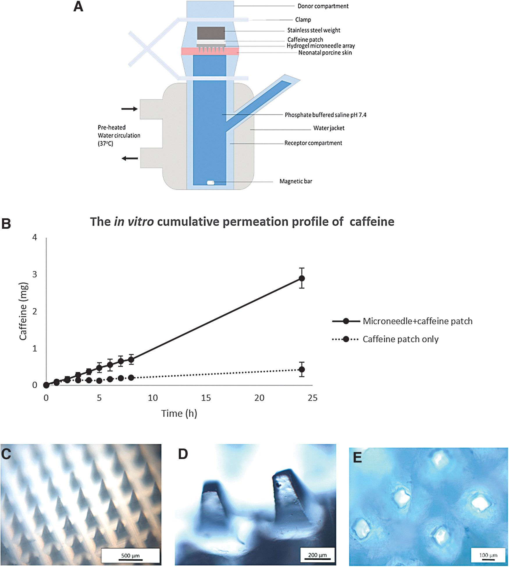
Only a caffeine patch without microneedle was placed on the skin for the negative control groups. Both donor and receptor compartments were clamped, and the donor compartment's opening was sealed with parafilm. Figure 5A provides a schematic diagram of the modified FDC model for the in vitro experiment. At fixed time points, a 250 μL sample was taken using the sampling port of the FDC and substituted with an equal amount of PBS. 16 At the end experiment, morphological changes in the hydrogel MN and skin samples (methylene blue stained) were visualized under a digital microscope. Caffeine from collected in vitro samples was quantified using UV-visible light spectroscopy at the wavelength of 273 nm.
Kinetic release
Five mathematical models (zero-order, first-order, Hixson–Crowell, Higuchi model, and Korsmeyer–Peppas model) were applied to determine the in vitro drug release pattern of caffeine. For every model, a graph was plotted based on the theoretical equation, as presented in Eq. 9 (zero-order), Eq. 10 (first-order), Eq. 11 (Higuchi model), Eq. 12 (Korsmeyer–Peppas model), and Eq. 13 (Hixson–Crowell model). The best-fit equation was examined based on the coefficient of determination (R
2
) values calculated from the graphs.
35
where Qi is the initial amount of caffeine loaded in the patch, Qt is the amount of caffeine permeated at a time point, k0 is the zero-order rate constant, k1 is the first-order constant, k is the constant reflecting the design variables of the system. Qt/Q∞ is the fraction of drug released over time, n is the release exponent, kHC is the Hixson–Crowell rate constant, and t is the time.35,36
Statistical analysis
One-way analysis of variance and Tukey's HSD post hoc test was used to assess the significance of the differences in swelling kinetic parameters of hydrogel films prepared from different foaming agents. Two samples t-test was used to compare the means of two different groups. In cases where equal variance is not assumed, the Games–Howell test was used. Results with p < 0.05 were considered to be statistically significant.
Results and Discussion
Swelling studies and network characteristics of PEG crosslinked PMVE/MA hydrogels
The result presented in Figure 1 shows the % swelling of PMVE/MA hydrogel formulations at different time points. The % swelling at 24 h for formulation C (2779.31% ± 226.89%) was significantly higher than formulation A (1359.26% ± 155.95%) (p < 0.001). This shows that an increase in the concentration of NaHCO3 significantly increases the hydrogel swelling, as similarly reported by Raj Singh et al. 25 It was explained that the release of CO2 from carbonates causes pore formation in the hydrogels. A higher concentration of NaHCO3 increases CO2 produced, resulting in higher swelling.11,25 The swelling curve for formulation D is not shown as these hydrogels were disintegrated when placed in PBS.

Percentage swelling of PEG 12,000 crosslinked Gantrez hydrogel films prepared from Gantrez AN-139 or Gantrez S-97 with different concentrations of NaHCO3 (mean ± SD, n = 3) (A). A t/S versus t swelling graph of different formulations of Gantrez hydrogels crosslinked by (B). NaHCO3, sodium bicarbonate; PEG, polyethylene glycol.
Meanwhile, the % swelling at 24 h for formulation B (2303.57% ± 221.67%) was significantly higher than formulation A (p < 0.005), showing that the higher MW Gantrez polymer results in a significant increase in hydrogel swelling. High MW polymers are composed of longer polymer chains, and hydrogels prepared from longer polymer chains exhibit a larger degree of swelling. 37 Similarly, Reena and Kumar reported a greater swelling of polyacrylamide gel when a polymer with a greater MW was used owing to a larger water holding capacity of longer polymeric chains in high MW polymers. 38
The swelling characteristics of PMVE/MA hydrogels are given in Table 2. There was no significant difference in initial swelling rate (ri) between all the formulations prepared, showing that the hydrogels absorbed water at a similar rate. A balance between the osmotic pressure of the external environment and elasticity within the gel network structure results in an equilibrium stage within the swelling phenomenon, Seq. 39 The highest % Seq was recorded for formulation C, 2825.05% ± 241.23%. Meanwhile, the % Seq of formulation B (2341.92% ± 218.22%) was significantly higher than formulation A (1381.22% ± 154.55%) (p < 0.05). However, there was no significant difference in the % Seq of formulations B and C. Hydrogels prepared from formulation D were disintegrated in PBS medium. This could be owing to an extremely weak crosslinked network when a high MW PMVE/MA (Gantrez S-97) polymer is mixed with 4% w/w of NaHCO3.
Dynamic Swelling Characteristics and Swelling Mechanisms of Different Formulations of Polyethylene Glycol–Crosslinked Gantrez Hydrogels (n = 3)
ri, initial rate of hydrogel swelling; ks, constant for swelling rate; Seq%, percentage of equilibrium swelling; n, diffusion exponent; k, gel characteristic constant.
The diffusion exponent (n) for hydrogel A was 0.96, indicating a non-Fickian (anomalous) type diffusion (Table 2). During anomalous diffusion, the drug release behavior is directed by both diffusion and swelling, whereby the relative solvent diffusion rate is less than the relative chain relaxation rate. The polymeric chains gradually reorganize as diffusion occurs rapidly, resulting in a time-dependent anomalous process. Meanwhile, hydrogels B and C had n values of 1.04 and 1.09, showing Super Case II diffusion. Super Case II diffusion is an extreme diffusion model where the polymeric networks dissociate owing to a high rate of solvent diffusion, leading to a greater solvent permeation. The external layer of hydrogel encounters the glassy nucleus and creates compression on the nucleus until the breaking point is reached. 40
As given in Table 3, the volume fraction of polymer, Ø, is highest in hydrogel A, formulated with a lower MW PMVE/MA and lower concentration of NaHCO3. As the volume fraction of polymer decreases, the volume available for solvent diffusion increases resulting in greater swelling. The average MWs between crosslinks, Mc, of formulation B (491,682,277 g/mol) was significantly higher than formulation A (52,986,275 g/mol), which implies an increase in the distance between two crosslinking points. The crosslink density, Ve, of formulation A was significantly higher than formulation B. This shows that the average MW between crosslinks increases owing to lower crosslink density when a higher MW of PMVE/MA was used in the hydrogel formulation.
Network Parameter of Different Formulations of Polyethylene Glycol–Crosslinked Gantrez Hydrogels (n = 3)
Ø, polymer volume fraction in the swollen condition; x, Flory–Huggins polymer–solvent interaction parameter; Mc, average molecular weight between crosslinks; Ve, crosslink density.
A significantly lower crosslink density was also recorded for formulation C than formulation A, indicating that increasing the concentration of NaHCO3 from 3% w/w to 4% w/w significantly reduced the crosslinks between the polymer chains of the hydrogel. Therefore, increasing the MW of PMVE/MA and concentration of NaHCO3 resulted in greater MC values owing to lesser Ve of hydrogel in the swollen condition. Besides, the polymer–water interaction parameter (X) decreased when the concentration of NaHCO3 increased. In polymer–water systems, a lower value of X indicates a strong interaction between polymer and water. Hence, greater crosslinks increased the interaction between the polymer and water. The increasing MW of PMVE/MA and concentration of NaHCO3 reduce the network structure rigidity. Comparatively, such systems can imbibe more solvent molecules. 28
Fabrication and morphological examination of PMVE/MA hydrogel MNs
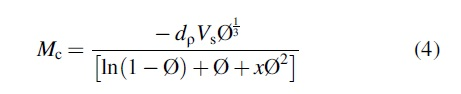
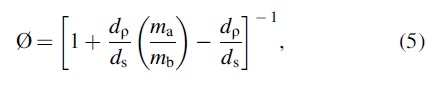
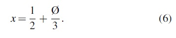
Figure 2 illustrates the morphology of MN arrays in xerogel state and after equilibrium swelling in PBS pH 7.4. After fabrication, the individual microneedle height was 727.15 ± 3.04 μm (n = 5) (Fig. 2A). A 10% reduction in needle height was recorded compared with the master mold needle height of 800 μm. This decrease was owing to water evaporation during the formulation process leading to a volumetric contraction, as similarly reported by Koh et al. 41 In general, the MNs were physically hard in the xerogel state, but their structural integrity is reduced in the swollen state, making them too soft for reapplication. This feature prevents the risk of diseases transmission among consumers by hindering the possibility of sharing or reusing the same MN patch. 15
Light micrographs of hydrogel microneedles in xerogel state
However, MN fabricated from formulation C were too flaccid and fragile because of very high water content in the swollen state (Fig. 2D). MN arrays prepared from formulation B (Fig. 2C) had higher swelling than formulation A (Fig. 2B) and better mechanical strength than formulation C as the MNs in the array maintained their upright position at equilibrium swelling. Therefore, formulation B was selected for further evaluation as it exhibited better swelling and physical properties than the other two formulations.
Ex vivo skin insertion study
Effect of insertion method on the penetration of hydrogel microneedles
Figure 3 provides the effect of different application methods in the penetration of MNs in neonatal porcine skin. The number of MNs penetrated following application of thumb pressure were lower than the other two application methods with a PE of 11% (Fig. 3A). Meanwhile, 100% penetration was recorded for both the applicator and the applicator and thumb pressure combined. The visualized micropores on the neonatal porcine show that MNs are mechanically hard enough to penetrate the SC in the xerogel state to facilitate drug delivery.

Comparison of MN insertion methods using thumb pressure
The average diameter of micropores created by applicator mediated and a combination of both applicator and thumb pressure MN application were 62.94 ± 14.21 μm and 100.53 ± 16.06 μm, respectively (Fig. 3B, C). The diameter of micropores produced by applicator-aided MN application is significantly lower than the combined method, showing that the latter method resulted in greater penetration. A higher MN penetration in applicator-aided application than thumb pressure shows that the impact force is crucial to overcome the viscoelastic property of skin for penetration. A previous study reported that the approximate force applied by thumb pressure was ∼20 N/array. 42 In this study, the force applied by thumb pressure was controlled between 15 and 20 N/array.
Although the force applied by the applicator (1.6 N) is less than thumb pressure, our results highlight the importance of impact force to penetrate the keratinized layer of skin. Similar results were previously reported for solid and dissolving MNs.20,23 Studies have reported that a lower force at a higher speed pierced SC but, a greater insertion force at a lower speed (as in thumb pressure) hardly penetrated it. Verbaan et al. reported that the manual application method with a force of 50 N/array resulted in an unsuccessful penetration of MN (300-μm needle length) on dermatomed human skin; meanwhile, impact applicator with a speed of 3 m/s enabled successful penetration. 43
It was explained that when MN was inserted without an impact force using the manual applicator or thumb pressure, the viscoelastic property of the skin offsets the penetration of MN. 44 In this study, following successful disruption of SC of the skin by the applicator impact force, the penetration is further enhanced by applying mechanical thumb pressure at a greater force. Therefore, combining both impact force and mechanical pressure is essential for better penetration of hydrogel MNs.
Effect of number of MNs on the penetration of hydrogel microneedles
Histological cross-sections in Figure 4 show that the penetration depth increases with a lower MN number. Figure 4A provides intact SC and the epidermal layer of neonatal porcine skin without MN treatment. For the arrays with 225 MNs/array, no penetration of the SC layer is observed but a mere bending of the skin (Fig. 4B). As for 169 MNs/array, the SC layer is disrupted, but the epidermal layer is not permeated (Fig. 4C). Meanwhile, for 100 MNs/array, complete penetration of the epidermal layer is observed (Fig. 4D). This finding shows that a higher penetration depth is achieved by lowering the MN number/array. Although the spacing between needles and the force applied is constant, the force will be distributed among the needles. With a smaller MN number, the force distributed to each MN increases, thus, resulting in a greater penetration depth. A similar result was described as the “bed of needle effect,” whereby a greater MN density and shorter interneedle distance decreases the penetration ability of MNs.45,46
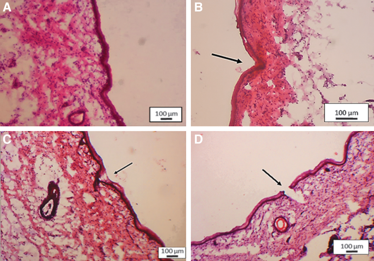
Observation of cryosectioned neonatal porcine skin before
In vitro caffeine delivery via hydrogel microneedle
As given in Figure 5B, caffeine displayed a first-order release profile with ∼2.9 ± 0.27 mg delivered in 24 h. As the caffeine patch contains an average loading of 14 mg caffeine, ∼20.7% of caffeine was released over 24 h. However, only 0.43 ± 0.2 mg (∼3.07% of total loading) was delivered for the negative control group without MN. The amount of caffeine delivered by hydrogel MN treatment was significantly higher than the negative control group (p < 0.001), showing that hydrogel MN treatment effectively enhanced the transdermal permeation of caffeine in in vitro. A 20.7% of drug release over 24 h shows that hydrogel MN would sustainably deliver caffeine and reduce the frequency of drug administration compared with the rapid release as in conventional administration.
Schematic illustration of the modified Franz cell diffusion model used for in vitro investigation of hydrogel MN-mediated transdermal delivery of caffeine
In standard clinical practice, for the management of apnea of prematurity, 5–10 mg/kg of caffeine is administered every other day through enteral or parenteral routes to maintain the drug level within the therapeutic window. 47 This could be owing to the rapid absorption of caffeine when administrated orally or intravenously as it reaches peak plasma in less than 1 h, followed by the distribution and elimination phase after 1 h, thus, necessitating frequent administration. 48 The sustainable release of caffeine using the hydrogel MN approach would prolong the absorption phase and ensure the plasma concentration of caffeine is maintained at the therapeutic level for an extended period. Hence, the drug administration frequency could be reduced from a daily basis to alternative days.
As ∼2.9 mg of caffeine was delivered through hydrogel MN over 24 h, it is suggested that maintaining the hydrogel MN for up to 48–72 h would deliver caffeine within the maintenance dose range of 5–10 mg/kg. 47 Moreover, increasing the size of the MN array would be ideal for providing the caffeine within the maintenance dose range of caffeine for the management of apnea.49,50 In this study, 10 × 10 MN arrays with a cross-sectional area of 0.5 cm2 were used for the pilot-scale development and evaluation. A therapeutically effective dose of caffeine can be administered through hydrogel MN by scaling-up the MN array size, as previous article reported that the MN patch size of up to 100 cm2 is feasible.51,52 Besides that, the multiple MN arrays can also be applied to achieve therapeutic dose. It was demonstrated that applying multiple hydrogel MN patches was feasible in in vivo studies to increase where more drug release is required to overcome the effect of metabolism and excretion and maintain a steady dose.11,16
Figure 5C and D shows the hydrogel MN before insertion and successfully swollen hydrogel MN at 24 h, respectively. After insertion, the MNs imbibe PBS from the receptor compartment underneath the skin and undergo swelling. This property enables the hydrogel MNs to mediate drug delivery. The microchannels created by MNs are crucial for drug transport across the skin because more MNs create more microholes for transdermal drug transport. The visualized micropores (Fig. 5E) on the neonatal porcine skin showed that MNs were mechanically hard enough to penetrate the skin in the xerogel state.
However, upon removal, the swollen tips of the hydrogel microneedles remain in the micropores. On the contrary, previous articles reported that PEG crosslinked PMVE/MA hydrogels can be removed intact without leaving any remains.11,14,53 In our case, the microneedles could experience lateral resistance owing to viscoelastic property of the skin. Despite that, the needle tips could be easily removed from the microchannels by rinsing the skin with water. Because the PMVE/MA hydrogel MNs are made up of biocompatible polymers, they do not cause any toxic reactions to the skin, as reported by previous studies.14,19
After 24-h treatment, the externally integrated caffeine patch was invisible owing to complete dissolution. Therefore, it can be said that the hydrogel microneedle was able to transport a sufficient amount of PBS across its matrices for the dissolution of the caffeine patch. Similar observations were reported in previous studies where complete dissolution of drug reservoirs attached to the hydrogel MN was noted at the end of 24 h in vitro experiment.31,54 The treatment proves that sustainable delivery of small compounds such as caffeine can be achieved using hydrogel MNs as 20% of the drug is released to the receiver medium up to 24 h. It is suggested that extending the treatment period would enable more drug permeation as the drug concentration has not yet achieved a plateau over the 24-h treatment period.
Analysis of kinetic release
Table 4 provides the coefficient of determination (R 2 ) value obtained from all the kinetic model graphs plotted from the in vitro caffeine permeation result. All the graphs showed linear relationships to different degrees. From the data, the drug release pattern of hydrogel was best fitted into the Korsmeyer–Peppas model with the highest R2 value (R 2 = 0.9962), which describes the drug release from the hydrophilic matrix. This R2 value was followed by the zero-order, Hixson–Crowell, first-order, and Higuchi models.
Coefficient of Determination Determined from the Plotted Graph Corresponding to Different Kinetic Release Models
Furthermore, the value of the release exponent of Korsmeyer–Peppas (n) was 1.002, indicating a Super Case II drug release kinetics. This release kinetic pattern is in accordance with the Super Case II mechanism of transport of swelling medium displayed by the hydrogels from formulation B in the swelling experiment. In Super Case II release kinetics, a combination of diffusion and erosion in the polymeric matrix could control the caffeine release from the integrated hydrogel MN. Moreover, this drug release mechanism is also associated with tension and state transformation in the hydrophilic semi-crystalline polymers during the sorption process in biological fluids.55,56
Conclusion
In this study, an optimized hydrogel microneedle system for the transdermal delivery of caffeine was developed. We have highlighted the synergistic effect between high MW of PMVE/MA copolymer and the concentration of NaHCO3 on the swelling and mechanical integrity of the hydrogels. The hydrogels produced from 3% w/w NaHCO3 with higher MW PMVE/MA copolymer swell at a higher rate and achieve high swelling at equilibrium. However, at 4% w/w the concentration of NaHCO3 with low MW PMVE/MA, the integrity of the hydrogel was compromised, resulting in weaker hydrogels although the swelling was high. A combination of 4% w/w with high MW PMVE/MA resulted in the disintegration of the formulation in the swelling medium.
Furthermore, we have also demonstrated the effect of different application methods in hydrogel MN permeation. Compared with thumb pressure, a greater skin PE was recorded when the applicator was used for MN application. In addition, a larger diameter of microporations was observed when a combination of both applicator and thumb pressure was used. Besides, the penetration depth increases as the MN number/array reduces. The optimized hydrogel MN formulation, design, and skin insertion methods were incorporated in an in vitro experiment. The in vitro result showed that the hydrogel MN significantly improved transdermally delivered hydrophilic drug–caffeine compared with the MN untreated group. In the future, optimization approaches described here can be incorporated on MN patches with a larger size to increase further the amount of drug delivered via transdermal manner.
Footnotes
Acknowledgment
The authors thank the School of Graduate Studies, Universiti Putra Malaysia, for the services and facilities.
Authors’ Contribution
R.C.: Methodology, investigation, formal analysis, and writing of original draft. E.R.M.T.: Conceptualization, resources, review and editing, supervision, project administration, and funding acquisition. J.S.: Validation, resources, visualization, and supervision. N.S.: Validation, visualization, review and editing. T.M.T.M.: Conceptualization, methodology, and supervision.
Disclosure Statement
None of the authors of this article has any competing interests to declare.
Funding Information
This project was funded by the Fundamental Research Grant Scheme (FRGS/1/2015/SKK08/UPM/02/8) from the Ministry of Higher Education, Malaysia.