
Abstract
Recent advances in the field of tissue engineering and regenerative medicine have contributed to the repair of damaged tissues and organs. Renal dysfunctions such as chronic kidney disease (CKD) are considered intractable owing to its cellular heterogeneity. In addition, the absence of definitive treatment options other than dialysis or kidney transplantation in advanced CKD. In this study, we investigated therapeutic effects of a three-dimensional (3D) bio-printed omentum patch as treatment source. Because omentum contains a lot of biological sources for immune regulation and tissue regeneration, it has been used in clinic for >100 years. By using autologous tissue as a bio-ink, the patch could minimize the immune response. The mechanically micronized omentum without any additives became small enough to print, but the original components could be preserved. Then, the 3D printed omentum patch was transplanted under renal subcapsular layer in unilateral ureteral obstruction (UUO) rat model. After 14 days of patch transplantation, the kidneys were analyzed through bulk RNA sequencing and histopathological staining. From the results, decreased tubular injury was observed in the omentum patch group. In addition, the omentum patch significantly altered biological process of gene ontology such as fibrosis-related gene and growth factors. RNA sequencing confirmed the antifibrotic effect by inhibiting fibrosis-inducing mechanisms within PI3K-AKT and JAK-STAT pathways. In conclusion, the omentum patch showed the effect of antitubular injury and antifibrosis on UUO kidneys. In particular, the omentum patch is expected to protect the organ from further degeneration and loss of function by inhibiting the progression of fibrosis. The omentum patch can be a novel therapeutic option for renal dysfunction.
Impact statement
Many studies and clinical trials are being conducted to develop new treatments for kidney disease. However, there are no newly developed renal replacement therapies. In this study, we developed a new treatment that can ameliorate renal interstitial fibrosis using three-dimensional (3D) bio-printed autologous omentum patch. The 3D printer enables precise patch printing, and the bio-ink made of autologous tissue minimizes the immune response after transplantation. The whole kidneys were analyzed by RNA sequencing and histopathological staining 14 days after transplantation. From the results, the omentum patch had the effect of relieving tubular injury in the injured state. Also, the omentum patch significantly altered biological process of gene ontology. In particular, genes related to fibrosis were observed to be downregulated by the omentum patch. RNA sequencing confirmed that the antifibrotic effect was owing to inducing mechanisms of PI3K-AKT and JAK-STAT pathways. The findings reported in this study represent a significant advancement in the application of 3D bio-printer to damaged organ treatments, especially fibrosis-related diseases.
Introduction
Chronic kidney disease (CKD) is defined as a progressive loss in renal function. It affects 8–10% of adults worldwide. Millions of people die prematurely each year from complications associated with CKD.1,2 The US Centers for Disease Control and Prevention (CDC) predicts that 47% of people over the age of 30 will develop CKD during their lifetime. 3 In the 11% of people with stage 3 CKD, lack of proper treatment often drives CKD progression to end-stage renal disease (ESRD), which requires dialysis or a kidney transplant. CKD is also one of the strongest risk factors for cardiovascular complications.4,5 The cost of treating CKD ($49 billion) is more than double the cost of ESRD ($23 billion). 6 The occurrence of CKD and the progressive renal fibrosis are difficult to treat because of complex signaling pathways and the chronic disease duration.
Therefore, it is important to inhibit the progression for disease via appropriate regulation and mitigation of disease-related stimuli. No definitive treatments are available to abrogate the severely altered histopathological lesions, especially in progressive CKD. 1 To suppress multiple signaling pathways in CKD, several strategies have been attempted including TGF-β inhibitor,7–9 BMP-7,10–12 galectin-3 inhibitor,13–16 and chemokine CCL2.17–19 However, the effect of treatment was limited owing to poor suppression of fibrosis and adverse effects.20–22 Therefore, novel treatment options are needed to regulate renal fibrosis and CKD progression.
Omentum has been reported to contain abundant biological sources for immune regulation and tissue regeneration, including anti-inflammatory and antibacterial cytokines.23,24 It is also large peritoneal fold that hangs from the stomach and wraps around the abdominal organs. Immune cell clusters called “milky spots” in omentum play a protective role by adhering to areas of inflammation, and absorbing contaminants for the local immune response. The milky spots of the omentum are clustered between adipocytes and mesothelial cells with leukocytes, mesothelial cells, B cells, macrophages, dendritic cells, T cells, and ILC2. 25 The physiological role of the omentum is not entirely clear, but it has been widely used in surgery for >100 years such as regenerative and reconstructive surgeries. 26
Several attempts to wrap omentum in the injured area have demonstrated its safety and effectiveness. For example, after lymphorenal disconnection for chyluria, omental wrapping around the renal pedicle increased blood supply and minimized morbidity and recurrence. 27 In gastrointestinal surgery, the omentum surrounding the anastomosis prevented leakage. 28 In gastric perforation, the omental patch promoted wound healing through the combined effects of angiogenesis, granulation, scaffolding, and fibrosis to regenerate. 29
Three-dimensional (3D) bio-printing is considered as one of the important tools for the scaffolds fabrication to repair or replace damaged tissues or organs. With the principle of layer-by-layer lamination, it is possible to quickly print in a desired shape using autologous biomaterials as bio-ink including extracellular matrix (ECM) and stromal cells with uniform density and porous structure. In particular, use of the ECM as bio-ink has been highlighted in tissue engineering field because of the cellular microenvironment and form of 3D network. 30
In this study, we evaluated the antifibrotic effects of omentum patch to resolve renal fibrosis induced by unilateral ureter obstruction (UUO) in rat models. To investigate such effects, we fabricated omentum as a bio-ink. Omentum bio-ink was transplanted to subcapsular space of the kidney in an animal model of CKD after 3D bio-printing. Although we evaluated this new approach using 3D bio-printed autologous omentum patch, which can be potentially used to treat CKD as an alternative to pharmacologic therapies associated with side effects.
Materials and Methods
Animals and experimental design
All animal procedures were conducted according to the Institute of laboratory animal research guide for the care and use of laboratory animals and approved by the Institutional Animal Care and Use Committee (IACUC) of Helixmith Co., Ltd. (VIC-21-10-004; Seoul, Korea). A total of 12 male Sprague-Dawley (SD) rats (average weight, 220 ± 20 g) were purchased from Raonbio, Inc. (Yongin, Korea). The rats underwent UUO and were divided into four groups: sham operation with fibrin patch (n = 3), sham operation with omental patch (n = 3), UUO with fibrin patch (n = 3), and UUO with omental patch (n = 3). To create a UUO model, the left flank was incised by ∼2 cm, and the left ureter was ligated. A position 0.5 to 1 cm distal to kidney was ligated with a 4.0 suture. Three days after the UUO model development, the autologous omentum was extracted and printed using 3D bio-printer. Then the patch was transplanted in the renal subcapsular layer. All experimental animals were killed 2 weeks after patch transplantation. Rats were anesthetized in a sealed chamber using 5% enflurane in oxygen and maintained by face mask of 2% enflurane.
Omentum processing and 3D bio-printing
The printing patches were performed by 3D bio-printer (Dr. INVIVO, ROKIT Healthcare, Inc., Seoul, Korea). For bio-ink preparation, autologous omentum was extracted from individual animals and processed using medical grade Dr. INVIVO AI Regen Kit (#ARK-001; ROKIT Healthcare, Inc., Seoul, Korea). All processes were performed while maintaining a sterile state in the operating room. In addition, all surgical devices were sterilized including a 3D printer. We used fibrin glue (Beriplast P, CSL Behring GmbH, Marburg, Germany) for thickening. The optimal ratio of omentum patch components was tested. The bio-ink concentration for the patch is indicated hereunder.
Bio-ink 1: mixture of micronized omentum and 90 mg/mL of fibrinogen
Bio-ink 2: 500 IU/mL of thrombin
We optimized the ratio between omentum and fibrin glue. From different ratios, omentum:fibrinogen:thrombin = 2:4:4 (20% of omentum), 6:2:2 (60% of omentum), and 8:1:1 (80% of omentum), 60% of omentum was selected to be a bio-ink. For the omentum patch printing, we used AI Regen, 31 an AI application that uses computer vision and machine learning technology to automatically generate 3D bio-printable patches. The omentum patch was printed layer-by-layer with bio-inks 1 and 2. The patch was transplanted into renal subcapsular space. As a control, the fibrin patch group was printed by diluting 90 mg/mL fibrinogen, 500 IU/mL thrombin, and saline to 60% volume to match the same concentration as the omentum patch.
Histopathological analysis
To analyze renal histology and immunohistochemistry, kidneys were embedded in paraffin and sectioned to 4 μm thick specimens. The sectioned tissue was deparaffinized and rehydrated. For Masson's Trichrome (MT) staining, the sectioned tissues were treated with Weigert's iron hematoxylin solution for 10 min to stain the nuclei. Collagen, cytoplasm, and muscle fibers were stained with reducing Biebrich scarlet acid fuchsin solution. Tissues were reacted with phosphomolybdic-phosphotungstic acid solution and stained with aniline blue for 10 min without washing. The nonspecific stained area was removed with 1% acetic acid. For sirius red (SR) staining, slice was stained with picro SR for 60 min at room temperature after incubation with hematoxylin for 10 min.
The tissue slides were placed in distilled water, dehydrated, and mounted. As given in Supplementary Table S1, semiquantitative analysis was performed on whole kidney slides in each group. Depending on the degree of assign, the severity of the change was represented in scores as None (−), Mild (+), Moderate (++), and Severe (+++). 32 Tubular injury score was assessed by tubular dilatation through 5 random fields. Interstitial fibrosis score was randomly evaluated as percentage of MT and SR stained area in 10 fields (each 3 animals) of renal cortical region at 100× magnification using ImageJ software (V1.8; NIH, Bethesda, MD). Fibrosis was examined by a pathologist.
4,6-diamidino-2-phenylindole Staining
The extracted omentum was stained with 4,6-diamidino-2-phenylindole (DAPI; Sigma-Aldrich, St. Louis, MO) following micronization method as described previously. Native and micronized omentum was fixed with 4% paraformaldehyde (PFA) and stained with 2 mg/mL DAPI. The stained tissue was imaged with a fluorescence microscope (Eclipse Ts2-FL; Nikon, Tokyo, Japan) after whole mounting.
RNA isolation, library preparation, and sequencing
After isolation of total RNA from rat kidney sample of all groups, DNA contamination was removed using DNase. To construct cDNA libraries with the TruSeq Stranded mRNA LT Sample Prep Kit (Illumina, San Diego, CA), total RNA was used. The protocol consisted of polyA-selected RNA extraction, RNA fragmentation, random hexamer primed reverse transcription, and 100 nt paired-end sequencing by Illumina NovaSeq 6000 (Illumina, Inc.). The libraries were quantified using qPCR according to the qPCR quantification protocol guide and qualified using an Agilent 2100 Bioanalyzer (Agilent Technologies, Inc., Palo Alto, CA).
Bioinformatics analysis
Raw reads from the sequencer were preprocessed to remove low-quality and adapter sequence before analysis and processed reads were aligned to the Rattus norvegicus using HISAT v2.1.0.33,34 The reference genome sequence and annotation data were downloaded from the NCBI. Then, the transcript assembly of known transcripts was processed by featureCounts v1.6.0. 35 Based on that result, expression abundance of transcript and gene was calculated as read count or fragments per kilobase of exon per million fragments mapped (FPKM) per sample. The expression profiles were used to do additional analysis such as differentially expressed gene (DEG). It was analyzed by substituting the group average FPKM value, and an absolute fold change (FC) cutoff value of 0.585 (≥1.5 FC) in log2 scale was utilized to assess dysregulated genes in each group.
In groups with different conditions, DEGs or transcripts can be filtered through statistical hypothesis testing. For differential expressed genes, gene ontology (GO) and Kyoto Encyclopedia of Genes and Genomes (KEGG) were performed with clusterProfiler v 3.18.1 in R v 4.0.3, 36 which supports statistical analysis and visualization of functional profiles for genes and gene clusters. Heatmap package was used to construct heatmap.
Statistical analysis
Data were statistically analyzed using mean (±standard error of the mean). Difference between repeated measurements of each group was statistically analyzed using analysis of variance or Student's t-test, and ***p < 0.001, **p < 0.005, and *p < 0.05 were considered statistically significant values. All statistical analyses were performed by Prism 8 (GraphPad, San Diego, CA).
Results
3D Bio-printing of omentum patch and its transplantation
For the 3D bio-printed omentum patch, autologous omentum of rats was extracted and micronized. Omentum has been used in clinic because of its abundant biological sources. 24 The reticulum-like morphology of blood vessels and fat structures was observed in the rat omentum (Fig. 1A). In a previous study, we confirmed that omentum tissue had become smaller through micronization, enabling printing. In addition, mechanical micronization allowed maintenance of tissue components such as ECM and cells (Fig. 1B). Because the milky spot (yellow arrow in the hematoxylin and eosin image), the dense cell area, is in omentum, there were concerns that the effect of omentum may be uneven if the omentum is used as it is. Cells were distributed unevenly in native omentum, but uniformly distributed cells were observed in the micronized omentum (Fig. 1C).

The process of the patch preparing and transplantation.
We developed an omentum and fibrin glue mixture at different ratios of omentum [omentum:fibrinogen:thrombin = 2:4:4 (20% of omentum), 6:2:2 (60% of omentum), and 8:1:1 (80% of omentum)] and discharged it with a syringe (Fig. 1D). When the content of micronized omentum was >80% by volume, it was difficult to maintain a solid shape for application to the damaged kidney. On the contrary, when the content of the micronized omentum was <20% by volume, although it was easy to maintain a patch-like solid shape, the biodegradability was low owing to the high content of fibrin glue. Therefore, we decided to use 60% micronized omentum, which was suitable for printing and had a good biodegradability.
We implanted the 3D printed patch into the subcapsular space of the kidney (Fig. 1E). In this process, we used AI Regen Kit and an AI application that uses computer vision and machine learning technology, to automatically generate 3D printed therapeutic patch. 31 The kidney was photographed, and the patch file was created by hand drawing. After sending the file to Dr. INVIVO, the omentum therapeutic patch was printed and transplanted.
Reduced tubular injury in UUO rat model by the omentum patch
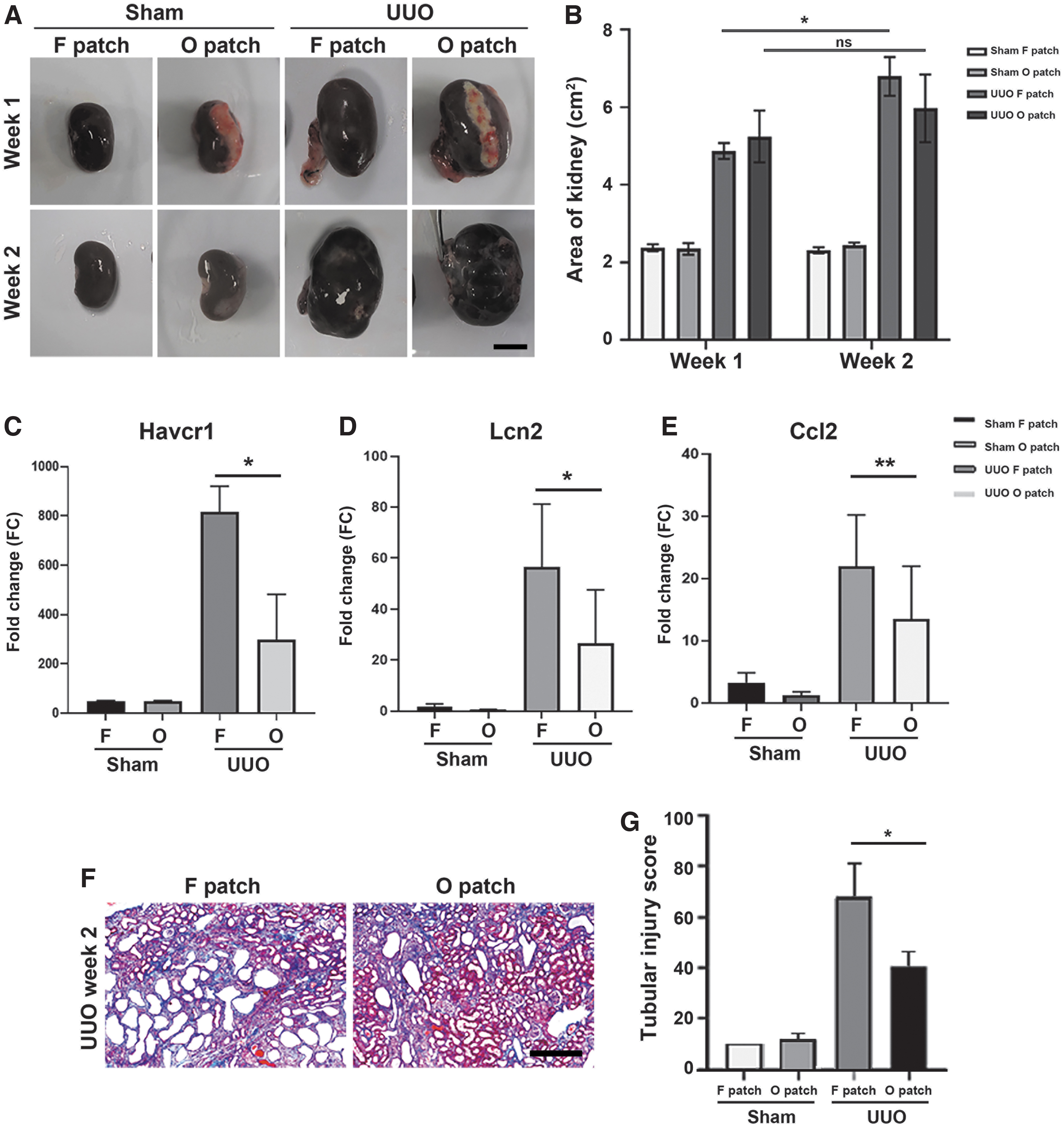
A rat UUO model of CKD was created by ligation of ureter. Significant renal hemodynamic and metabolic changes are induced owing to ureteral obstruction. 37 Severe tubular dilation, interstitial expansion, hydronephrosis, and interstitial fibrosis were observed in the UUO group with fibrin patches compared with the UUO group with omentum patches (Supplementary Table S1). Although it is difficult to confirm the functional effect, the UUO can be easily modeled via ureteral ligation, and has been widely used to represent various CKD histopathological features.38,39 To validate the surgery, a sham group was included as a control. We observed that kidneys of the UUO group enlarged after the operation by ligated urine pressure (Fig. 2A).
Reduced tubular injury by the omentum patch in the UUO rat model.
In addition, after 2 weeks of patch applying, the form of patch disappeared. The kidney area (width × length) in the fibrin patch group increased quantitatively up to week 2, whereas the area in the omentum patch group increased until week 1, but there were no significant (ns) changes in week 2 (Fig. 2B). After 2 weeks of UUO, the renal tubule damage-related genes such as Havcr1 (encoding KIM-1), Lcn2 (encoding NGAL), and Ccl2 (chemokine) were significantly increased in the fibrin patch group, but the omentum patch group had lower gene expressions (Fig. 2C–E). As given in Figure 2F, histopathological analysis (MT staining) demonstrated that UUO-induced tubular injury was histopathologically shown to be tubular dilatation. In the fibrin patch group, tubular dilatation significantly expanded in the renal cortex and medulla.
On the contrary, less tubular dilatation was observed in the omentum patch group. Tubular injury was scored using individual five random fields in each kidney sample 40 (Fig. 2G). In the sham groups, there were no significant differences between the fibrin patch and the omentum patch. However, in the UUO groups, tubular injury score was 0.59 times lower in the omentum patch group than in the fibrin patch group. The omentum patch decreased tubular injury in the UUO model, but there were no changes in the sham groups. Therefore, we concluded that the omentum patch had the effect of relieving tubular injury in the injured state.
Regulation of biological process by the omentum patch
To investigate GO enrichments of the omentum patch, the gene expression level of the kidney in each group was analyzed using bulk RNA sequencing. The GO was selected for biological processes by comparing fibrin versus omentum patch groups after UUO. The predominant 10 up- or downregulated pathway was analyzed (Fig. 3). Of interest, many genes related to epithelial cell proliferation or migration were upregulated in the omentum patch group (Fig. 3A). Because the tubular epithelial cells are responsible to constitute the basic functional units of kidney, the proliferation or recruitment of epithelial cells are important to recovery injured kidney. 41 In addition, the genes related to renal tubule development was upregulated in the omentum patch group compared with that in the UUO control group. The reconstruction of renal tubule plays critical roles in kidney tissue regeneration. 42

GO analysis of biological processes in omentum patch group after UUO. Top 10 of the predominant upregulated (red)
On the contrary, there were downregulated genes in the omentum patch group (Fig. 3B). For example, gene expression of organic anion transport was downregulated. Organic anion transporter expressed in renal proximal tubules cells is known as a role for elimination of metabolic waste or toxins. 43 Moreover, mononuclear cell differentiation and response to toxic substance for anti-inflammatory response were downregulated in the omentum patch group. These genes changes occurred only in the UUO groups, and did not appear in the shame groups. It indicates that this is not a gene expression of omentum cells, but rather a change in kidney cells owing to the active component of omentum. Therefore, we concluded that the omentum patch regulates biological processes to recover damaged kidneys.
KEGG pathway in the omentum patch group
Transcripts were visualized by pathway analysis through KEGG. We performed pathway enrichment analysis on the contraregulated DEGs identified from “UUO or sham with fibrin patch” versus “UUO with fibrin or omentum patch” (Fig. 4). In the differentially expressed genes analysis of UUO fibrin patch versus sham fibrin patch, it was observed that ureteral obstruction induced 6561 gene upregulation (Fig. 4A). On the contrary, 1440 genes were downregulated by applying the omentum patch.

Pathway analysis of contraregulated DEGs using KEGG.
In conclusion, 488 of the genes increased by UUO were lowered by the omentum patch. Top 15 enriched KEGG pathways were listed. Counts of matched genes represent spot sizes, and the color gradient represents the p-value in the analysis pathway (n = 3). From the results, fibrosis signaling including PI3K-AKT, JAK-STAT, and MAPK, which were raised by UUO, were observed to reduce after applying the omentum patch. In Figure 4B, 612 of the 3329 genes decreased by UUO were upregulated by the omentum patch. All data suggest that the omentum patch helps kidney recovery by regulating the valence of genes that are abnormally up- and downregulated by UUO.
Effects of renal interstitial fibrosis induced by UUO
Because a decrease in fibrosis signals was observed, we analyzed for the anti-interstitial sclerosis effect of the omentum patch on the kidneys by bulk RNA sequencing. To evaluate the interstitial sclerosis, we selected fibrosis-related genes including Acta2, Ctgf, Smad3, Cxcl6, Ccl2, Havcr1, Lcn2, Sox9 (SRY-box 9), and Maff, 44 as well as profibrotic transcription factors including Irf1, Nf-kb1, Stat3, and Tgfb1 45 as gene sets. From the heat map, we observed that profibrotic or fibrotic-related genes were upregulated in the fibrin patch group, but not in the omentum patch group (Fig. 5A). From the result of quantitative analysis, the expression of fibrosis markers such as Acta2, Ctgf, Tgfb1, Smad3, and Cxcl6 was significantly lower in the omentum patch group compared with that in the fibrin patch group (Fig. 5B).

Effect of omental patch on renal interstitial fibrosis induced by UUO in rats.
However, the endothelial cell marker, Pecam1, was 1.6 times higher in the omentum patch group compared with that in the fibrin patch group. MT staining (fibrosis; blue) and SR staining (fibrosis; red) were performed to evaluate interstitial fibrosis (Fig. 5C). Fibrosis accumulation was histopathologically observed in kidney cortex after 2 weeks of UUO. Renal fibrosis on the transverse sectioned kidney over all area was scored by a histologist. As given in Figure 5D and E, the MT and SR stained areas increased 3.06-folds in the fibrin patch group and 2.1-folds in the omentum patch group of UUO kidney compared with that in the sham group, respectively. From the results, fibrosis scores were higher in the fibrin patch group compared with those in the omentum patch group. Together, these findings suggest that the omentum patch played a role in lowering renal fibrosis in the rat UUO model.
Discussion
In this study, we demonstrated that the micronized omentum was used as a bio-ink for kidney treatment. Through physical micronization, the omentum tissue could be used as a bio-ink by reducing the size and removing fibers. In addition, because there was no chemical treatment for ink preparation, cellular components and micro size ECM remained in the ink without affecting cell viability. 46 It is known that micronization of adipose tissue can lead to mechanical disaggregation, and extraction of components including cells and growth factors that promote cell viability, proliferation, and differentiation of the natural matrix after transplantation.47–49 Therefore, we utilized micronization for processing omentum, and the micronized omentum was applied to 3D printing with adequate viscosity (Fig. 1).
After applying the ometum patch to kidneys, we analyzed the whole transcriptome. Among 38,000 genes, the differences in gene expression of each group were observed. In addition, the biological functions and pathways were identified by whole transcriptome analysis combined with bioinformatics analysis. We observed that genes related to proliferation of fibroblast such as Ccl2, Myc, Relb, and Sox9 were upregulated in UUO groups compared with that in the sham groups. In addition, the expression of CKD-related genes including Acta2 (a-SMA), Ctgf, Tgfb1, Smad3, and Cxcl6 were increased after ureteral obstruction. Shunsakju et al showed increased expression of Havcr1 and Lcn2 in several forms of CKD, and their expression levels were correlated with tubular interstitial fibrosis and tubular cell damage. 44 Of interest, despite the obstruction, the FPKM values of fibrosis-related genes were greatly reduced in the omental patch group.
It is known that a rarefaction of peritubular capillaries in kidney occurred in fibrosis. 50 In Figure 5B, we observed 1.6 times higher expression of endothelial cell marker, Pecam1, in the omentum patch group compared with that in the fibrin patch group. This indicates that the omentum patch could inhibit the rarefaction in peritubular capillaries. To confirm the effect of omentum on capillary rarefaction, imaging analysis such as IHC or IF is required for further study. GO results showed that while many genes were expressed in the omentum patch group with UUO, the omentum patch applied to shame model did not induce gene expression. Therefore, we assume that the valid ingredient of omentum is activated by signals from injured kidneys, and that the ingredient as a paracrine factor regulates genes for kidney recovery, such as inhibiting fibrosis-related genes.
Because tubular injury is caused by tubular dilatation, interstitial dilatation, and hydronephrosis in the injured kidney after UUO, we analyzed signaling focusing on renal injury and fibrosis. We observed some upregulated fibrosis signaling in UUO with the fibrin patch group including PI3K-AKT, 51 JAK-STAT, 52 and MAPK. 53 However, in the omentum patch group, the fibrosis signaling was not activated much even after UUO. Then, we investigated that the recovery effect of the omentum patch after renal injury was related to the PPAR signaling pathway through KEGG analysis of the transcriptome. PPAR gamma is known to be expressed in the collecting duct of the kidney, podocytes, mesangial cells, and vascular endothelial cells.54,55 Furthermore, it is known to play an important role in kidney function, especially in antifibrotics. 56 In the results, we observed upregulation of PPAR signaling-related genes including Me1, Cpt1b, Rxrg, Acox2, and Pck1 in the omentum patch group compared with the fibrin patch group after UUO.
Finally, we observed that genes related to retinol metabolism, such as Cyp1a1, Aldh1a7, Aox1, Adh7, and Ugt1a9 were upregulated in the omentum patch of UUO group than in the fibrin patch of UUO group. Retinoids have been shown to exert both protective and preventive effects in various kidney disease animal models through improving podocyte injury, including differentiation of kidney progenitor cells, and attenuating inflammation and apoptosis of the kidney cells. 57 All data suggest that the omentum patch regulated the role in inhibiting renal injury in UUO model.
This study has limitations. The number of animals used in each group was not sufficient for a more robust statistical analysis of the data collected. Nevertheless, we suggest the omentum patch efficiency in the UUO model with demonstrated gene set and histopathology analysis. The kidney is composed of a complex vascular mass and a dense structure; it is difficult to deliver therapeutic substances using traditional delivery methods. A possible mechanism of recovery is that stem cells from the kidney capsule layer are recruited as paracrine effects of the omentum. There is a stem cell niche in the renal capsule layer, and the omentum patch can play a role in boosting cells through paracrine effects. 58
In this study, it can be manufactured within 30 min in various shapes and thicknesses and can be freely customized and modeled according to the characteristics of tissues and organs. Furthermore, the type of patch manufactured in this study enabled for controlled release of the omentum component. When transplanting cells for regeneration to damaged tissue, encapsulation is required to maintain cell viability and function 59 and therefore, the transplantable patches that use micronized omentum are a new approach for therapeutic omentum.
In conclusion, we demonstrated that omentum patch transplantation for damaged kidneys can ameliorate renal fibrosis in the UUO model. The transplanted omentum patch can help cellular repair and antifibrotic effects, thereby protecting the kidney. In addition, it can prevent ESRD progression and block fibrosis by reducing fibrotic factors. As a novel approach to attenuating fibrosis in patients with CKD, the autologous 3D bio-printed patch technology has shown therapeutic potential in tissue repair and regenerative medicine. Further studies are required to demonstrate a more minimally invasive method and a CKD model to confirm renal function. These findings suggest the potential use of autologous 3D omentum patches for renal fibrosis and other organs.
Footnotes
Authors' Contributions
H.J., B.Y.C.: study design, executing experiments, writing the article; G.J.: collecting and analyzing data; J.P.L., J.L.: study design, analyzing data; A.C., B.K.: executing experiments; J.H.P., Y.H.K.: analyzing data; J.R.: study design, writing the article.
Statement of Ethics
Animal experiments conform to internationally accepted standards and have been approved by the Institutional Animal Care and Utilization Committee of Helixmith, Inc., (Seoul, Korea).
Disclosure Statement
H.J., B.Y.C., B.K., and J.R. are employees of ROKIT Healthcare, Inc.
Funding Information
This research was funded by ROKIT Healthcare Inc., located in Seoul, Republic of Korea.
References
Supplementary Material
Please find the following supplemental material available below.
For Open Access articles published under a Creative Commons License, all supplemental material carries the same license as the article it is associated with.
For non-Open Access articles published, all supplemental material carries a non-exclusive license, and permission requests for re-use of supplemental material or any part of supplemental material shall be sent directly to the copyright owner as specified in the copyright notice associated with the article.