
Abstract
Mesenchymal stem cell and 3D printing-based bone tissue engineering present a promising technique to repair large-volume bone defects. Its success is highly dependent on cell attachment, spreading, osteogenic differentiation, and in vivo survival of stem cells on 3D-printed scaffolds. In this study, we applied human salivary histatin-1 (Hst1) to enhance the interactions of human adipose-derived stem cells (hASCs) on 3D-printed β-tricalcium phosphate (β-TCP) bioceramic scaffolds. Fluorescent images showed that Hst1 significantly enhanced the adhesion of hASCs to both bioinert glass and 3D-printed β-TCP scaffold. In addition, Hst1 was associated with significantly higher proliferation and osteogenic differentiation of hASCs on 3D-printed β-TCP scaffolds. Moreover, coating 3D-printed β-TCP scaffolds with histatin significantly promotes the survival of hASCs in vivo. The ERK and p38 but not JNK signaling was found to be involved in the superior adhesion of hASCs to β-TCP scaffolds with the aid of Hst1. In conclusion, Hst1 could significantly promote the adhesion, spreading, osteogenic differentiation, and in vivo survival of hASCs on 3D-printed β-TCP scaffolds, bearing a promising application in stem cell/3D printing-based constructs for bone tissue engineering.
Impact statement
The 3D printing scaffolds are considered ideal materials to support cell growth for bone tissue engineering. Its clinical application is limited partially due to the poor interaction between mesenchymal stem cells and 3D-printed scaffolds. In this study, we introduced histatin-1 (Hst1) to coat 3D-printed β-tricalcium phosphate scaffold for the improvement of adhesion and osteogenic activity of human adipose-derived stem cells on it. The results indicate promising application of Hst1 in stem cell/3D printing-based constructs for bone tissue engineering.
Introduction
In recent years, the 3D printing technique appears to be promising to provide ideal scaffold materials for repairing large-volume bone defects (LVBD). Three-dimensional printing enables the production of patient-personalized and anatomically matched scaffolds and constructs with high tunability and complexity. 1 Various 3D printing techniques, such as material extrusion and powder bed fusion, have been developed to print CaP-based bone scaffold. 2 Such printed scaffolds often need postprinting heating and sintering to get sufficient mechanical strength and/or remove the organic phase, which precludes the potential for simultaneous incorporation of cells, proteins, or heat-labile bioactive molecules. Therefore, the thereby-produced 3D scaffolds lack intrinsic pro-osteogenic elements so that they cannot be applied alone to heal LVBD. 2
One promising method to confer pro-osteogenic elements is to combine multipotent mesenchymal stem cells (MSCs) with 3D-printed scaffolds.3,4 Despite the high cost and regulatory issues, stem cell-based therapy has distinct advantages in bone tissue engineering compared with growth factor-based therapy, which is limited by uncontrolled release and concerns of ectopic bone formation upon implantation. The success of the MSC/3D printing-based bone tissue engineering technique is largely dependent on the interactions between stem cells and 3D-printed scaffolds 5 since optimal cell–substrate interactions are critical to promote cell survival, proliferation, and differentiation so as to sustain bone regeneration.6,7 Also, transplanted cell survival is significantly affected by anoikis when cells detach from their substrate after implantation.8,9
Approaches to increase the survival rate of the transplanted MSCs have been explored to improve the effectiveness of cell-based therapies, which include modulating cell metabolism by altering the repair environment with biomaterials to promote cell adaptation to a harsh implantation environment. Consequently, a large variety of techniques have been developed by improving the surface properties of scaffolds, such as preimmobilization of the RGD peptide on substrate 10 and a coating of extracellular matrix (ECM)-derived polymer. 11 However, most of these substrate-targeting techniques do need premodifications of scaffolds with physical or chemical reactions and introduce additional components, which inevitably increases the complexity of the constructs and makes it more difficult to pass through regulatory control to clinical application. Consequently, it is highly needed to develop a new strategy that can promote cell–substrate interaction without the need for presurface modifications.
One promising agent to improve cell adhesion to scaffolds is histatin-1 (Hst1) that belongs to a large human saliva-derived histidine-rich peptide family 12 expressed in the saliva of humans and higher primates. 13 In the past decade, Hst1 has been recognized as a potent cell-activating agent to stimulate a series of cell activities. 13 For example, Hst1 can promote the adhesion, spreading, and migration of various types of cells, such as epithelial cells, fibroblasts, osteogenic cells, and endothelial cells, on bioinert glass surface, titanium sandblasted and acid etched surface, and hydroxyapatite.14–18 Furthermore, Hst1 also significantly enhances cellular metabolic activity by subcellular targeting to mitochondria and endoplasmic reticulum.19,20 In our recent study, we also show that Hst1 significantly enhances the osteogenic differentiation of preosteoblasts. 17 All these findings indicate that Hst1 can promote cell–substrate interaction without the need for presurface modification of biomaterials.
In this study, we aimed to develop an Hst1 coating strategy and explore its application potential in promoting the interaction between MSCs and 3D-printed scaffolds for bone tissue engineering. We hypothesized that using Hst1 to coat β-tricalcium phosphate (β-TCP) scaffolds can improve cell attachment, which can further improve the cell survival after implantation of MSC/3D printing-based constructs. Aiming this, we first assessed the time-course and dose-dependent effects of Hst1 on the adhesion and spreading of human adipose-derived stem cells (hASCs) on 3D-printed β-TCP scaffolds so as to select the optimal condition of Hst1. Thereafter, we investigated Hst1's effects on the proliferation, osteogenic differentiation, and in vivo survival of hASCs on β-TCP scaffolds. We also adopted specific inhibitors to explore the potentially involved signaling pathways.
Methods
Peptide synthesis and purification
The lyophilized linear Hst1 (DSPHEKRHHGYRRKFHEKHHSHREFPF-YGDYGSNYLYDN) and scrambled Hst1scr (SDHSRHEEFKPRFHYHGGDYYRGRSKNFYHLEYKDHNH) peptides with a purity of 95% were purchased from Hangzhou Huibo Science and Technology Company. Hst1scr was utilized as a negative control. The lyophilized peptide was dissolved in 10 mM of ultrapure water to create a histatin stock solution, which was then kept at −20°C until needed. The peptide solution was further diluted with phosphate-buffered saline (PBS).
Cell culture and chemicals
After being purchased from Cyagen Biosciences Technology (Guangzhou, China), the hASCs were isolated from the adult adipose tissue and cultured in hASC complete growth medium (HUXMD-90011; Cyagen Biosciences Technology). Cyagen carried out all of the routine characterization assays (including osteogenic, adipogenic, and chondrogenic differentiation assays) as a quality control. Trypsin was used to detach cells after they reached 70–80% confluence in culture flasks. An incubator at 37°C with 95% air and 5% CO2 combination was used to culture cells. ASCs at passage 4–6 were used for all experiments.
Measurement of cell adhesion and spreading on glass surface
ASCs were seeded on glass coverslips at a density of 1.2 × 10⁴ cells/well in 48-well plates and exposed to 0–50 μM of the Hst1 or Hst1scr for 1.5 h. ASCs were then fixed with 4% paraformaldehyde at room temperature for 10 min, permeabilized using 0.1% TritonX-100 in PBS for 10 min, and then blocked with 1% bovine serum albumin (BSA) at room temperature for 1 h. Subsequently, samples were stained with FITC-phalloidin in the dark at room temperature for 2 h and then counterstained with DAPI for 5 min. Samples were washed more than five times with PBS and then imaged with a Leica TCS SP8 laser scanning confocal microscope (Leica Mikrosysteme Vertrieb GmbH, Germany). The attached number and surface area of hASCs were counted according to four randomly captured images using ImageJ.
Adhesion of hASCs on 3D-printed β-TCP scaffold surface
Three-dimensional printing TCP scaffold fabrication was performed according to our previously published method. 4 The 3D-printed TCP scaffolds were precise and consistent in their structure, with a diameter of 5 mm, a thickness of 1 mm, and a pore size ranging between 350 and 400 μm. Cell suspension was performed as described in the Measurement of Cell Adhesion and Spreading on Glass Surface section. Subsequently, cells were labeled using the PKH-26 Red Fluorescent Cell Linker Kit (Sigma-Aldrich) in accordance with the manufacturer's instructions. Labeled cells were seeded on β-TCP scaffold surface in 48-well plates at a density of 1.2 × 10⁴ cells/well, treated with 0, 0.1, 1, 10, or 50 μM Hst1 for 1.5, 3, and 24 h. Cells on scaffolds were stained with Hoechst 33342 to stain nuclear.
The images were captured with the Leica TCS SP8 laser scanning confocal microscope. On the micrographs, the data (the attached number of cells, cell area, cell width, and cell length) were performed using ImageJ software.
Cell viability and proliferation on 3D-printed β-TCP scaffold surface
Cells were seeded on β-TCP scaffold surface according to the Adhesion of hASCs on 3D-Printed β-TCP Scaffold Surface section. Using the PrestoBlue cell viability reagent from Invitrogen, the viability and proliferation of the hASCs on the scaffold surface in the presence of vehicle or 10 μM Hst1 were assessed after treatment for 1, 3, 5, and 7 days. After mixing 10 μL of PrestoBlue solution with 90 μL of cell culture medium and incubating for 30 min, the absorbance was measured at the wavelength of 600 and 570 nm, respectively. The cell viability was calculated as per the kit protocol.
Alkaline phosphatase staining and quantification
After cell seeding on the surface of the β-TCP scaffolds according to the instructions in the Adhesion of hASCs on 3D-Printed β-TCP Scaffold Surface section, they were cultured in the osteogenic induction medium (hASC complete growth medium with 50 μg/mL of
Animal surgery
The Animal Ethics Committee of General Hospital of Southern Theater Command examined and approved the animal study (No. 2019051702). After being treated with or without 10 M Hst1, PKH26-labeled hASCs were seeded onto 3D-printed-TCP scaffolds and incubated for 3 h. Five male nude mice (5–6 weeks old) had 1% pentobarbital intraperitoneally injected into their backs while under general anesthesia. Two dorsal subcutaneous pockets were made in each animal, and cell–scaffold constructions were carefully placed inside each one. One nude mouse randomly received one sample prepared in the presence of Hst1 on the left side and one sample in the absence of Hst1 on the right side. After implantation, the skin was closed using standard nonresorbable suture materials. Fluorescent images were acquired by Living Image Software IVIS Lumina LT Series III system (PerkinElmer) over 5 weeks. The background signal was removed from two-dimensional pictures.
The multicolor scale used to depict fluorescence intensity ranged from red (least intense) to yellow (most intense). For anatomical representations, signal intensity pictures were superimposed over grayscale reference images. In each experiment, signals were gathered from a predetermined ROI using the contour ROI tool, and Living Image Software 4.5 was used to measure the total radiant efficiency ([p/s]/[M/cm2]).
Immunofluorescence staining
ASCs grown on coverslips at a density in 48-well plates were exposed to 10 μM Hst1 for 1.5 h. Afterward, they were fixed with 4% fresh paraformaldehyde for 10 min, permeabilized with 0.1% TritonX-100 in PBS for 10 min, and then blocked with 1% BSA at room temperature for 1 h. Cells were exposed to primary antibodies produced in blocking solution against talin (1/100, ab71333; Abcam), vinculin (1/100, ab129002; Abcam), or paxillin (1/100, ab32084; Abcam) overnight at 4°C. Following washing, the cells were treated for 1 h at room temperature with Alexa Fluor 594-conjugated secondary antibodies. Then cells were washed in PBS and stained with FITC-phalloidin to reveal F-actin and counterstained with DAPI. Immunofluorescence images were captured using the Leica TCS SP8 laser scanning confocal microscope. The fluorescence intensity was analyzed using ImageJ software.
Signaling pathways for the effects of Hst1
As per the cell adhesion experiment, the signaling pathway-specific inhibitors of ERK1/2 (U0126; TargetMol, China), JNK (SP600125; TargetMol), and p38 MAPK (SB203580; TargetMol) were added to the medium to examine the role of potential signaling pathways in the effects of Hst1 on the spreading of hASCs. hASCs were pretreated with or without 10 μM of inhibitors for 2 h in serum-free medium before seeded on glass coverslips in 48-well plates at a density of 1.2 × 10⁴ cells/well. Cell adhesion and spreading assays were performed as described in the Measurement of Cell Adhesion and Spreading on Glass Surface section.
Statistical analysis
All quantitative data in this study represent the mean value ± standard deviation. Data were analyzed using GraphPad Prism (GraphPad Software version 9.1, La Jolla, CA). To compare cell adhesion and spreading at selected time points, one-way analysis of variance (ANOVA) with Bonferroni's multiple comparisons test was applied. For the data of cell proliferation from different groups at different time points, a two-way ANOVA with Bonferroni's multiple comparisons test was applied. Other experiments were analyzed by paired t-tests. A value of p < 0.05 was considered statistical significance. *p < 0.05; **p < 0.01; ***p < 0.001.
Results
Effects of Hst1 on the adhesion and spreading of hASCs on bioinert glass surface
We first evaluated the dose-dependent effect of Hst1 on the adhesion and spreading of hASCs on the bioinert glass surface. Fluorescent images showed that the cell surface area was significantly larger with the increased concentration of Hst1 (Fig. 1A). Quantitative analysis showed that Hst1 dose dependently increased the surface area of hASCs by 1.13, 1.19, 1.27, and 1.41 times for 0.1, 1, 10, and 50 μM of Hst1, respectively. In contrast, 10 μM of Hst1scr did not cause significant changes in the cell surface area (Fig. 1B). The numbers of attached cells in the groups of 10 and 50 μM were significantly higher (1.40 and 1.37 times, respectively) than the control, which was not found for Hst1 at other concentrations or 10 μM of Hst1scr (Fig. 1C).

Hst1 enhances hASC adhesion and spreading on glass surface. Cells were seeded at 1.2 × 10⁴ cells/well and cultured in the presence of vehicle, 0.1–50 μM Hst1, or 10 μM Hst1scr for 1.5 h.
Effects of Hst1 on the adhesion and spreading of hASCs on 3D-printed β-TCP scaffolds
At 1.5 h postseeding, the cell numbers in the groups of 10 (2.23 times) and 50 μM of Hst1 were significantly higher than the control group (Fig. 2A, B). In contrast, Hst1 at other concentrations did not significantly change the cell number. Three hours postseeding, Hst1 dose dependently increased the numbers of hASCs on 3D-printed β-TCP scaffolds (Fig. 2A, C). However, no significant difference in cell number was found between 10 and 50 μM of Hst1. Twenty-four hours postseeding, in comparison with the control, a dramatic increase (2.14 times) in cell surface area was found in the group of 10 μM of Hst1. This effect was also more significantly profound than that at the 50 μM of Hst1 (Fig. 3B).

Hst1 enhances hASC adhesion on 3D-printed β-TCP scaffold surface.
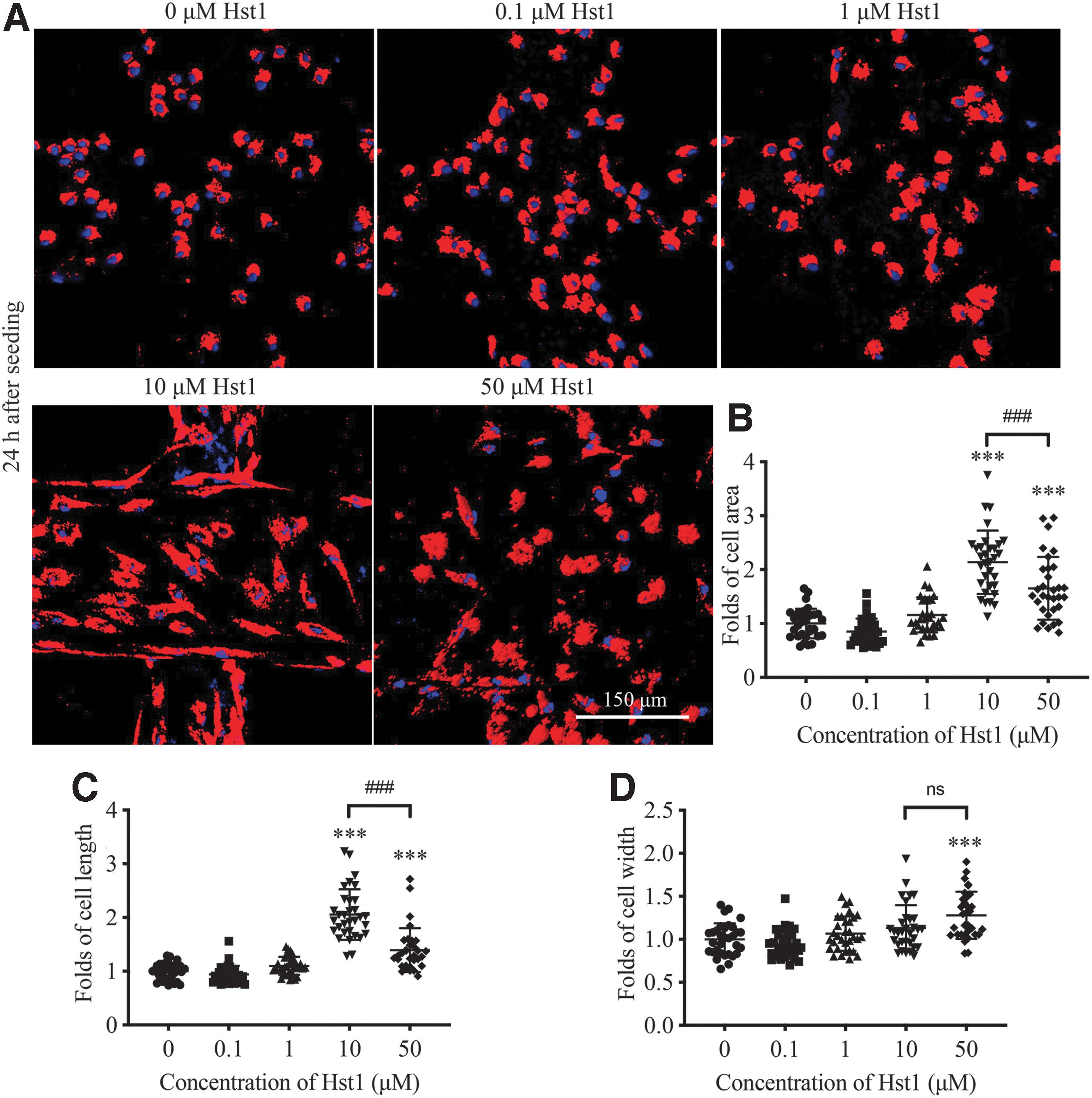
Hst1 enhances hASC spreading on 3D-printed β-TCP scaffold surface.
A similar trend was found in the length of hASCs (Fig. 3C). In contrast, only cells at the concentration of 50 μM of Hst1 had a significant higher value of cell width (Fig. 3D). According to these results, we chose 10 μM of Hst1 in the following experiments.
Effects of Hst1 on the proliferation and osteogenic differentiation of hASCs on 3D-printed β-TCP scaffolds
We used the PrestoBlue reagent to analyze the effect of Hst1 on the proliferation of hASCs. One, 3, 5, and 7 days postseeding, the cell metabolic activity in the group of 10 μM was always significantly higher than that in the control group (Fig. 4A). Four days postseeding, much stronger ALP-positive signals were detected in the group of 10 μM of Hst1 than the control (Fig. 4B). Quantitative analysis showed that the ALP-positive area in the group of 10 μM of Hst1 was significantly larger (3.12 times) than that in the control group (Fig. 4C).

Hst1 enhances hASC proliferation and osteogenesis on 3D-printed β-TCP scaffold surface.
Effects of Hst1 on the survival rate of hASCs on 3D-printed β-TCP scaffolds in vivo
Luminescence-labeled hASCs seeded on the 3D-printed β-TCP scaffolds in the presence or absence of 10 Hst1 were subcutaneously implanted in nude mice and imaged over 5 weeks. Immediately after subcutaneous implantation, bright spotty fluorescence was detected at the implantation site. The fluorescence intensities decreased gradually with time (Fig. 5A). At each time point, the fluorescent intensities in the Hst1 group were significantly higher than those in the controls (Fig. 5B).
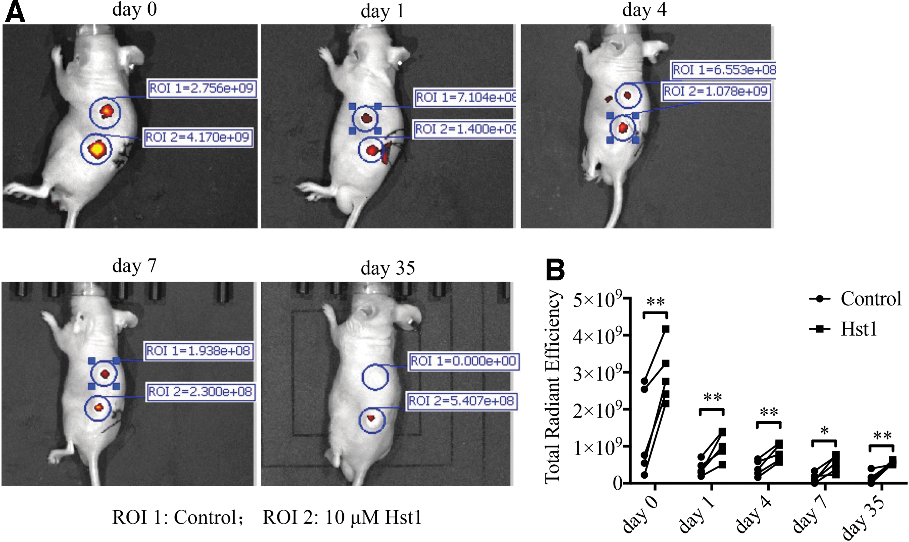
Hst1 enhances the survival rate of hASCs on 3D-printed β-TCP scaffolds in nude mouse models.
Effects of Hst1 on the expression and distribution of focal adhesion complex-associated proteins
The bidirectional mechanical communication between cells and substrate is mediated by integrin-based focal adhesion complexes. These complexes allow cells to adhere to the ECM or biomaterials, transmitting endogenous contractile forces and sensing the ECM rigidity. 21 To investigate how Hst1 influenced the adhesion and spreading ability of hASCs, we analyzed the expression and distribution of focal adhesion complex-associated proteins, such as vinculin, paxillin, and talin. Quantitative analysis showed that the mean gray values of talin (Fig. 6B), vinculin (Fig. 7B), and paxillin (Fig. 8B) staining in the Hst1 group were similar to the control group. However, the integrated fluorescence intensities per cell of talin (Fig. 6C), vinculin (Fig. 7C), and paxillin (Fig. 8C) in the Hst1 group were significantly higher than those in the control groups.

Hst1 increases cell–ECM adhesion strength of hASCs. Cells were plated on the glass coverslips for 90 min. The cells then were fixed and stained with talin antibodies and anti-F-actin antibodies.
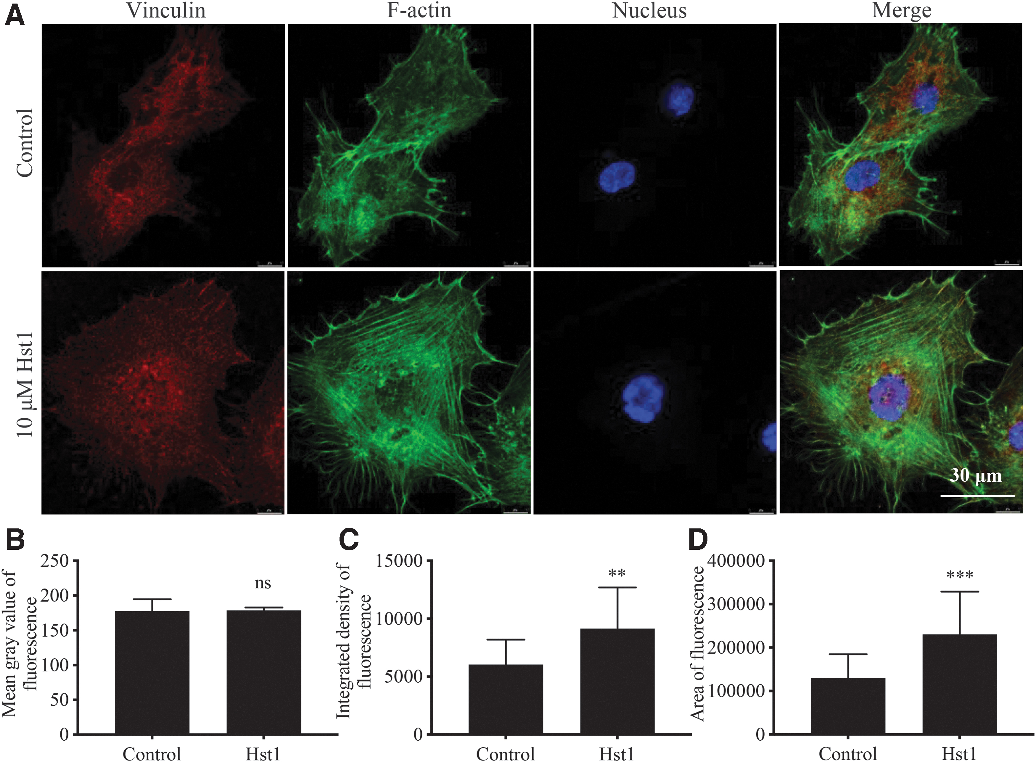
Hst1 increases the cell–ECM adhesion strength of hASCs. Cells were plated on the glass coverslips for 90 min. The cells then were fixed and stained with vinculin antibodies and anti-F-actin antibodies. Fluorescence for vinculin and F-actin is shown in red and green, respectively. Hoechst nuclear staining is represented in blue. The images were captured using a 63 × objective lens.
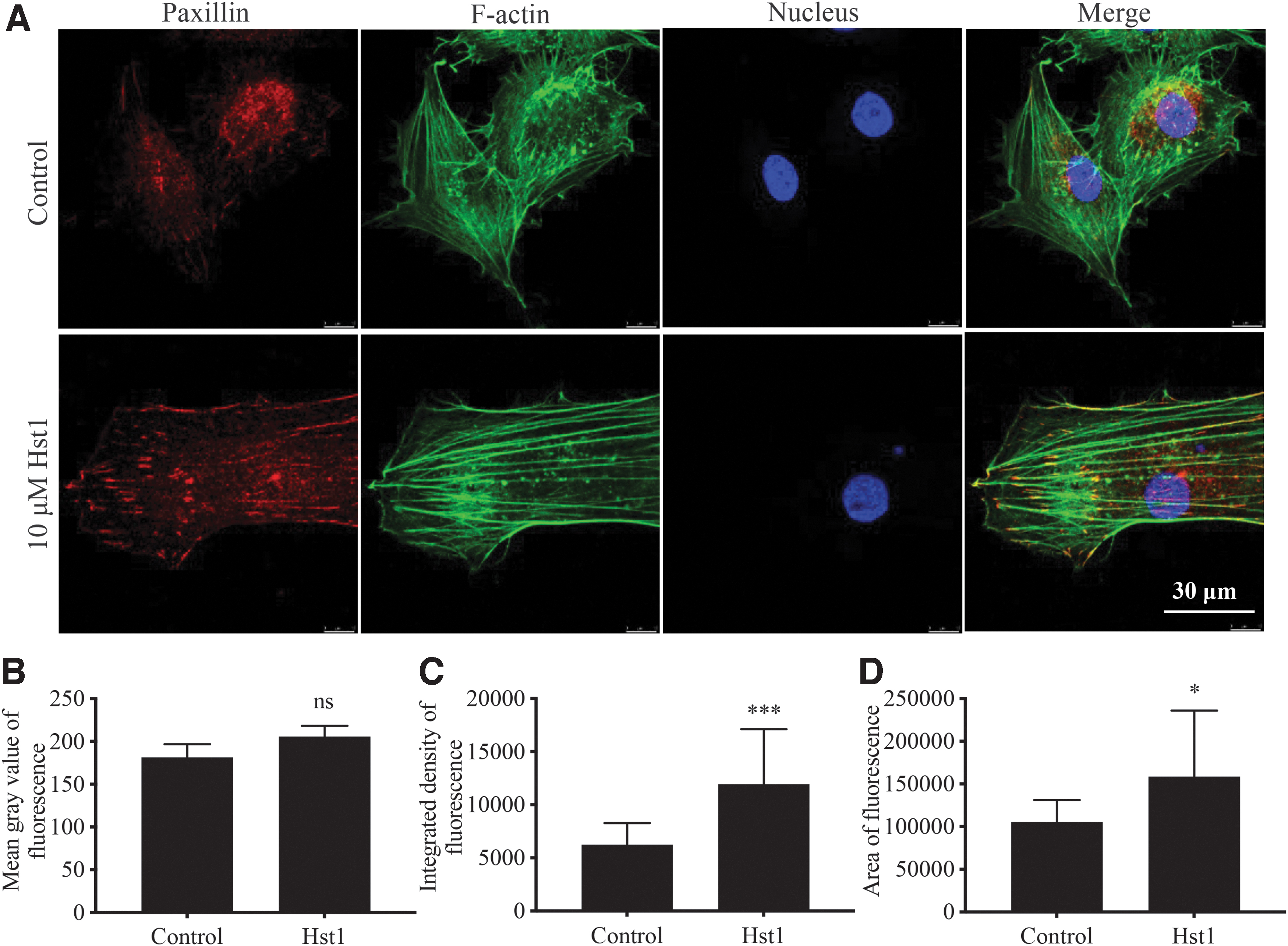
Hst1 increases cell–ECM adhesion strength of hASCs. Cells were plated on the glass coverslips for 90 min. The cells then were fixed and stained with paxillin antibodies and anti-F-actin antibodies. Fluorescence for paxillin and F-actin is shown in red and green, respectively. Hoechst nuclear staining is represented in blue. The images were captured using a 63 × objective lens.
Furthermore, the areas of fluorescence signals of talin (Fig. 6D), vinculin (Fig. 7D), and paxillin (Fig. 8D) in the Hst1 group were also significantly higher than those in the control groups.
Effects of specific inhibitors of p38, JNK, and ERK1/2 signaling on Hst1-induced adhesion and spreading of hASCs on bioinert glass surface
To reveal the potential signaling pathways involved in the effect of Hst1 on the adhesion and spreading of hASCs, we used several specific inhibitors, such as p38 MAPK inhibitor (SB203580), JNK inhibitor (SP600125), and ERK1/2 inhibitor (U0126) to investigate their effects on cell adhesion and spreading. As shown in Figure 9C, the fold change of total cell number for Hst1/No Hst1 was significantly higher than that for (Hst1+SB)/SB (Fig. 9A) and (Hst1+U)/U (Fig. 9C). This indicated that p38 MAPK and ERK1/2 signaling pathways are involved in the effect of histatin on the cell adhesion to scaffolds. However, the fold change of total cell number of Hst1/No Hst1 was significantly lower compared with SP, which implied that the JNK signaling pathway might not participate in the cell–scaffold interaction with histatin (Fig. 9B). Meanwhile, none of the inhibitors significantly changed the cell surface area (Fig. 9).
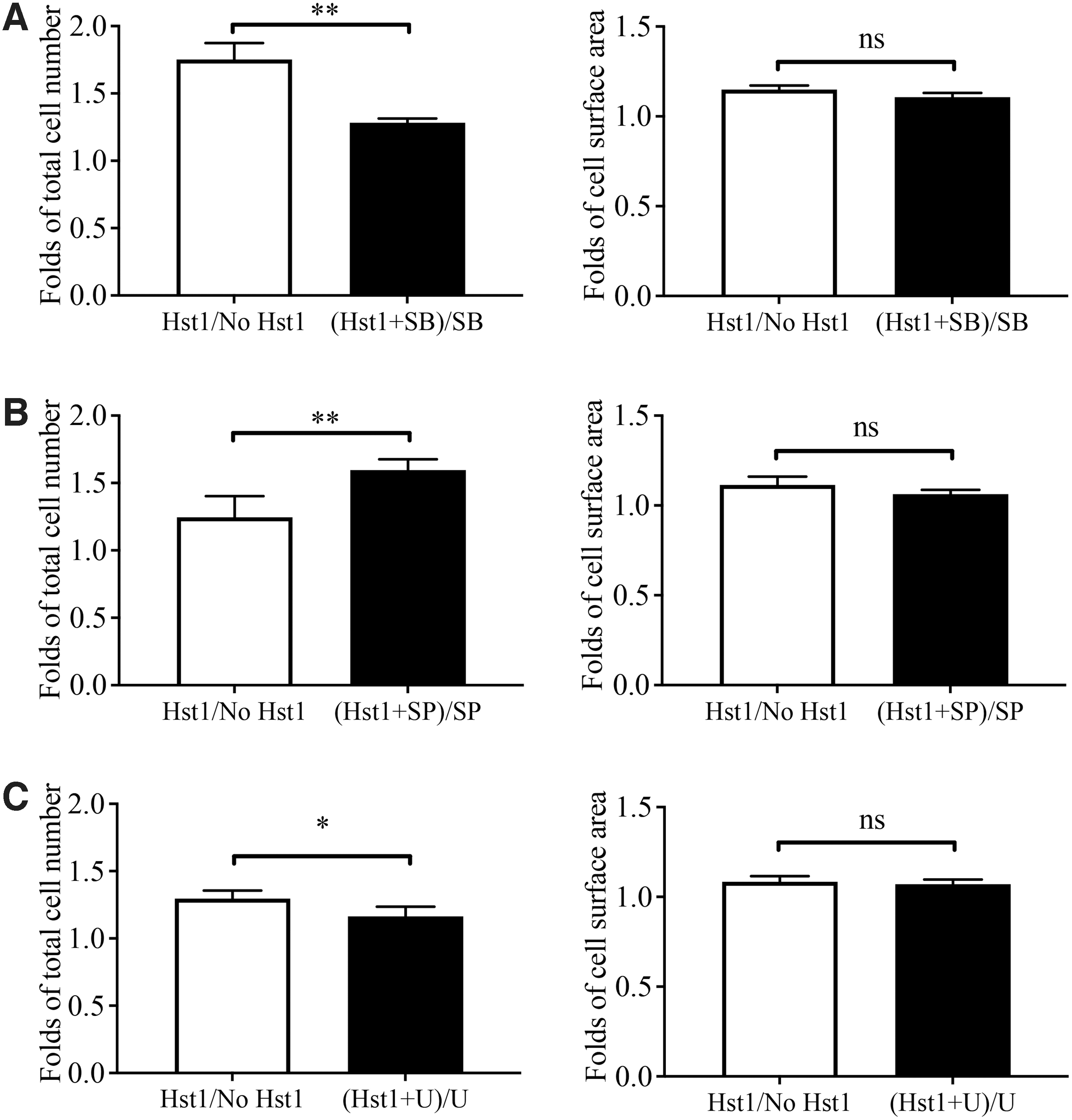
Effect of MAPK signaling on Hst1-induced cell adhesion and spreading. Treating the cells with 10 μM inhibitors of MAPK 2 h before seeding.
Discussion
Cell–scaffold interaction is critical for the success of MSC/3D printing-based bone tissue engineering. In this study, we investigated the feasibility and efficacy of applying Hst1 to enhance the adhesion and osteogenic differentiation of hASCs on 3D-printed bioceramic scaffolds. We showed that Hst1 significantly promoted the adhesion, spreading, and osteogenesis of hASCs on 3D-printed β-TCP scaffolds. Furthermore, Hst1 enhanced the in vivo survival activity of hASC-based constructs. Our data suggested a promising application of using Hst1coating strategy in stem cell/3D printing-based bone tissue engineering.
Our recent in vivo study shows that Hst1 significantly promoted BMP2-induced ectopic osteogenesis and angiogenesis 22 in rats and the healing of osteochondral defects of temporomandibular joint in rabbits, which suggests that Hst1 may be also active on MSCs. However, hitherto, there is still a lack of substantial evidence to prove the effects of Hst1 on the adhesion and osteogenic activities of MSCs. In this study, we first evaluated the effects of Hst1 on the adhesion and spreading of hASCs on both bioinert glass surfaces and 3D-printed β-TCP scaffold surfaces. Consistent with previous studies,17,18,23,24 the optimal concentration of Hst1 was 10 μM. Cell morphological analysis further showed that 10 μM of Hst1-induced cell spreading on 3D-printed β-TCP scaffold surface was more attributed to the increase of cell length but not cell width.
This phenomenon is different from the behavior of another osteogenic cell type MC3T3-E1 preosteoblast that tends to stretch isotropically on bioinert surface in the presence of 10 μM Hst1. 18 The discrepancy in cell behavior may be due to the different cell types and substrates.
The mechanism of protein adhesion to scaffold surfaces is of great importance to explore the optimal strategy to load proteins, such as the optimal protein concentration, incubation time, and preincubation with or without serum. This, however, was not explored in the present study. According to previous studies, protein adherence to ceramics mainly depends on electrostatic adsorption and hydrogen bond.25,26 We presume that histatin adhering to 3D-printed ceramics utilizes a similar mechanism. Aiming this, it is worth quantitatively assessing histatin adhesion to β-TCP scaffolds using labeled histatin and investigating the adsorption kinetics of histatin using the Langmuir adsorption isotherm model in the future.
In the following study, we used PrestoBlue cell viability reagent to analyze the effect of Hst1 on cell metabolic activities. The metabolic activities of hASCs in the group of Hst1 were significantly higher after cell seeding (varying from 1.15 to 1.68 times) compared with the control group. Four days postseeding, the ALP staining of the cells treated with Hst1 was also significantly higher (3.12 times) than the control group. Our results further showed that the Hst1-treated hASC/3D-printed β-TCP construct bore significantly higher numbers of viable hASCs than the non-Hst1-treated hASC/3D-printed β-TCP constructs. This suggested that the Hst1 facilitated the cell adhesion to 3D-printed β-TCP scaffolds and further significantly contributed to the high number of osteogenic cells in vivo. Further study in an orthotopic site should be performed to see the in vivo healing efficacy of the Hst1-treated hASC/3D-printed β-TCP constructs.
A previous study showed that endocytosis-related signaling of the Ras and Rab interactor 2 (RIN2)/Rab5/Rac1 signaling axis plays a critical role in Hst1's effect. 15 In this study, we showed that the integrated intensities of fluorescence per cell and the distribution area of focal adhesion complex-associated proteins, such as paxillin, vinculin, and talin, were significantly enhanced under the treatment of Hst1. However, the mean gray values of these proteins in the Hst1 groups were similar to those in the control groups. These findings indicated that the promoting effect of Hst1 on the spreading of hASCs was not attributed to the strengthening of focal adhesion as substrate-targeting strategy. 27
Although the effect of Hst1 shows a cell-type-independent property, the involved signaling pathways differ in various cell types. For example, the promoting effect of Hst1/Hst2 on the migration of epithelial cells could be nullified by U0126 (inhibitor of ERK signaling) and PTx (inhibitor of GPCR signaling) but not by SB203580 (inhibitor of p38 signaling).28,29 Similarly, Hst1 induces ERK phosphorylation and two different ERK inhibitors prevent stimulation of gingival fibroblast attachment by Hst1. 16 However, our recent study showed that U0126 and p38 but not PTx significantly inhibited the Hst1-induced spreading of MC3T3-E1 preosteoblastic cells. In this study, we found that all inhibitors, including SB203580, U0126, and SP SP600125, did not significantly affect the spreading of hASCs. However, U0126 and SB203580 significantly inhibited Hst1-induced adhesion of hASCs to 3D-printed β-TCP constructs. It seems that Hst1 activates different signaling pathways to affect the cell behavior.
In summary, our current study showed that Hst1 could significantly promote the adhesion, spreading, proliferation, osteogenic differentiation, and in vivo survival of hASCs on 3D-printed β-TCP scaffolds, which indicated a promising application of Hst1 in MSC/3D printing-based constructs for bone tissue engineering.
Footnotes
Authors' Contributions
D.W.: methodology (lead); formal analysis (equal); investigation (equal); data curation (equal); writing—original draft preparation (equal); and funding acquisition (equal). H.W.: methodology (lead); formal analysis (equal); investigation (equal); data curation (equal); writing—original draft preparation (equal); and funding acquisition (equal). Y.Y.: methodology (lead); formal analysis (equal); investigation (equal); data curation (equal); and writing—original draft preparation (equal). N.W.: methodology (support) and investigation (equal). R.T.J.: methodology (support). W.C.: methodology (equal). X.L.: methodology (equal). S.L.: methodology (equal). Y.Q.: visualization (support). F.H.: conceptualization (lead); writing—review and editing (equal); visualization (equal); and supervision (equal). H.L.: conceptualization (support); writing—review and editing (equal); visualization (equal); and supervision (equal). G.W.: conceptualization (support); writing—review and editing (equal); visualization (equal); supervision (equal); and funding acquisition (equal). All authors have read and agreed to the published version of the article.
Disclosure Statement
No competing financial interests exist.
Funding Information
This research was supported by the Shenzhen Science and Technology Innovation Committee (JCYJ20210324105408022), High-End Foreign Expert Recruitment Plan of China (No. G20200216024), Key Research and Development Plan of Zhejiang Province (No. 2021C04013), Department of Education of Guangdong Province (No. 2019KQNCX121), Guangzhou Municipal Health Commission (No. 20201A010067), and Guangzhou Medical University (No. 2020A046).