
Abstract
The management of large amounts of bio-wastes, such as bovine femurs from kitchens and slaughterhouses, has long been a challenging issue. However, through the utilization of a hydrothermal process, it is possible to transform these bio-wastes into valuable products. In this study, we focused on extracting hydroxyapatite (HAp), the primary inorganic component of bovine femurs, for potential use in bone tissue engineering scaffolds. By subjecting the femurs to hydrothermal treatment at varying times and solvents, we successfully decomposed and removed the organic matter present, resulting in the extraction of HAp. To comprehensively evaluate the properties of the extracted HAp, we employed several characterization techniques that provided valuable insights into the structure, morphology, and elemental composition of the extracted HAp. Furthermore, we conducted a Cell Counting Kit-8 assay, which confirmed the favorable biocompatibility of the extracted HAp. Overall, this study highlights the potential of hydrothermal treatment as an environmentally friendly and cost-effective method for handling bio-waste, specifically bovine femurs. The extracted HAp exhibits promising characteristics, making it suitable for a wide range of biomedical applications. This research contributes to the sustainable utilization of bio-waste and underscores the importance of resourceful exploitation for environmental protection.
Impact statement
Waste bovine femur bone represents a valuable resource with rich hydroxyapatite (HAp) content, making it an attractive candidate for recycling purposes. HAp holds significant importance in the biomedical field due to its remarkable biological activity and other favorable properties. In this article, HAp was extracted from bovine femur waste by a hydrothermal method. The study extensively investigated the properties of HAp under varying reaction conditions, including particle size, morphology, and the presence of trace ions. The results from cellular experiments demonstrated the immense potential of the extracted HAp as a valuable biomaterial for various applications. This work opens up new avenues for utilizing waste bovine femur bone and contributes to the advancement of biomedical materials research.
Introduction
Hydroxyapatite (HAp) has emerged as a highly desirable material for bone tissue engineering in recent years.1,2 It plays a critical role in the structure of bone, where collagen fibers mineralize onto the surface of HAp. 3 HAp possesses several advantageous properties, including noninflammatory and biologically active characteristics, 4 excellent biocompatibility, 5 nontoxicity, 6 nonimmunogenicity, 7 and osteogenic potential. 8 These attributes have spurred extensive research on the application of HAp in various biomedical fields. 9 Consequently, HAp has made significant strides in applications such as bone defect repair,9,10 bone tissue engineering scaffolds,11,12 implant coatings, and controlled drug delivery systems. 13
HAp can be prepared by various synthetic methods, including dry methods like mechanochemical methods,14,15 high-temperature processes such as roasting, 16 and wet methods like chemical precipitation,17,18 emulsion methods, 19 hydrothermal method,20,21 and sol–gel method.22,23 Synthetic methods have been developed to synthesize HAp in various morphologies; however, each method has its pros and cons. Sol–gel methods offer precise control over particle morphology and size, but have a low particle yield. Chemical precipitation methods are cost-effective and allow control over the reaction conditions to obtain different morphologies, but additional roasting may be required for improved properties. Some chemical methods may form toxic intermediates during the synthesis of HAp, posing challenges in achieving desired properties. While synthetic HAp is commonly used, it has limitations in terms of synthesis flaws and cost.
In addition, HAp can be obtained from natural resources or bio-wastes such as seashells,24–26 bovine bones,27,28 pig bones, 29 and fish bones,30–33 which have shown enhanced bioactivity compared with synthetic HAp. 34 Natural HAp contains additional elements, such as Na+, Mg2+, and F−, in addition to those present in the HAp structure, which mimics the ionic replacement structure found in mammalian bones.35,36 These additional ions play an important role in osseointegration, inducing cell differentiation and stimulating bone tissue growth. Synthetic HAp may suffer from unstable morphology and long synthesis cycles, making natural HAp an excellent candidate for bone tissue engineering applications.37,38
Nowadays, bio-waste has become a significant contributor, accounting for ∼40% of the total waste. Faced with the heavy pressure of bio-waste on environmental protection, the resourceful exploitation of bio-waste has attracted widespread attention. Many researchers have tried to extract HAp from biological waste, mainly by high-temperature roasting and hydrothermal methods. Both methods have been shown to be effective in obtaining pure HAp. Hydrothermal technology, in particular, has emerged as a promising and environmentally friendly method for treating biological waste. It offers the advantage of removing unwanted elements like fat, bone marrow, and other adhesions from the bone without the use of organic solvents. Bovine femur bones, one of the primary natural sources of HAp, are morphologically and structurally similar to human bone. Studies have revealed that bovine femur bones contain the highest amount of HAp per unit weight, with ∼1.6 kg of bone capable of extracting 1 kg of HAp. By harnessing high-temperature roasting and hydrothermal methods, the extraction of HAp from bovine femur bones becomes feasible.
However, the handling time and temperatures were changed within a wide range in previous studies, and the biological evaluation of the extracted HAp was not conducted.
This study aims to convert waste bovine bone into a product of high biomedical value using an efficient and cost-effective method, thereby mitigating its environmental impact and fulfilling biomedical demands. Herein, natural HAp was extracted from bovine femur bones by hydrothermal reaction. The hydrothermal process involved varying solvents and holding times. Through this method, the organic components of the bones were decomposed and removed to obtain pure HAp. Characterization of the extracted HAp was conducted using X-ray powder diffraction (XRD), thermogravimetric analyzer (TG), and Fourier transform infrared spectroscopy (FTIR). The results confirmed that longer holding times led to more effective extraction of pure HAp from bovine bones. Further analysis using infrared spectroscopy and X-ray diffractometry (XPS) examined the presence of carbonates as well as elements such as sodium and magnesium in the obtained HAp. Morphological analysis was performed using scanning electron microscopy (SEM). Additionally, the toxicity of the obtained materials was evaluated using the Cell Counting Kit (CCK)-8 assay.
Materials and Methods
Materials
The bovine femur bone was sourced from a commercial slaughterhouse in Hangzhou, Zhejiang Province, China. Sodium hydroxide (NaOH, 96.5% purity) was obtained from Aladdin Reagent (Shanghai) Co., Ltd. Ultrapure water was prepared in the laboratory. The cells used in the article were purchased from the cell bank of the Chinese Academy of Science (Shanghai, China). CCK-8 was purchased from Beyotime Biotechnology (Shanghai, China). Live–Dead Cytotoxicity Assay Kit was obtained from MesGenBiotech (Shanghai, China). Dulbecco's modified Eagle's medium (DMEM), fetal bovine serum, penicillin, and streptomycin were purchased from Thermo Fisher Scientific (China).
Methods
HAp was extracted from bovine femurs through hydrothermal treatment at various holding times in various solvents. The properties of the extracted HAp under different conditions were characterized using XRD, TG, and FTIR. Additionally, the elements present in the obtained HAp were analyzed using energy-dispersive spectroscopy (EDS) and XPS to detect the presence of trace elements. Morphological analysis of the obtained materials was conducted through SEM. The toxicity was assessed through cell experiments using the CCK-8 method. All the animal experiments were conducted under the guidelines approved by the Regional Ethics Committee for Animal Experiments (Zhejiang Sci-Tech University, China).
Experiment
Preparation of bone powder
Large pieces of tissue were removed from the bone surface using a knife. Then the bones were cut into small pieces using a bone saw. The pieces were boiled in water for 3 h, and then the bone marrow and other soft tissue were removed. Subsequently, the samples were rinsed with ultrapure water several times, and dried in an oven at 90°C for an hour. After that, the dried samples were manually ground into finer powders using a mortar. Finally, the bone powder was obtained after sieving.
Extraction of HAp
Natural HAp in bone powder was obtained by hydrothermal treatment.39–41 The ground bone powder was loaded into a reactor containing sodium hydroxide solution (NaOH, 25wt%) at a ratio of 1:4. 42 The hydrothermal reactors were tightly sealed and heated in an oven. The hydrothermal experiment was conducted at a temperature of 250°C for 5 and 12 h, respectively. The same hydrothermal process was also conducted in ultrapure water. The untreated bone powder (raw bovine femur powder [RAW]) served as a control, and its properties were compared with those of the extracted HAp in different groups (UPW-5 h, UPW-12 h, NaOH-5 h, and NaOH-12 h). Figure 1 shows the flow chart of HAp extraction from bovine femur.
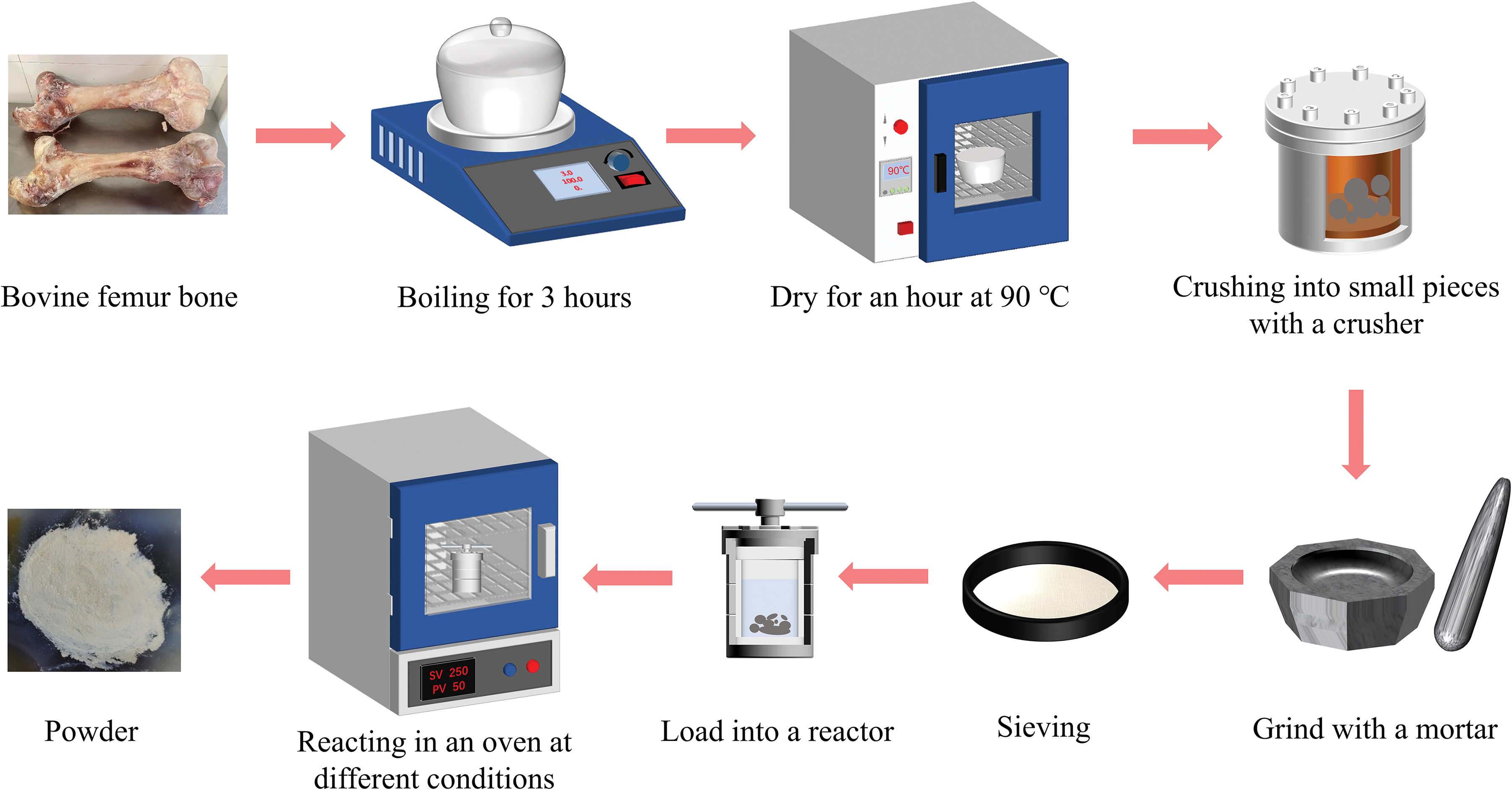
Flow chart of HAp extraction from the bovine femur. HAp, hydroxyapatite. Color images are available online.
Characterizations
Thermogravimetric analysis (TGA)
TGA is a thermal analysis technique that measures the relationship between the mass of a substance and its temperature under programmed temperature control. The mass loss of different sample was analyzed using a thermal analyzer (TGA, METTLER TOLEDO, TGA/DSC2).
XRD analysis
An X-ray powder diffractometer (XRD, Bruker AXS, A8 Advance) was used to examine the physical phase of the powder in the 2θ range of 20° to 65°.
FTIR analysis
FTIR (Thermo Fisher Scientific, Nicolet™ iS50) was used to investigate the functional groups contained in different samples within the wave number range of 4000 to 400 cm−1. The powder and potassium bromide (KBr) are mixed and ground in a ratio of 1:100, and the mixture is pressed into translucent flakes for testing.
XPS analysis
An X-ray photoelectron spectrometer (XPS, Thermo Fisher Scientific, K-ALPHA) was utilized to determine the elemental composition of the powders. EDS was employed to measure the powder elemental composition.
Morphological analysis
The microstructure and particle size of the samples were investigated using a SEM (Hitachi S-4800). Transmission electron microscopy (TEM, JEOL JEM-2100) was used to analyze the particle size of the treated powders.
CCK-8 assay
The biocompatibility of the HAp extracted by the hydrothermal method was assessed using the CCK-8 assay. HAp was immersed in DMEM at the concentrations of 0.5, 1, 5, and 10 mg/mL for 24 h, followed by centrifugation to remove the precipitate to obtain the immersion solution. Human umbilical vein endothelial cells (HUVECs) and mouse fibrosis cell lines L929 (L929) cells were cultured in HAp immersion solution for 48 h, and the absorbance of the supernatant was measured at a wavelength of 450 nm.
Results
TGA analysis
The TGA spectra of HAp obtained by the hydrothermal procedure and raw bovine bone powder (RAW) are shown in Figure 2. There is a clear difference in weight loss between the raw bovine bone powder and the extracted HAp. For all samples, an initial weight loss occurs at 50°C to 200°C, which is attributed to the evaporation of residual adsorbed water from the surface of the material. Another stage of weight loss exists at 250°C to 500°C, which is related to the removal of organic matter contained in the sample at high temperatures. Bones are known to contain about 30% of organic matter such as collagen, and at this stage, RAW loses about 20% more than the powder after hydrothermal treatment. Finally, according to Olsen et al.,
43
the mass loss shown in the TG at temperatures higher than 500°C is caused by the release of carbon dioxide from the decomposition of carbonates in the structure, suggesting that the HAp extracted from bovine femur bone by the hydrothermal method also contains some amount of

TG spectra of RAW and extracted HAp extracted under different conditions. RAW, raw bovine femur powder; TG, thermogravimetric analyzer. Color images are available online.
The mass loss after treatment for all samples was as follows: 31.4% for RAW, 9.2% for samples treated with ultrapure water at 250°C for 5 h, 6.8% for UPW-12 h, 11.3% for NaOH-5 h, and 0.5% for NaOH-12 h. Previous studies have shown that mammalian bones contain 30% to 40% organic matter. Therefore, the hydrothermal method can effectively remove the organic matter from bone powder.
X-ray powder diffraction
Figure 3 displays the XRD spectra of the treated bone powder and RAW powder, respectively, including bone powder reacted with ultrapure water at 250°C for 5 and 12 h, and bone powder reacted with 25wt% NaOH solution at 250°C for 5 and 12 h. Comparing the spectra with the JCPDS standard card, it can be observed that the material obtained by the hydrothermal reaction clearly corresponds to the peaks of JCPDS No.09-0432, indicating the presence of HAp. This confirms that the hydrothermal treatment can remove organic compounds from bone powder without destroying the molecular structure of HAp. In contrast, the XRD peaks of the raw powder (RAW) are fewer and broader than those of the treated samples. This may be due to the presence of collagen-assembled fibers on the surface of HAp, which scatters the X-ray radiation.

XRD spectra of RAW and extracted HAp extracted under different conditions. XRD, X-ray powder diffraction. Color images are available online.
Notably, the intensity of HAp obtained through the hydrothermal process with NaOH as the solvent is relatively higher than that obtained with ultrapure water. The spectrum of HAp obtained through hydrothermal treatment with NaOH as solvent shows a closer resemblance to the standard HAp in terms of peak strength. This indicates that the hydrothermal process is capable of extracting HAp from bovine bone. Moreover, the HAp obtained after a longer reaction time is closer to the standard HAp. A higher intensity and closer planar spacing can be observed in the spectra of NaOH-12 h group compared with that of the other standard HAp.
Fourier transform infrared spectroscopy
Figure 4 illustrates the FTIR spectra of extracted HAp and RAW powder, respectively, which show the presence of carbonate groups (
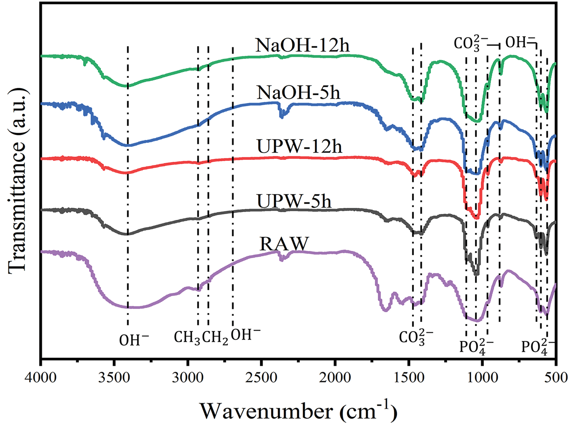
FTIR spectra of RAW and extracted HAp extracted under different conditions. FTIR, Fourier transform infrared spectroscopy. Color images are available online.
After the hydrothermal process, the intensity of these absorption bands is reduced or even absent in the spectra of the extracted HAp, indicating the removal of organic matter through hydrothermal treatment. The double splitting peaks around 1435–1470 cm−1 are associated with the asymmetric stretching of
It is known that HAp in natural bone contains about 3–8wt% of carbonate ions. The presence of a certain amount of
XPS analysis
Figure 5 indicates the XPS spectra of the HAp powder, which shows the bonding structure and the elemental composition. The wide scan spectrum indicates the main elemental composition of HAp and the chemical state. XPS analysis confirms the presence of other elements (e.g., K and Si) besides calcium (Ca), phosphorus (P), and oxygen (O) in HAp, which can have significant benefits for daily life. By fitting the narrow spectral peaks of XPS elements, it is observed that

XPS spectra of extracted HAp extracted under different conditions.
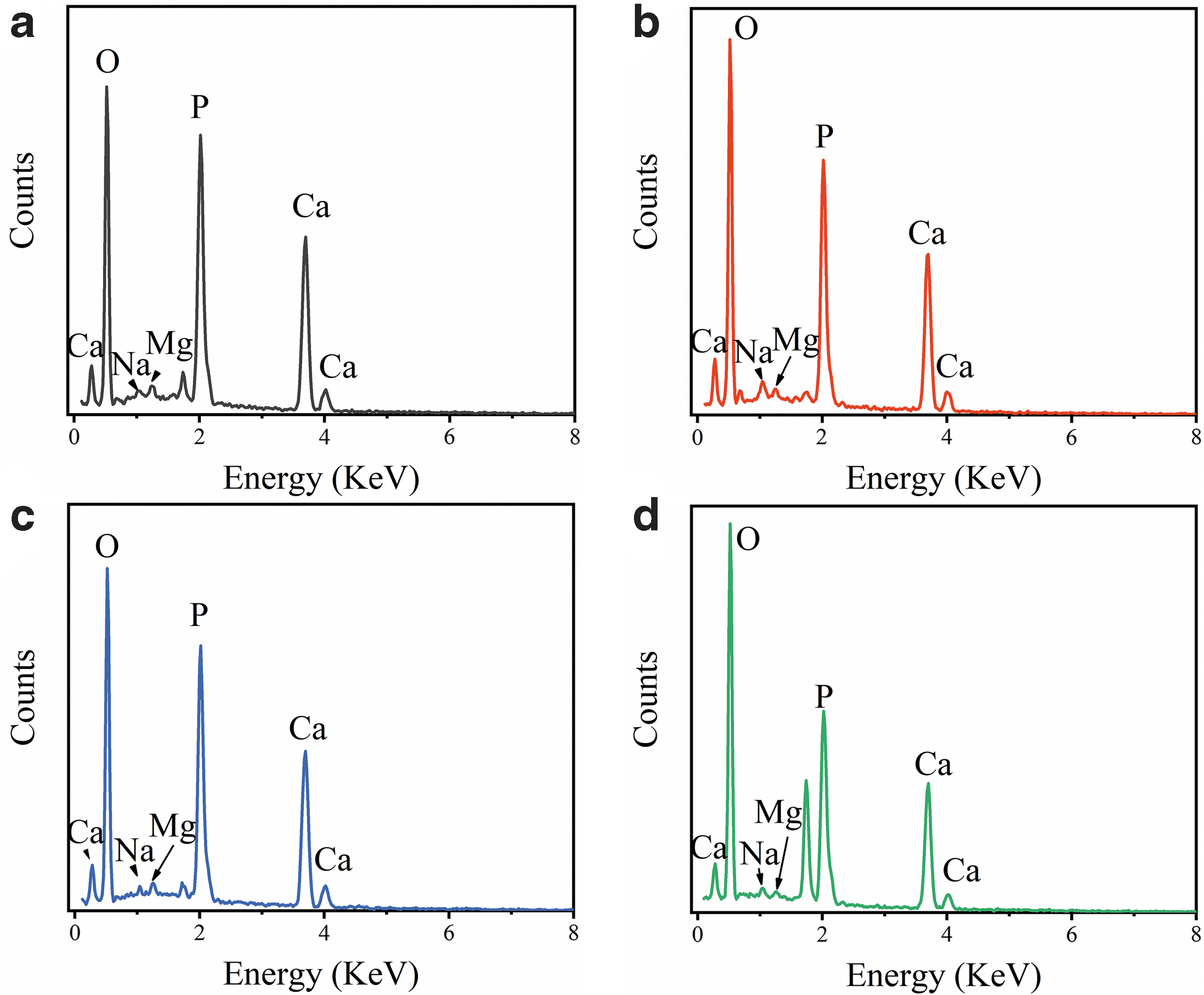
EDS spectra of extracted HAp extracted under different conditions.
Morphological analysis
To verify the influence of the hydrothermal method on the shape and size of the extracted HAp, the RAW and treated bone powder were investigated by SEM (Fig. 7). The SEM image of the powder treated with ultrapure water for 5 h (Fig. 7a) shows particles with irregular shapes and sizes, while the SEM image of the same solvent treatment for 12 h (Fig. 7b) shows that the particles are mainly in irregular lamellar structures with irregular edges and an increase in particle size compared with the 5-h treatment.
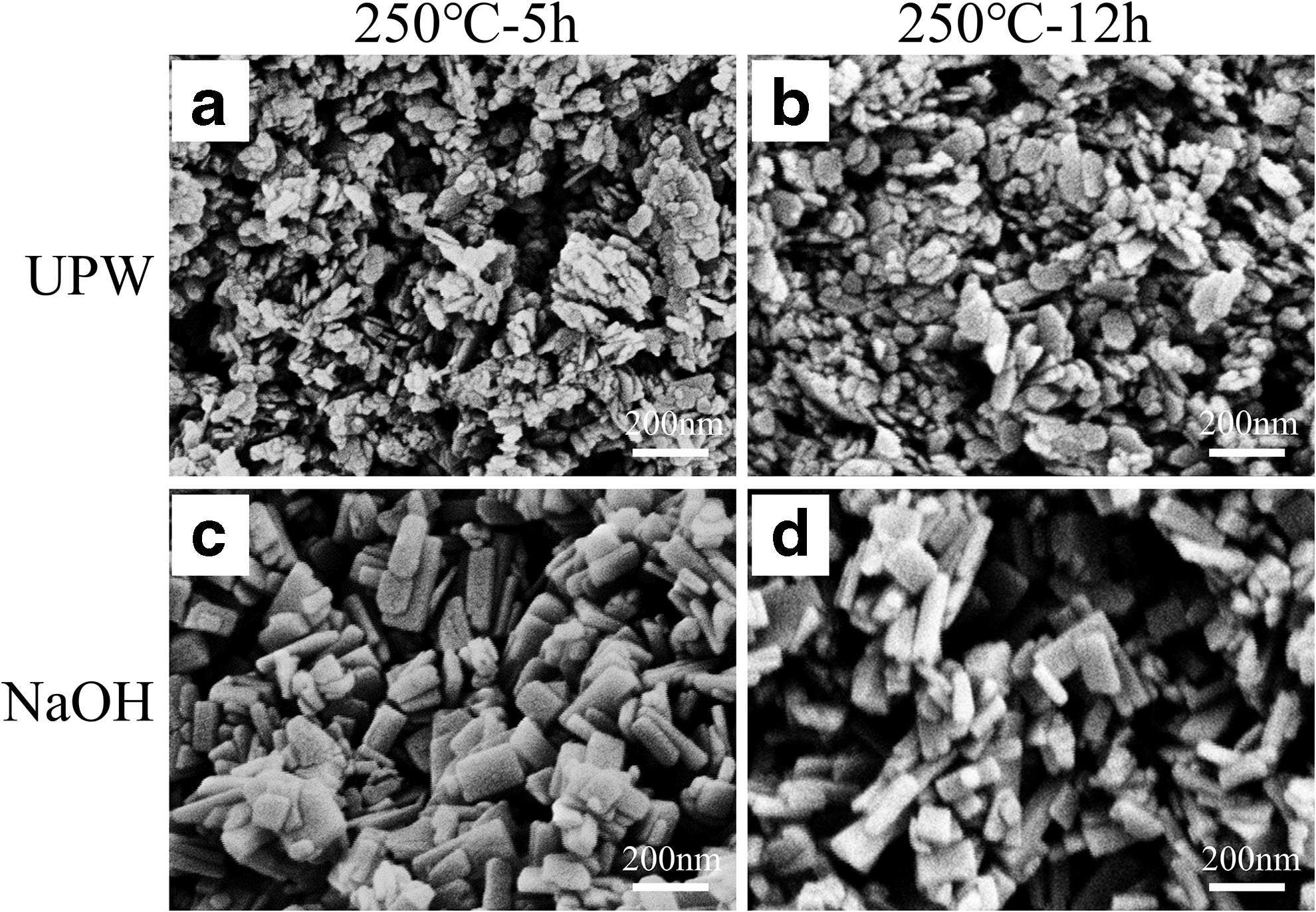
SEM images of HAp extracted under different conditions.
The SEM image of the powder treated with NaOH at 250°C for 5 h (Fig. 7c) exhibits predominantly rectangular lamellar morphology with more regular borders than the UPW-treated material. In contrast, the SEM image of the NaOH-treated sample for 12 h (Fig. 7d) shows a wide range of particle sizes, with most of the particles agglomerated and predominantly rod shaped, probably due to the grinding action. The particle sizes range irregularly from around 50 to 300 nm. No organic matter is observed, indicating the removal of organic components during the hydrothermal treatment. Similar changes are observed in the powder treated with ultrapure water at 250°C for 12 h (Fig. 7b).
Moreover, the morphology of HAp particles depends on the soak time of treatment and reaction temperature. The holding time and reaction temperature of the treated bone powder directly affect grain growth. As shown in Figure 7b, the grains of the particles are larger after a longer holding time of 12 h compared with 5 h. Under the high-temperature treatment at 250°C, the HAp particles showed significant changes compared with RAW. Grain growth is known to be associated with the absorption of heat energy, where higher temperature and longer holding time result in increased thermal energy absorption and more extensive grain growth, as suggested by Hoque et al. 45
In vitro biocompatibility
In Figure 8, the toxicity of HAp is evaluated through cell viability analysis using immersion solution culture. Figure 8a and b shows the quantitative data of cell activity of HUVECs and L929 cells, respectively, after incubation with HAp soaking solution at different concentrations for 48 h. The results indicate that the cell survival rate of HAp treated with NaOH is higher compared with HAp treated with ultrapure water after 48 h of incubation.
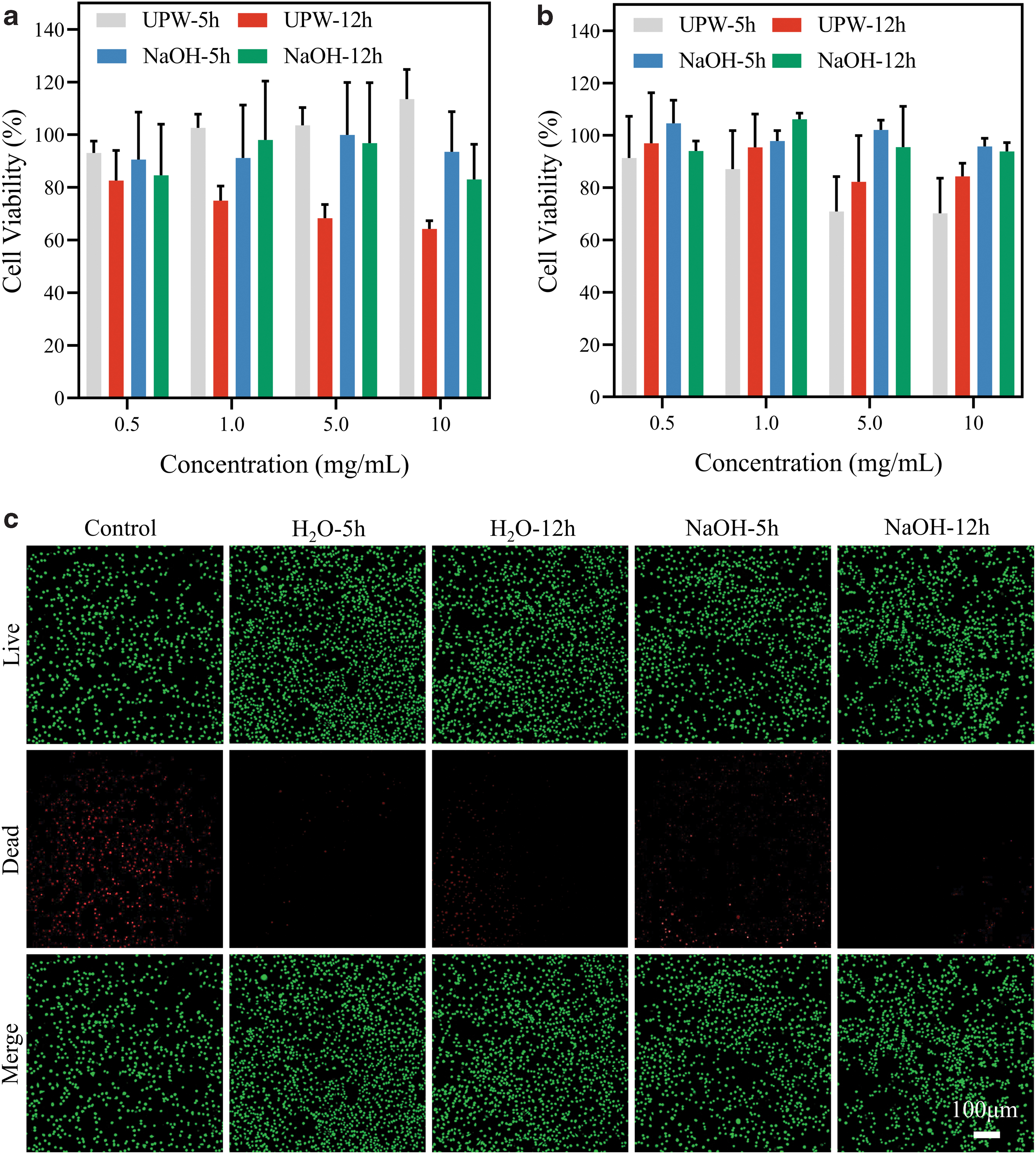
Quantitative detection of HUVEC
In Figure 8a, the toxicity of HAp extracted from bovine femur to HUVECs was not evident within the tested concentration range. However, the UPW-12 h group shows a gradual decrease in cell viability with increasing HAp concentration, possibly due to the presence of organic components that were not completely removed during the reaction with ultrapure water. The HAp does not exhibit significant toxicity in the concentration range of 0.5–1 mg/mL. Furthermore, the effect of different conditions of extracted HAP on cultured L929 cells is compared using confocal laser scanning microscopy at a concentration of 5 mg/mL (Fig. 8c). The effect of HAp immersion on cell activity is relatively minimal at this concentration.
The blank control group shows only a slight red fluorescence under the same culture conditions and for the same time, indicating a low number of apoptotic cells in the absence of external stimuli for 12 h, while the experimental groups are not significantly different compared with the control group.
Discussion
The treatment of biological waste has long been a pressing concern, and HAp extracted from bone waste holds great promise for biomedicine applications. While previous studies primarily employed the roasting method to extract HAp from bone waste, this study utilized waste bovine femur as the raw material and employed a hydrothermal process with varying reaction conditions to investigate the properties of the obtained materials.
The qualitative analysis of the obtained materials using XRD, FTIR, and XPS confirmed the main component to be HAp. However, depending on the experimental conditions, some of the materials contained small amounts of organic matter, and HAp extracted under a reaction time of 12 h exhibited higher crystallinity. A comparison of the reaction spectra at 5 h in the same solvent revealed that the XRD peak of HAp extracted at 12 h of reaction was sharper, and the TG plot showed a higher residual quality. The XPS and EDS results clearly demonstrated the presence of trace elements, such as Na and Mg, in addition to the major elements Ca, O, and P. This composition similarity to human bone suggests that the extracted HAp is not a stoichiometric ratio material and may have an important role in bone regeneration, making it a promising material for application. The SEM analysis revealed that the HAp extracted by the hydrothermal method predominantly took the form of rods. In vitro cell experiments demonstrated the excellent biocompatibility of the extracted HAp at a concentration of 5 mg/mL.
The immersion solution, tested on L929 cells using live/dead cell staining, showed no significant adverse effects compared with the control group, indicating good biocompatibility and nontoxicity. These findings highlight the potential use of the extracted HAp in biomedical fields, particularly for bone repair applications.
In this study, we focus on the utilization of waste materials to produce valuable substances, which is a crucial aspect of green and eco-friendly research. Hydrothermal treatment is a typical method to obtain inorganic substances. However, the processing parameters and procedures have significant impacts on the properties of materials. Here, only the hydrothermal method was used without secondary calcination, which is efficient and cost-effective. The HAp obtained by the single hydrothermal method has a lower degree of crystallinity, and the carbonate ions in the crystal structure are relatively more.
Although the procedure is simple, we verified the security and effectiveness of this method, which is suitable for actual production and biomedical application. Targeting important scientific and social issues, we used a simple and effective method to extract HAp from waste bovine femur bone, which is both eco-friendly and valuable, and the obtained HAp is biologically active for further biomedical applications. HAp is widely used as an artificial bone graft. HAp obtained by hydrothermal method can be combined with polymers to form microspheres for promoting bone repair.
Nano-HAp can encapsulate many bone implant materials to improve their bone restorative properties. Additionally, the structure of HAp makes it promising as an environmental remediation material, which can be used as a wastewater and soil treatment adsorbent. Moreover, HAp finds application in catalysis, including as photocatalysts and active carriers, further expanding its potential in various fields.
Conclusions
In this study, natural HAp was successfully extracted from bovine femur waste using a hydrothermal treatment method. The extracted HAp particles exhibit a rod-shaped morphology with sizes mainly ranging from 50 to 200 nm. The biocompatibility assessment demonstrates that the extracted HAp has no significant adverse effects on cell viability, indicating its potential for use in biomedical applications. The hydrothermal extraction method employed in this study is environmentally friendly, as it eliminates the use of organic solvents and utilizes bio-waste as a raw material. This approach not only transforms biological waste into a valuable product but also contributes to environmental protection through resourceful utilization. Overall, the successful extraction of pure HAp from bovine bone waste using the hydrothermal method highlights its promising potential as an economical and sustainable material for bone repair applications.
Footnotes
Authors' Contributions
Conceptualization: Y.Z., S.Y., and X.K.; methodology: Y.Z., S.Y., and F.K.; software: Y.Z.; validation: Y.Z. and F.K.; formal analysis: Y.Z., S.Y., and J.Z.; investigation: Y.Z. and D.W.; resources: D.W. and J.Z.; data curation: Y.Z.; writing—original draft preparation: Y.Z.; writing—review and editing: Y.Z., S.Y., and X.K.; supervision: X.K.; project administration: X.K.; funding acquisition: X.K. All authors have read and agreed to the published version of the article.
Disclosure Statement
No competing financial interests exist.
Funding Information
This research was financially supported by the Key Research & Development Program of Zhejiang Province (2021C01180, 2019C04020), Medical Science and Technology Project of Zhejiang Province (2023RC027), and the National Natural Science Foundation of China (51672250, 51902289).