
Abstract
The aim of this study was to analyze the effect of ozone (OZN) therapy on the dynamics of bone tissue in ovariectomized rats treated with zoledronic acid (ZOL). Female Wistar rats aged 6 months (n = 110) were subjected to bilateral ovariectomy (OVX). At month 3 post-OVX, 10 animals were euthanized to characterize the bone tissue architecture using microtomography (micro-CT). The remaining animals were divided into two groups: ZOL group, administered with ZOL (100 μg/kg body weight); saline (SAL) group (0.45 mL of SAL solution), both for 28 days. At month 3 post-treatment, 10 animals from each group were euthanized to characterize the bone architecture using micro-CT. The remaining animals were divided into the following groups: ZOL (n = 20), ZOL + OZN (n = 20); SAL (n = 20), and SAL + OZN (n = 20). The animals in ZOL + OZN and SAL + OZN groups were intraperitoneally administered with OZN (0.7 mg/kg body weight) once every 2 days. On days 30 and 60, six animals from each group were euthanized for analysis and structural characterization of bones in the femoral head and spine. Some samples of the femoral neck were subjected to biomechanical tests, while some samples were analyzed under a laser confocal microscope. The other samples collected from the femoral neck and spine were analyzed for area of neoformed bone and used for performing inflammatory cell and osteocyte counts. Data were submitted to statistical analysis considering a significance level of p < 0.05. Bone volume percentage and osteocyte and inflammatory cell counts were upregulated in the femoral head region of the ZOL + OZN group. Biomechanical analysis of the femoral neck revealed that the modulus of elasticity was similar between the ZOL and ZOL + OZN groups but differed significantly between the SAL and SAL + OZN groups. The positive areas for calcein and alizarin in the ZOL and ZOL + OZN groups were higher than those in the SAL and SAL + OZN groups. This suggested a positive synergistic effect of OZN and ZOL on the maintenance of bone mass and restoration of bone tissue vitality in ovariectomized rats.
Impact statement
Ozone therapy has the potential to improve the dynamism of gnathic bones and long bones due to the decompensation caused by zoledronate therapy in ovariectomized rats.
Introduction
Pathological conditions, such as systemic arterial hypertension, diabetes mellitus, 1 anemia, and malnutrition, significantly affect the metabolism of long-lived organisms. Consequently, senescent organisms exhibit pathological metabolic changes that require treatment and strategies to optimize the biological response. Bone is one of the tissues that undergoes the most changes due to aging and other injuries, especially in women owing to menopause-related hormonal changes.2–6
The climacteric stage promotes a change in osteoblast and osteoclast activities, increases bone remodeling activity, and decreases deposition activity, thus affecting the bone macroarchitecture and microarchitecture and resulting in an imbalance in the bone turnover process. Clinically, these changes lead to bone fragility and increased susceptibility to fractures, especially in regions of the femoral neck and spine.7–11
Some drugs are used to modulate the pathological changes in bone turnover. Antiresorptive drugs are often used to treat osteopenia, osteoporosis, and bone metastasis. Other classes of drugs produce similar therapeutic effects although by different mechanisms of action, such as bevacizumab (antivascular endothelial growth factor monoclonal antibody; used to treat bone metastases, 12 denosumab (the receptor activator of nuclear factor-kappa B ligand inhibitor), 13 Sunitinib (platelet-derived growth factor inhibitor), and temsirolimus (specific mTOR inhibitor) that act on bone tissue metabolism.14–17
Elucidation of the drug characteristics will provide useful insights into the mechanism of drug-induced osteonecrosis, which was initially diagnosed in the maxillofacial bones. 18 It has recently been observed in patients with necrosis of the long bones, tibia, and fibula who use antiresorptive drugs.19–21 In patients, who use antiresorptive drugs,19–21 necrosis of the long bones, tibia, and fibula has recently been observed.
Based on the physiological and pathological effects of drugs on bone tissue, several therapies have been developed, such as hyperbaric oxygen, laser, and medicinal ozone (OZN) therapies. 22 OZN, which is an allotropic form of oxygen with three oxygen atoms (O3), at room temperature is a gas and has a characteristic odor.23–28 Some studies have demonstrated that OZN therapy showed a promising result in improving bone metabolism and repair 29 through its antioxidant effects, by modulating immune responses and upregulating the release of growth factors; however, limited studies have examined the therapeutic effects of OZN therapy.
Elucidation of the mechanism of action of antiresorptive drugs and their ability to inhibit bone metabolism and the ability of OZN to optimize the dynamics of bone tissue are the novelty of this study, and the findings will be of importance for the purposes of clinical application. The study hypothesis, which was based on the data that were analyzed relative to the biological principles of OZN in tissues (H1), was that OZN therapy would improve bone metabolism in rats that had a low bone density, which received zoledronic acid (ZOL) for osteoporosis.
The aim of this preclinical study was to analyze the bone dynamics (femoral head and spine) of ovariectomized rats treated with ZOL and systemically administered OZN.
Methods
Animals
Female Wistar rats (Rattus norvegicus; n = 110), aged 6 months and with a mean weight of 250 g, were used in this study, after obtaining approval of the Institutional Ethics Committee on Animal Use (Protocol 00405-2019). The animals were housed in cages (four animals per cage) in a temperature stable environment (22°C ± 2°C) with a controlled light cycle. Standard diet and water were provided ad libitum, except for 12 h before surgery, when the animals had food restrictions. The experimental procedures were performed according to the norms established by the “Guide for the Care and Use of Laboratory Animals.” 30
Experimental design and sample collection
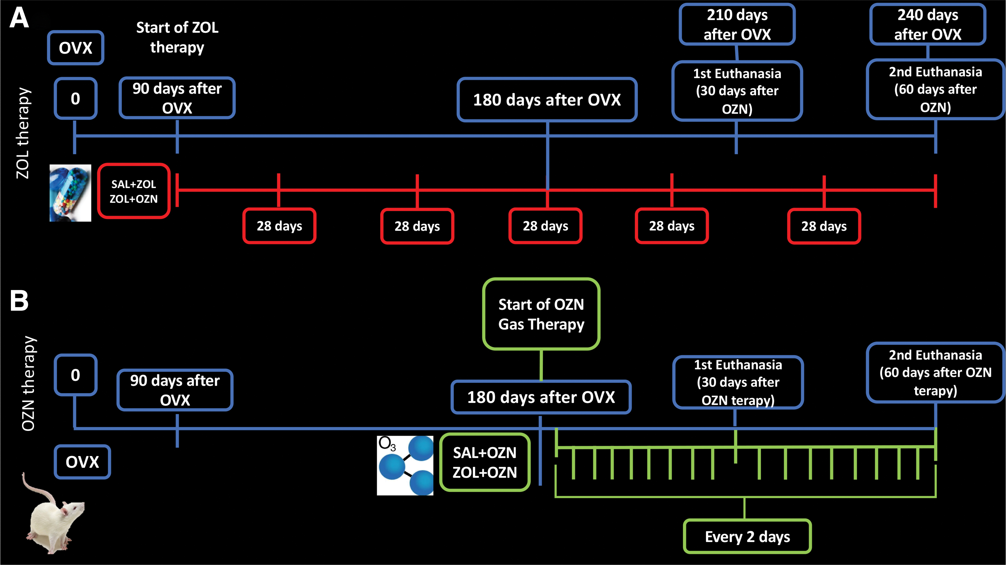
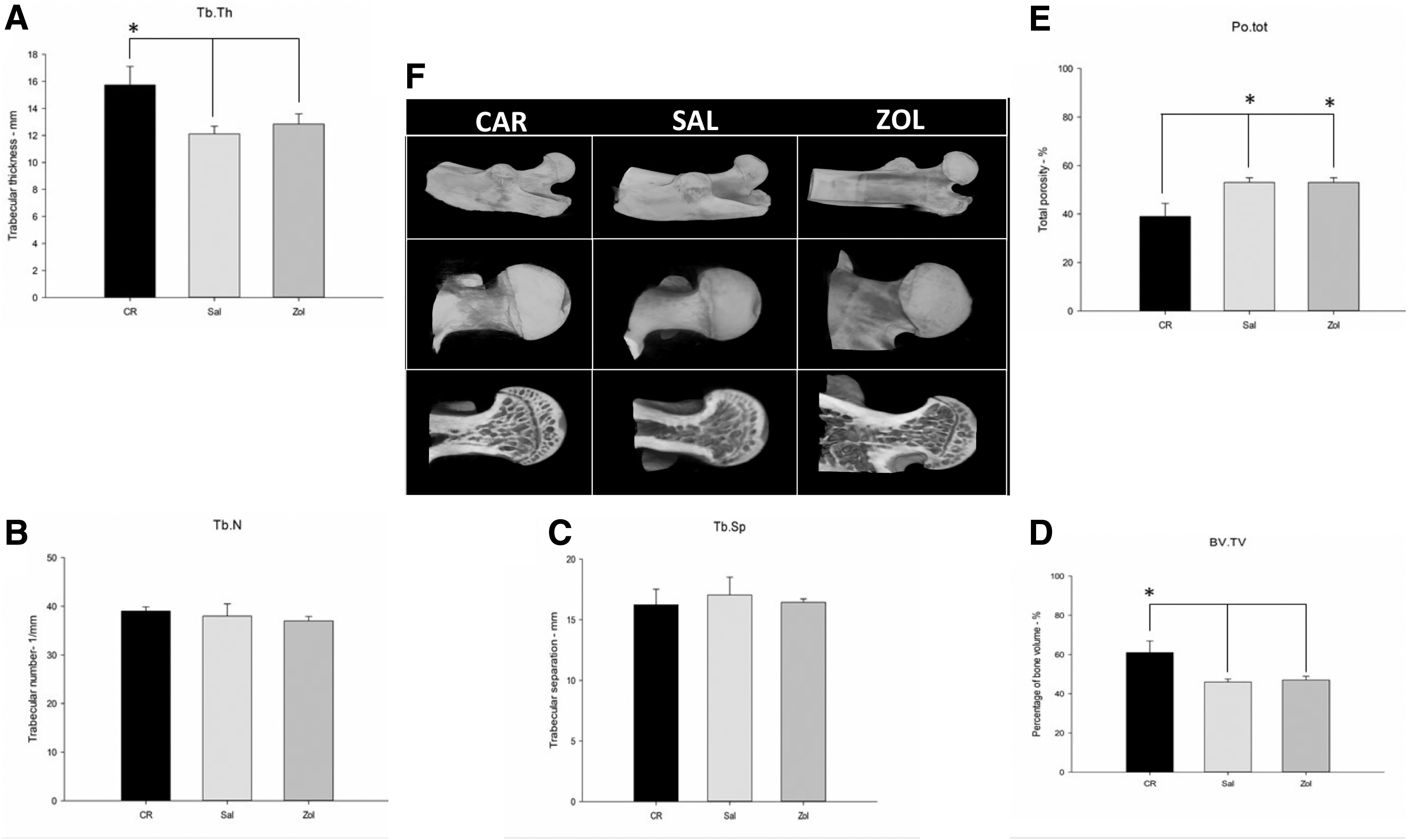
To perform the ovariectomy (OVX), all animals were subjected to sedation with a combination of ketamine (75 mg/kg-Dopalen®; Agribrans do Brasil LTDA) and xylazine (10 mg/kg-Anasedan®; Agribrands do Brasil LTDA). Subsequently, 10 animals were selected for the first characterization (CAR Group) (90 days after OVX). After they were euthanized, the femoral head samples were micro-CT scanned. The images showed a decrease in bone density at 3 months after OVX,31–33 which mimicked the epidemiological profile of osteoporosis in humans. Then, the remaining 100 animals were randomized into two groups: ZOL group (n = 50), treated with ZOL; and saline (SAL) group (n = 50), treated with SAL solution. At 180 days after OVX, 10 animals from each group were selected for a second characterization using micro-CT, to compare osteoporotic bone architecture with and without treatment. The remaining 80 animals were then subdivided into the following groups: ZOL group was divided into ZOL (n = 20) and ZOL + OZN groups (n = 20); SAL group was subdivided into SAL (n = 20) and SAL + OZN groups (n = 20) (Fig. 1A).

Experimental design of the study, observing the initial number of animals, the characterization periods using micro-CT, the division of the experimental groups, the euthanasia periods, and analysis and division of the experimental groups. Color images are available online.
ZOL administration
In humans, ZOL is usually intravenously administered at a dose of 5 mg, as a single dose annually.34,35 Thus, the same parameters were used for adjusting the dose and evaluating the average life span, body weight, and body surface of Wistar rats. The dose established was 100 μg/kg administered at every 28 days,36–39 until euthanasia (Fig. 2A).
Administration of OZN therapy
Medical OZN was administered at a dose of 0.7 mg/kg body weight with 8 mL per application based on previous studies.40–42 The animals were intraperitoneally (IP) injected once every 2 days until the end of the experiment (Fig. 2B).
Euthanasia and sample collection
The animals were anesthetized using the same protocol as described above. Cardiac perfusion was performed with SAL solution containing 0.1% heparin (100 mL), followed by administration of fixative solution (4% formaldehyde) in 0.1 M phosphate-buffered SAL (pH 7.4) at 4°C.
Characterization of bone microarchitecture
Bone microarchitecture was characterized using the micro-computed tomograph SkyScan (SkyScan 1176 Bruker Micro CT, Aartselaar, Belgium, 2003) and the following parameters: 8 μm sections, 90 kV, and 111 μA. The images were reconstructed with NRecon software (SkyScan, 2011; Version 1.6.6.0), and the images were reconstructed and positioned using the Data Viewer software (SkyScan, Version 1.4.4 64 bits). The region of interest was used to define the femoral head, and the following parameters were analyzed: bone volume fraction (BV/TV%), trabecular thickness (
Histological processing of samples
Semiserial histological sections 5-μm thick were obtained from the femoral head and spine, for staining with Hematoxylin and Eosin (HE), Picrosirius-red, and for the immunochemistry technique. The organs were analyzed under HE.
Histopathological and histomorphometric analysis
All analyses were performed by a blinded examiner and certified histologist (E.E.). Three samples of each femoral head and spine (L4) were collected in duplicate, and the histological HE slices were analyzed considering cortical and medullary bone tissue patterns. For histomorphometry, the slices were photomicrographed under a 6.3 × objective lens considering: bone tissue area, using “free hands” from ImageJ; and 100 × magnification considering: bone tissue vitality (osteocytes) and inflammatory cells (lymphocytes), using a 130-point grid produced by the ImageJ software. Each histological parameter coincident with a grid point was computed, and the percentage of vital bone relative to the total area was calculated. Finally, histological slices from organs (mesentery, lungs, liver, kidney, and brain) were stained with HE, and potential changes, such as presence of cellular infiltrates, hyperplastic tissues, metaplastic transformation, or dysplastic transformation, were investigated. 44
Birefringence analysis
The sections stained with Picrosirius red were captured under × magnification using a polarized microscope as previously described, 45 to evaluate the quality and quantity of bone collagenous content. The green, yellow, and red color spectra were defined following the RGB (red, green, blue) values. For quantification, the images were binarized for each color spectrum, and the number in pixel 2 of each color corresponding to each field was found. Subsequently, the average values in pixel 2 were calculated using the AxioVision (4.8) software and subjected to statistical analysis to calculate the average and standard deviation for each group in each experimental time interval. 46
Immunohistochemical and histomorphometric analysis
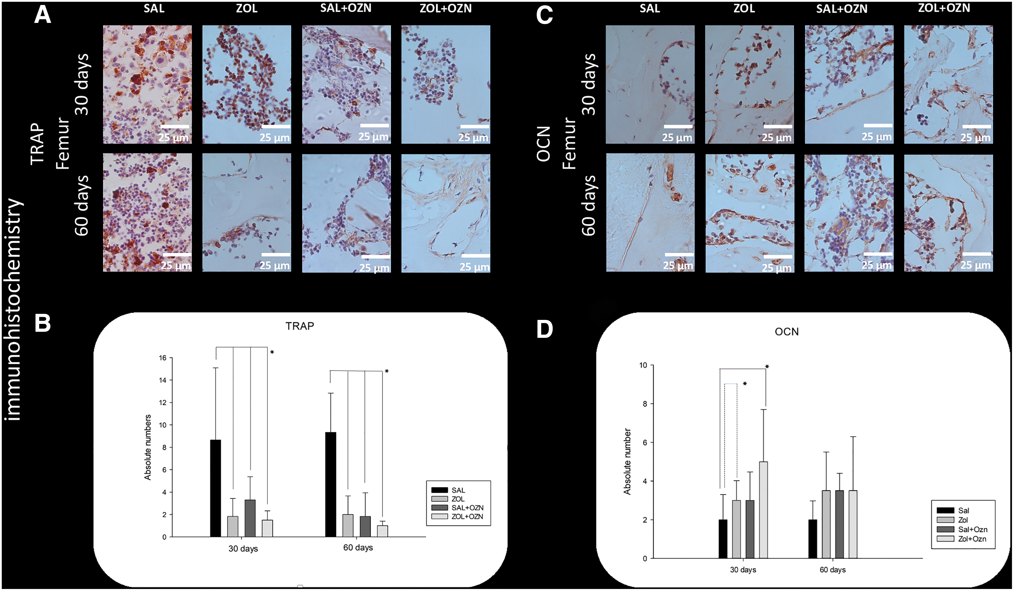
For immunohistochemistry, OCN and TRAP (SC-18319 and SC-30833, respectively; Santa Cruz Biotechnology®) were analyzed. 45 The negative control specimens were subjected to the same procedure. The target protein expression was semiquantitatively (ordinal qualitative analysis) evaluated. The analysis was performed using absolute numbers of cells per slide labeled with diaminobenzidine, with labeling being considered a positive result. Negative controls were used to assess the specificity of the antibodies.
Confocal laser microscopy
For analysis of the calcified tissues using laser confocal microscopy, the standardized times were 14 and 42 days from the beginning of OZN application (after the end of the OZN application). The fluorochromes calcein and alizarin were intramuscularly administered at a dose of 20 mg/kg 47 body weight. The femurs were subjected to laboratory processing and cut into pieces with a polisher until sections of ∼1 μm thickness were obtained. The sections were analyzed under a laser confocal microscope.
ImageJ software was used to analyze the images using the “color threshold” tool, and each image was standardized according to hue, saturation, and brightness. After detecting the fluorescence of the calcein-positive region, the “measure” tool was used to determine the area in pixel. 2 The same procedure was performed to analyze alizarin-positive regions to obtain the bone dynamics data.
Biomechanical analysis
After processing and fixing the femurs in a standardized acrylic resin framework, a vertical force was applied to the femoral head region until fatigue and fracture. Graphs were prepared, in which the displacement and deformation were represented. The modulus of elasticity and energy of deformation were analyzed from the graphs. The modulus of elasticity was obtained from the slope of the displacement-deformation curve, whereas the energy absorbed by the body was calculated using the area under the force-deformation curve.48,49
Statistical analysis
Statistical analysis was performed using Sigma plot. The values were initially subjected to the Shapiro–Wilk normality test. Bone tissue percentage data in the femoral head region were analyzed using two-way analysis of variance (ANOVA), followed by the Tukey test, whereas percentage data of bone tissue in the spine were analyzed using one-way ANOVA, followed by the Tukey test. Differences were considered significant at p < 0.05.
Results
Characterization of bone microarchitecture
Bone microarchitecture in the femoral head region was examined after OVX, ZOL, and SAL therapies. The BV/TV and trabecular thickness (
Histopathological analysis
In the femoral head, the photomicrographs showed that the ZOL Group had an extracellular matrix without osteocytes and osteoclast voids, representing the nonvital bone tissues, in both periods (Fig. 4A). The ZOL + OZN group exhibited regions of the extracellular matrix with osteocytes filling the laminae, representing bones with increased vitality (Fig. 4C, D). SAL + OZN and ZOL + OZN Groups exhibited enhanced inflammatory cell (lymphocyte) infiltration into the femoral head region (Fig. 6A, B). A similar inflammatory response was observed in the spinal region of the SAL + OZN and ZOL + OZN Groups.
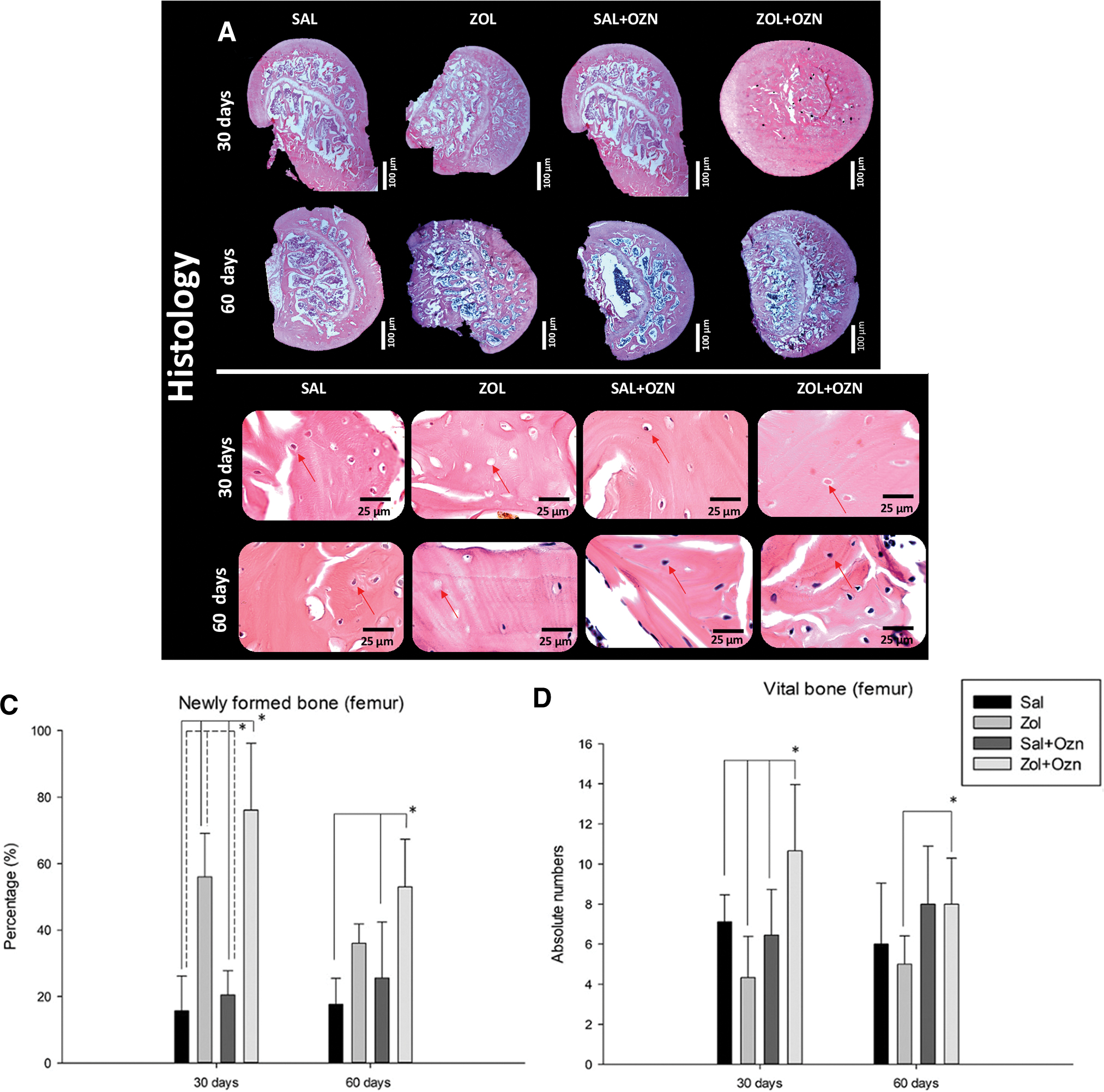
The photomicrographs of the experimental groups also showed that in both periods, there was an imbalance between the proportion of cortical and trabecular bone tissue, with a higher value of trabecular volume (Figs. 4A and 5A). In the most critical regions, represented by the medulla, the trabeculae were thin, with considerable spacing between them. The administration of ZOL clearly promoted a larger amount of cortical bone tissue to the detriment of trabecular bone, with greater thickness of the trabeculae specially at day 30.

Histomorphometric analysis
Bone tissue area
At day 30 and 60, ZOL + OZN Group (76.1 ± 20.10 and 52.98 ± 14.34) exhibited the highest percentage of bone area in the femoral head, followed by the ZOL (56 ± 13.07 and 36 ± 5.85), SAL + OZN (20.46 ± 7.35 and 25.56 ± 16.85), and SAL (18.27 ± 10.4 7 and 17.64 ± 7.82) Groups (Fig. 4B).
When the spine was analyzed, similar results were found. At day 30, ZOL + OZN Group (59.22 ± 8.48) revealed higher percentage of bone tissue area compared with the ZOL (42.86 ± 7.36), SAL + OZN (27.81 ± 18.50), and SAL Group (12.73 ± 5.44) (Fig. 5A–D).
Bone tissue vitality
At day 30, ZOL + OZN Group had the highest number of osteocytes in the femoral head (10.66 ± 3.31), followed by SAL and SAL + OZN Groups (7.11 ± 1.36 and 3.6 ± 1.87), and ZOL Group (4.33 ± 2.06). At day 60, the number of osteocytes had significantly increased in ZOL + OZN (8 ± 2.3) compared with ZOL (5 ± 1.44) Group.
No significant difference in the spine was observed between groups (Fig. 5D).
Assessment of the inflammatory profile
In the femoral head region, the number of inflammatory cells (lymphocyte) in ZOL + OZN (5.3 ± 4.38) and SAL + OZN Groups (3.6 ± 1.87) was higher than in SAL (2.3 ± 2.23) and ZOL Groups (2.22 ± 1.92) at day 30 (Fig. 6C).
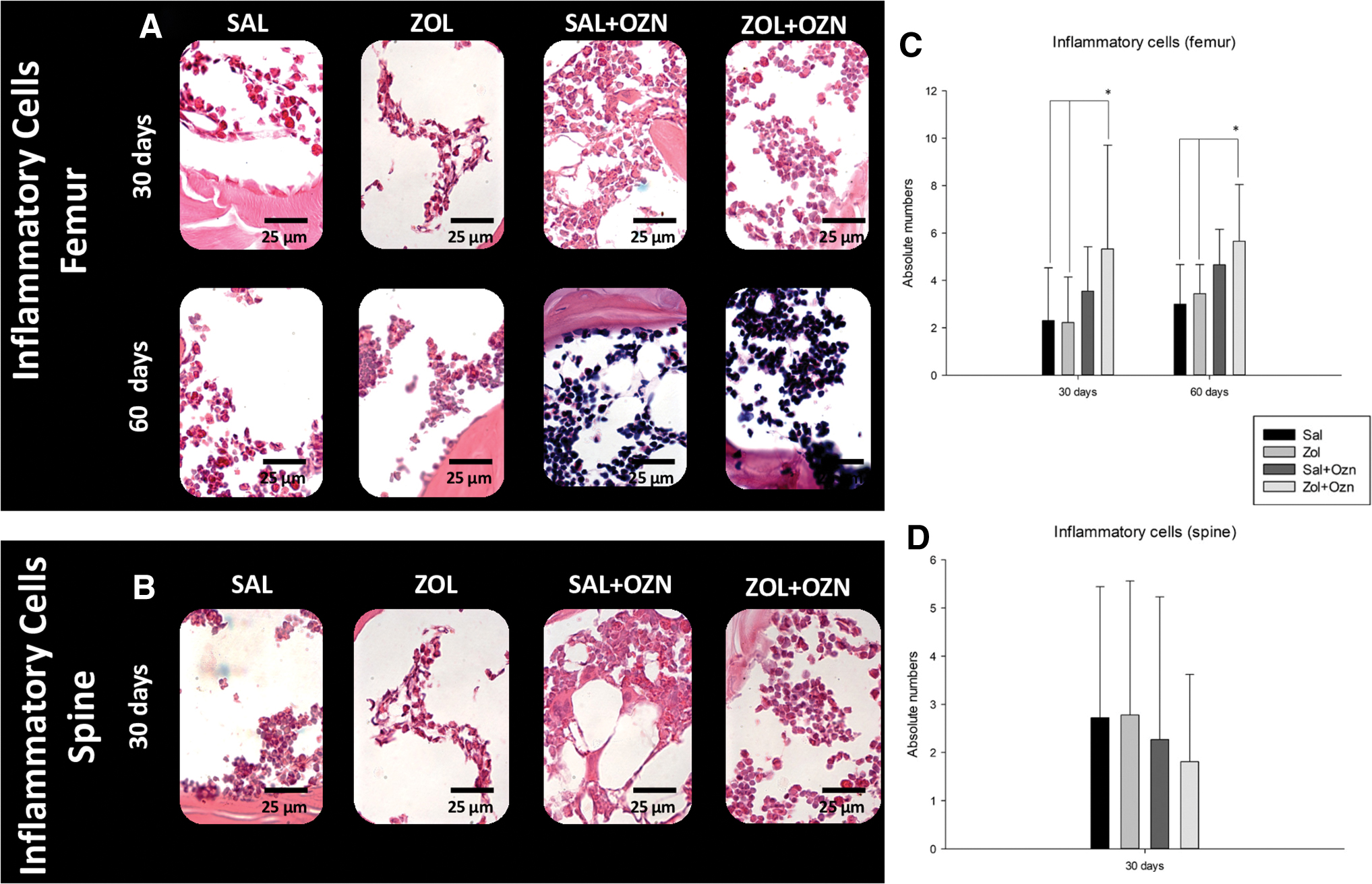
No significant difference in the spine was observed between groups (Fig. 6D).
Immunohistochemistry
TRAP immunolabeling revealed that the osteoclastic activity in the SAL Group (8.66 ± 6.43) was higher than it was in ZOL (1 ± 1.6), ZOL + OZN (3.3 ± 2.07), and SAL + OZN Groups (1.5 ± 0.83) at day 30 (Fig. 8A, B). Whereas the value of OCN+cell of ZOL + OZN Group (4.83 ± 2.71) was higher compared with SAL (2.5 ± 1.3) and ZOL (3.33 ± 1.03) Groups (Fig. 8C, D). No significant differences were detected at day 60.

Birefringence analysis
Green spectra fibers were significantly increased in SAL Group (0.55 ± 0.32 and 0.68 ± 0.24) compared with ZOL (0.17 ± 0.1 and 0.2 ± 0.08) at days 30 and 60 and in the ZOL + OZN Group (0.38 ± 0.1 and 0.38 ± 0.03) compared with ZOL (Fig. 9B).
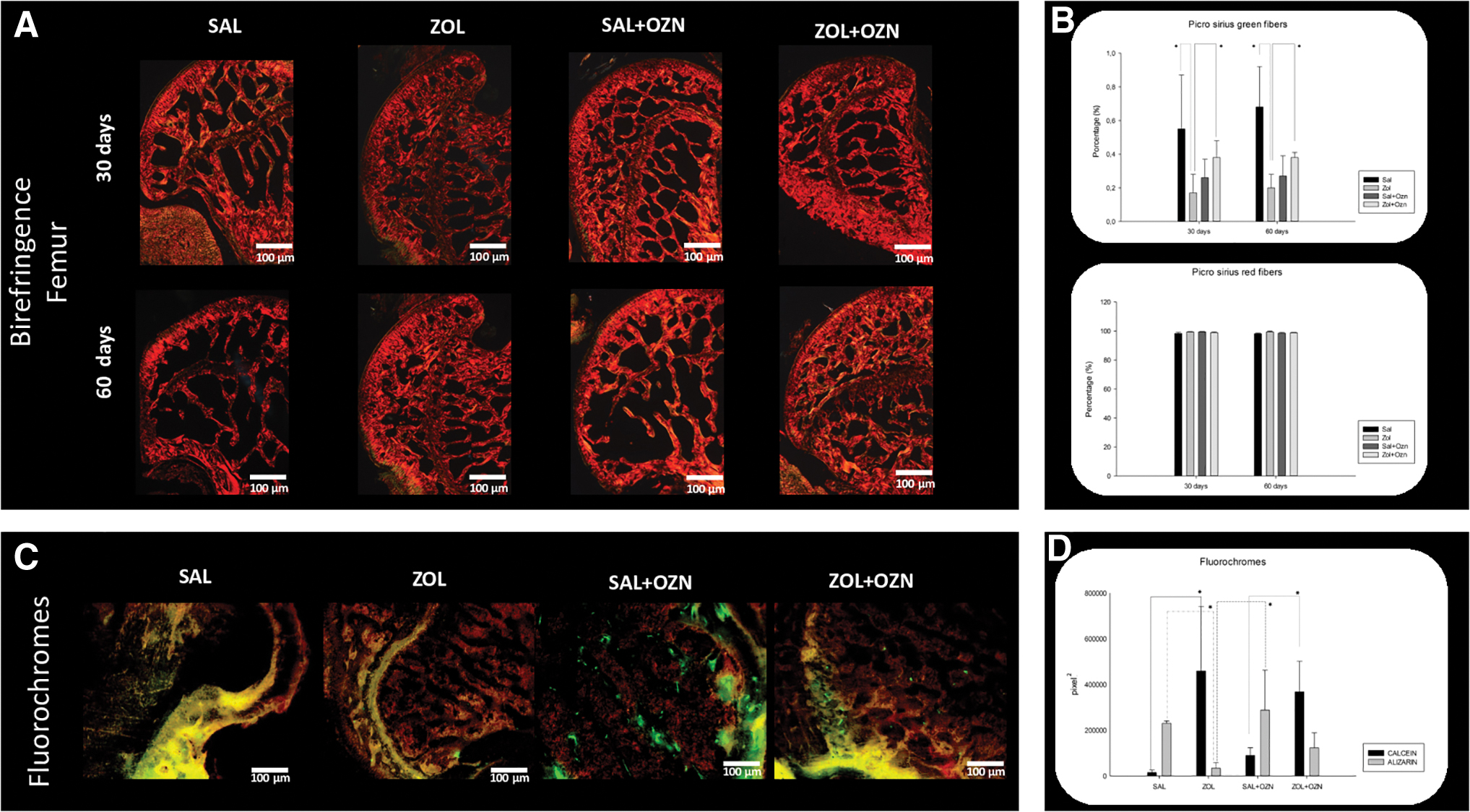
Considering the red fibers, no differences were detected between the experimental groups and time intervals (Fig. 9A, B).
Confocal laser microscopy
At day 14, the precipitation of calcium value (calcein-positive areas) in the ZOL (459,209 ± 119,264.7) and ZOL + OZN Groups (367,886.2 ± 104,836.3) was higher than it was in SAL (14,821.8 ± 6148.2) and SAL + OZN Groups (89,817.4 ± 18,191.56). The intensity of alizarin-stained regions, which represent neoformed bone (calcium precipitation over 42 days), in the SAL + OZN (287,997.8 ± 97,384.36) Group was significantly higher than in the ZOL (38,302 ± 17,853.75) and ZOL + OZN Groups (122,740.6 ± 50,417.25). Furthermore, alizarin staining intensity differed significantly between the SAL (230,040 ± 14,682.92) and ZOL Groups (Fig. 9C, D).
Histopathology of brain, liver, mesentery, lung, and kidney
Histopathological features of the organs obtained at day 60 did not reveal abnormalities with OZN therapy in comparison with the nontreated Groups (Fig. 7A).
Biomechanical analysis
The modulus of elasticity value of the femoral neck region was the highest in the ZOL + OZN Group (557.31 ± 146.16), followed by the SAL + OZN (272.13 ± 30.37), SAL, and ZOL Groups (p < 0.05) (order of statistical significance: SAL + ZOL = ZOL + OZN > SAL = ZOL) (Fig. 7B). The deformation energy, which is the energy necessary to deform the femoral neck by 0.3 mm, in the ZOL+ OZN group (26.56 ± 6.04), was significantly higher than that in the SAL (10.3 ± 1.1) and SAL + OZN (9.4 ± 3.61) Groups (p < 0.05) (Fig. 7B).
Discussion
The biomedical application of OZN has been associated with several benefits, such as improvement in renal function in patients undergoing hemodialysis, alleviation of skin diseases, and pain management in patients with herniated discs.50,51 The mechanism of action of ZOL has been well described and widely discussed in the literature. ZOL promotes significant changes in bone turnover, leading to a decrease in biological activity that can lead to bone necrosis.52–54 In this study, the number of osteocytes in the regions of the femoral head and spine were analyzed, which represents an alternative method to determine the vitality of bone tissue in these regions. The number of osteocytes in the ZOL + OZN Group was significantly higher than that in the ZOL Group, indicating that OZN had increased the number of osteocytes, which can be used as a determinant of the vitality of bone tissue.
However, the effect of OZN on bone tissue metabolism and its association with antiresorptive drugs has not been previously demonstrated. The hypothesis of this study was validated as was evidenced by the ZOL and ZOL + OZN Group exhibiting improved histological and histomorphometric parameters compared with the SAL, ZOL, and SAL + OZN Groups. The ovariectomized rats with decreased bone density exhibited the expected results. In particular, ZOL inactivated osteoclasts and consequently maintained bone quantity and density.
One study demonstrated the potential of OZN for stimulating the biological response with a significant increase in the formation of epithelium, deposition of extracellular matrix, proliferation of cells, 25 expression of endogenous enzymes, antioxidant effects, and the synthesis of oxygenase and downregulation of pro-inflammatory cytokines. 55 Another study evaluated the effect of OZN on bone metabolism and reported an increase in the number of osteoblasts in animals subjected to regular applications of OZN, 56 data that were consistent with the results of this study.
Immunohistochemical analysis revealed the upregulation of TRAP expression and downregulation of OCN expression in the SAL Group. In contrast, OCN was upregulated and TRAP was downregulated in the ZOL Group, which was expected due to the experimental model adopted in this study. These findings were consistent with those of histological analysis. The expression of TRAP and OZN was balanced in the SAL + OZN and ZOL + OZN Groups, which was an encouraging result as the balance between osteoclastic and osteoblastic activities is a primordial factor for maintaining bone tissue health. Another study that investigated the repair process of critical defects in the caps of diabetic rats using biomaterials, and analyzed the expression of BMP2 and OCN, revealed increased labeling of BMP2 and OCN in the groups treated with OZN, 57 validated using histological analysis. The total area value of neoformed bone was higher in the groups treated with OZN, which was consistent with the results of this study.
In this study, the femoral neck region was subjected to biomechanical analysis to validate the histological results, considering that this region is often affected in patients with severe osteoporosis. Moreover, are associated with serious comorbidities, and ZOL therapy has been one of the most recommended treatments.58–60 One study demonstrated that the administration of ZOL significantly improved the biomechanical characteristics of bones with low density, 61 consistent with the results of this study, as shown in the groups treated with ZOL.
The improvement in biomechanical characteristics, increase in number of osteocytes in the ZOL + OZN group, and decrease in osteoclast voids can be attributed not only to the modulatory characteristics of OZN with stimulation of oxygen metabolism and cellular energy production but also to the immunomodulatory, antioxidant, and microcirculation-promoting effects.23,26,42 Based on these previous findings in conjunction with the results of confocal laser microscopy analysis of this study, it could be speculated that the bone renewal in the SAL + OZN and ZOL + OZN Groups could be higher than they were in the SAL and ZOL Groups. Thus, OZN is capable of significantly modulating bone dynamics, stimulating the deposition of vital bone matrix, especially in ZOL-treated rats.
Some studies that have evaluated the effects of OZN under conditions of enhanced inflammation, infection, and decreased biological response have demonstrated the ability of OZN to modulate tissue response,26,41,57 showed areas of tissue necrosis in ZOL groups. OZN restored bone tissue dynamics and consequently stimulated the deposition of vital bone tissue.
ZOL has deleterious effects on bone tissue hemostasis irrespective of the dose and route of administration and has a half-life of 10–12 years.18,62 Thus, the antiresorptive activity of ZOL in bones exposed to increased mechanical overload such as the femur was higher than that in the spine. The spine of quadrupedal animals, such as those used in this study, is assumed to be exposed to less mechanical impact and undergoes a natural remodeling process without the presence of any external traumatic or bacterial aggressor. Therefore, even at lower doses and short treatment periods, ZOL effectively decreased osteoclastic activity and consequently promoted dynamic changes in the entire skeleton. It is, therefore, important to understand how OZN will act in the physiological bone regeneration of tissues in the face of simple traumas such as walking 63 .
Limited studies have examined osteonecrosis associated with bisphosphonates in long bones, such as the tibia and femur, which are constantly exposed to mechanical overload.19–21 Thus, the number of patients with osteonecrosis is expected to increase as ZOL treatment is increasingly widespread. The application of OZN may be an alternative therapeutic strategy for these patients as it stimulated the rapid deposition of vital bone matrix in the ZOL-treated animals. OZN is postulated to exert synergistic effects with ZOL to maintain bone mass in ovariectomized rats and induce the deposition of new bone tissue due to its ability to stimulate angiogenesis and modulate bone metabolism.40,56,64 Therefore, a combination of these therapies may be favorable for reducing the onset of drug-related osteonecrosis.
Adverse effects of OZN have also been reported. 65 OZN exerts a molecular effect and alters the oxidation of amino acids and fatty acids, resulting in toxicity and contributing to systemic adverse effects. However, the effects of continuous OZN administration through inhalation in animals and humans have been discussed. In contrast, OZN was IP administered in controlled and periodic doses in this study. The metabolism and absorption-related organs exhibited healthy morphological and microscopic structures. Therefore, the controlled administration of OZN did not have any deleterious effects on the tissues analyzed.
This study demonstrated that OZN produced systemic effects on oxygen metabolism and antioxidant effects. 25 Furthermore, the inflammatory cell and lymphocyte counts were upregulated in the femoral head and spine of animals treated with OZN. This can be explained by the mechanism of action of OZN in tissues. Peroxides produced by OZN regulate antioxidant molecules in red blood cells and promote the release of cytokines, growth factors, and autacoids, which favor metabolism.25,66 It can be postulated that OZN acts by stimulating the hematopoietic system. This could explain the upregulation of inflammatory cell counts.
However, OZN did not produce deleterious effects since the metabolism and absorption-related organs did not exhibit structural aberrations. This hypothesis was reinforced by the work of Soares et al., 25 who administered OZN subcutaneously to rats harboring skin wounds. The authors reported the expression of FGF-2, which is related to the proliferation of fibroblasts and blood vessels, in the OZN-treated group. Observing the command capacity that OZN presents to the organism, Picrosirius red staining analysis was chosen to determine the effect of OZN on collagen metabolism. OZN stimulated collagen maturation in the ZOL + OZN group, data that were consistent with the study 25 in which it was reported that OZN stimulated the production of fibroblasts and consequently increased collagen production.
This study had several limitations, especially related to the time of ZOL administration and the differences in the treatment of osteoporosis between experimental and clinical studies. The results were obtained in a short period in this study. As antiresorptive drugs have a half-life of at least 10 years, future studies must examine the synergistic activities of OZN and ZOL over a long period.
The clinical application of systemic OZN was also a limiting factor as intraperitoneal administration is associated with morbidity and may result in poor patient compliance. Moreover, dose adjustment must be studied to ensure that patients have the same synergistic effect as that observed in this study. Furthermore, the results obtained using the animal model in this study cannot be extrapolated to humans. This is because the quadrupedal gait model differs from that of the human model. Therefore, the drug response may vary, especially in the spine.
Further studies are needed to understand the activity of OZN in combination with ZOL in experimental models of systemic disease. The activity of OZN and its long-term synergic effects when combined with alternative drugs should be analyzed to determine the routes of administration to ensure decreased morbidity. Finally, this study only analyzed the behavior of bone tissue without any type of injury. Therefore, future studies must evaluate the mechanisms using bone fracture models and tooth extraction sites.
Conclusion
Thus, OZN produced a positive synergistic effect with ZOL in rats with low bone density. OZN stimulated dynamic changes in the bones of the femoral head and spine. The osteocyte and inflammatory cell counts and the percentage of bone volume in the ZOL + OZN group were higher than those in the ZOL group.
OZN therapy was not associated with deleterious changes in the metabolism or absorption-related organs and tissues. Therefore, intraperitoneal administration of OZN was demonstrated to be safe in the experimental model.
Authors' Contributions
T.J.L.N.: Review and editing; conceptualization. L.A.D.: writing—original draft; formal analysis; writing—review and editing. M.E.S.: writing—original draft; formal analysis; writing—review and editing. M.A.M.: Software; writing—review and editing. M.J.Q.L.: Software (lead); writing—review and editing. J.A.S.: Writing—original draft (supporting); Writing—review and editing. E.E.: Methodology; writing—review and editing. L.P.F.: Conceptualization (supporting); Writing—original draft (supporting); Writing—review and editing (equal).
Footnotes
Disclosure Statement
The authors deny conflict of interest. The funders had no role in the design of the study; in the collection, analyses, or interpretation of data; in the writing of the article; or in the decision to publish the results.
Funding Information
Coordination for the Improvement of Higher Education Personnel (CAPES) Financing Code 001 and Fundação de Amparo a Pesquisa do Estado de São Paulo (FAPESP) (Process: 2019/19445-9).
The authors express gratitude for support received from the Coordination for the Improvement of Higher Education Personnel (CAPES) Financing Code 001, through a scholarship to the author Lima-Neto, T.J. The authors are also grateful for the support of the Fundação de Amparo a Pesquisa do Estado de São Paulo (FAPESP) (# 2019/19445–9) to the author Simon, M.E; and the Conselho Nacional de Desenvolvimento Cientifico e Tecnologico (CNPq, Brazil) grants #309970/2022–9 and #311368/2019-0 for the financial support to L.P.F. and J.A.S, respectively.