
Abstract
Synthetic hydroxyapatite (HA) is a widely studied bioceramic for bone tissue engineering (BTE) due to its similarity to the mineral component of bone. As bone mineral contains various ionic substitutions that play a crucial role in bone metabolism, the bioactivity of HA can be improved by adding small amounts of physiologically relevant ions into its crystal structure, with silicate-substituted HA (Si-HA) showing particularly promising results. Nevertheless, it remains unclear how distinct material characteristics influence the bioactivity due to the intertwined nature of surface properties.
A coculture methodology was optimized and applied for in vitro quantification of the biological response. Initially, HA and Si-HA samples were produced and characterized. To compare the bioactivity of the samples, a method was developed to measure interactions in an increasingly complex environment, first including fibronectin (FN) adsorption and subsequently cell adhesion in mono and coculture using primary human osteoblasts (hOBs) and human dermal microvascular endothelial cells (HDMECs), with and without FN precoating. An experimental set-up was designed to assess to what extent different surface features of the samples contribute to the induced biological response. An 8-nm gold sputter coating was applied to eradicate the electrochemical differences and polishing and abrading was used to reduce the differences in surface topographies.
Overall, 1.25 wt% Si-HA exhibited most nanoscale variations in surface potential. In terms of bioactivity, 1.25 wt% Si-HA samples induced the highest osteoblast attachment and vessel formation. Additionally, in vitro vessel formation was established on Si-HA surfaces using a hOB:HDMEC cell ratio of 70:30 and a methodology was established that enabled the assessment of the relative effect of topographical and electrochemical features induced by silicon substitution in the HA lattice on their bioactivity. It was found that the difference in the amount of protein attached to HA and 1.25 wt% Si-HA after 2 h was affected by topographical differences. Conversely, electrochemical differences induced different vessel-like structure formation in coculture with a FN precoating. Without an FN precoating, both topographical and electrochemical differences dictated the differences in angiogenic response. Overall, 1.25 wt% Si-HA surface features appear to induce the most favorable protein adsorption and cell adhesion in mono and coculture with and without FN precoating.
Impact Statement
Hydroxyapatite (HA), a synthetic bioceramic with structural similarities to bone mineral, has garnered significant attention in bone tissue engineering (BTE). However, optimizing its bioactivity remains a challenge due to the intricate interplay between physical and electrochemical surface properties. Our work pioneers the exploration of Si-HA’s bioactivity using an in vitro coculture system using hOBs and HDMECs. These insights inform the design of advanced bioceramics, bridging material science and clinical applications. Notably, electrochemical differences between Si-HA and HA were observed to be responsible for the differences in cell behavior on both materials, whereas topographical variations were found to dictate protein adsorption.
Introduction
Surface properties of a biomaterial are known to have a significant influence on the biological response upon implantation, but predicting tissue engineering outcomes based on these features remains complicated due to the complex nature of the biological response. Upon implantation, various steps occur before cells arrive at the surface, including the formation of a water/ion layer and subsequent protein adsorption. Proteins enable cellular attachment, with fibronectin (FN) being the most dominant protein modulating osteogenesis. This high-molecular-weight glycoprotein preferentially interacts with osteoblasts and mediates their adhesion, differentiation, and matrix mineralization. 1 At a later stage, cells arrive at the surface and will consequently, indirectly sense the surface material through the water/ion and protein layer, which can significantly change depending on the surface properties of a biomaterial. Furthermore, while the main cell type involved in bone regeneration is osteoblasts, a complex network of blood vessels made up of endothelial cells is required in addition for the osteoblasts to survive. It is, thus, important to have a biomaterial that accommodates favorable protein adsorption and suits the requirements of both osteoblasts and endothelial cells.
Hydroxyapatite (HA) is one of the most widely investigated biomaterials for bone regeneration applications due to its similarity in composition to the mineral component of bone, nevertheless, many additional ions that are involved in bone metabolism are lacking.2,3 Ion-substituted synthetic HA can provide physiologically relevant ions, which may increase the bioactivity of HA. Si-HA, has been previously found to significantly enhance the osteoblast attachment and upregulate their proliferation and differentiation. 3 Ions can affect the bioactivity of materials in multiple ways either directly, as a result of their chemical properties or through dissolution, or indirectly by altering the physical properties of a material. While some articles suggest a direct effect of silicate to more likely influence its bioactivity, there remains much uncertainty regarding this matter. 4 Fundamental knowledge on how these substitutions influence the protein and cell response would provide invaluable information for future optimization studies.
While a significant amount of research has been carried out in monoculture on Si-HA, in vitro, research using coculture is more limited. However, as explained by Kirkpatrick et al., heterotypic cell–cell contact in vivo, can significantly change the behavior of the separate cell types as upregulation of the production of matrix proteins may occur in cells through a process known as heterotypic cellular crosstalk. 5 Osteoblasts (OBs), for example, are known to increase their Vascular Endothelial Growth Factor (VEGF) production when in contact with endothelial cells (ECs), subsequently promoting the latter cell type to start forming blood vessels. ECs, on the other hand, produce bone morphogenetic protein-2, which is an important protein stimulating the differentiation and proliferation of OBs.6,7 It is, therefore, important to assess this type of interaction to better understand how certain material properties may improve or reduce the biological response. This study aims to establish a coculture of primary human osteoblasts (hOBs) and human dermal endothelial cells (HDMECs) on HA and Si-HA surfaces with systematically altered surface properties to assess the effect of different physicochemical surface properties on the angiogenic response to these biomaterials. To achieve this, HA and Si-HA samples were produced, and their surface properties thoroughly examined. Biological analysis was subsequently carried out by systematically increasing the complexity of the system, starting with a monoculture with and without FN preadsorption, followed by hOB:HDMEC cell ratio optimization for coculture analysis. The HA and Si-HA surfaces were then altered using a thin gold layer and/or abrasion to discern electrochemical from topographical cues enabling assessment of their effect on vessel formation in coculture.
Materials and Methods
Substrate fabrication
HA and 1.25 wt% Si-HA powders were produced in-house through the wet precipitation method using calcium hydroxide (Sigma-Aldrich, UK) and orthophosphoric acid (H3PO4, Fisher Scientific, UK) as described previously. 8 The amounts of reactants for both HA and Si-HA are listed in Table 1, according to results obtained by Friederichs et al. 8 The theoretical amount of 1.5 wt% silicate inside the HA lattice was found to result in 1.25 wt% silicate substitution according to a study previously carried out in our laboratory. Therefore, 1.25 wt% is used to define the Si-HA assessed in this article (hereafter, 1.25 wt% Si-HA). In short, calcium carbonate was thermally decomposed to form calcium oxide, which is subsequently dissolved in distilled water to obtain calcium hydroxide [Ca(OH)2]. Orthophosphoric acid (H3PO4, Fisher Scientific, UK) was then added in a drop-wise manner to the Ca(OH)2. For Si-HA, the appropriate amount of tetraethyl orthosilicate was added to the Ca(OH)2 solution before adding orthophosphoric acid to obtain a theoretical Si amount of 1.5 wt%. The pH of the mixture was maintained above 10.5. The precipitate was filtered overnight using 11 µm pore size Whatman TM filter paper (Sigma-Aldrich, UK), dried and subsequently ball-milled for 4 h to obtain a powder with a median particle size of 17 µm. Calcium phosphate powders were calcined at 900°C in a humidified argon atmosphere using a tube furnace (Lenton Thermal Design, LTS, model LTF 16/75/450) and compressed at a pressure of 124 MPa for 1 minute, using a manual hydraulic press (Specac). The produced discs were, subsequently, subjected to sintering at 1200, 1250, and 1300°C in a tube furnace (Lenton Thermal Designs, LTS, Model LTF 16/75/450) under a protective humidified argon atmosphere. A heating rate of 5°C/min, a dwell time of 60 min, and a cooling rate of 4°C/min were handled.
Appropriate Amounts of Reactants for HA and Si-HA Powder Production through the Wet Precipitation Method and Their Respective Theoretical Molar Ratio. Adapted from 8
General surface characterization
X-Ray fluorescence
X-ray fluorescence (XRF) was carried out by AMG Analytical Services (Rotherham, UK) on Si-HA and HA powder batches to identify the exact composition of each powder and enable the quantification of the Ca/P ratio. For XRF analysis with a lower reporting limit of <0.02, a panalytical PW2404 wavelength dispersive XRF was used by AMG Analytical Services, which requires 1 g of a sample fused with 10 g lithium tetraborate into a glass bead. Using the XRF results, Ca/P ratios were calculated for each powder batch.
Density measurements
The green and sintered density of the discs were obtained before and after sintering, respectively, by measuring geometric dimensions and sample mass of the discs. Density measurements were carried out in triplicate using three different samples produced using the same conditions. The obtained mean densities are reported as the percentage of the known full theoretical density of phase-pure HA, 3.156 g/cm3, ± standard deviation. 9
Scanning electron microscopy
Scanning electron microscopy was used to analyze the grain size and the porosity of all samples (5800 JEOL Electron Microscope at an operating voltage of 10 kV). Discs were mounted on scanning electron microscopy (SEM) stubs using carbon tape to ensure electrical conductivity. Samples were sputter-coated with platinum for 1 minute at 40 mA. The average grain size was calculated using the line intercept method at seven random locations using Gwyddion software.
Kelvin probe force microscopy
Surface roughness and surface potential of all samples were analyzed through Frequency Modulated-Kelvin Probe Force Microscopy (FM-KPFM, Dimension icon pro, Bruker) using a conductive platinum–iridium-coated tip (SCMPIT, Bruker, resonant frequency 75 kHz, spring constant 2.8 N/m) under ambient conditions. Few 3 × 3 µm scans were obtained at a scan rate of 0.250 Hz and 512 samples/line. All analyses were carried out using Gwyddion software. Root mean square (RMS) roughness (Rq) was measured on two different locations on the disc using an image size of 3 × 3 µm. Height profiles and potential profiles were extracted from three random locations on the image and were compared with assess topography-correlated artifacts, as described in Supplementary Appendix A.
Contact angle measurement
To assess the surface energy (SE) of all samples, contact angle measurements were carried out using a Drop Shape Analyzer system (Kruss DSA100, Germany). Static contact angles of water were measured on polished biomaterials in triplicate. Before the measurement, the needles and syringes were cleaned thoroughly. Contact angles were determined at 20°C, and images were taken using Kruss DSA1 control and evaluation software on contact with the surface. Care was taken to place the scratches induced on the PS and PS-C samples in the same position relative to the camera to avoid variation as a result of thermodynamic hysteresis. Experiments were carried out at the Department of Earth Sciences at the University of Cambridge with the kind help from Dr. Belinda Fonseka and Beatrice Anna Maria Boggio Robutti.
The static contact angle can be used to obtain an approximation of the balance of surface tension between the liquid and solid (
Deconvoluting Nanoscale variations in potential and surface roughness
An extensive methodology was created to measure the deconvoluted effect of topographical and electrochemical properties on protein and cell attachment, involving reduction of the differences in topography for one set of samples and removing the electrochemical differences for the others. As seen in Figure 1, in this article, four sample groups were assessed, the first one being the untreated (UT) 1.25 wt% Si-HA and HA discs, the second one the polished and scratched/abraded (PS) samples to obtain similar surface topographies, and the third one the coated (C) samples to create equal electrochemical properties. Lastly, the fourth set of samples is polished, scratched/abraded, and subsequently coated (PS-C) and serves as a control.
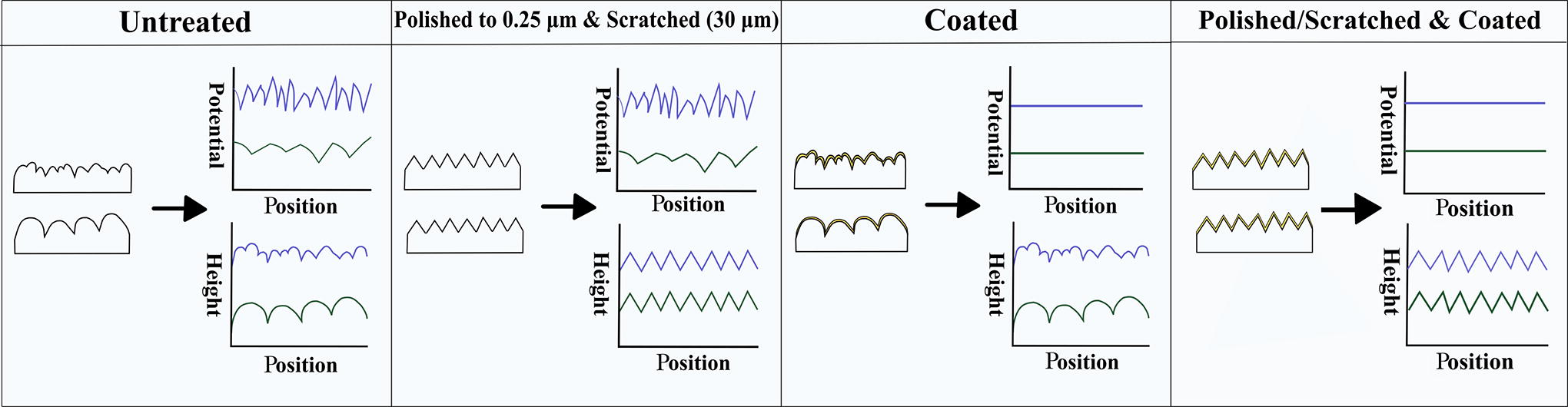
Schematic showing the methodology to deconvolute between topographical and electrochemical properties using a schematic representation of the effect of the different treatments on surface properties and their effect on the potential profile.
To validate that the polishing was effective in reducing the topographical differences between the surfaces SEM was carried out to compare the surface topographies of the different samples as described in Section Scanning Electron Microscopy. In addition, as surface roughness can affect the water contact angle, assessment of the surface energy was carried out to examine if gold-coated and abraded surfaces induce a comparable hydrophobicity. To validate if the gold coating was able to effectively remove the electrochemical differences between the surfaces while leaving the surface topography untouched a KPFM analysis was carried out as described in Section Kelvin Probe Force Microscopy. Samples were coated using a sputter coater at 20 mA to create 6-, 8-, or 9-nm-thick coatings. Height profiles and potential profiles were extracted from three random locations on the image, to assess which gold coating successfully removed the nanoscale potential variations across the surfaces without affecting the topography. The RMS on all samples was measured based on a 1.5 × 1.5 µm area sampled from random locations across the materials. All analyses were carried out using Gwyddion software.
Experiment: in vitro assessment
Protein adsorption
For the assessment of protein adsorption direct conjugation of FN was used, which directly tags the protein with a fluorescent dye. To tag the FN, first, 500 µL of dimethyl sulfoxide (DMSO) was added to 1 mg of the Atto 565 NHS Ester BioReagent (Merck, Germany) in an Eppendorf tube. To prevent alteration of the positive charge on FN, it was aimed to tag approximately 10 lysine groups out of the total number of 76 on the protein. The required amount of dye was calculated using the following equation:
To ensure deprotonation of the lysine groups on the protein, the FN solution was diluted in a 0.1M bicarbonate buffer. The calculated volume of the fluor was then added to the protein buffer solution and conjugated on a stirrer for 2 h in the dark. A dialysis cassette (Slide-A-LyzerTM Dialysis Cassette, Thermo Fisher, UK) was prepared to purify the protein solution using tris-buffered saline (TBS). The protein solution was subsequently stored at −7°C in aluminum foil.
Based on the protein concentration obtained after conjugation measured through the BCA analysis, a dilution was prepared to obtain a protein concentration of 2.5 µg/mL. Initial experiments conducted in our laboratory demonstrated that, at this specific concentration, FN successfully adhered to both surfaces without causing saturation enabling clear assessment of the differences in affinity to FN adsorption on both surfaces. All samples from each group (UT, PS, C, and PS-C) were then incubated for 2 h with FN solution at 37°C. After adsorption, samples were washed three times with 700 µL TBS and subsequently stored in 700 µL TBS at 4°C overnight until image analysis. Fluorescent analysis was carried out using a Carl Zeiss fluorescence microscope (Carl Zeiss AG, Oberkochen, Germany). The images were analyzed using the analyze histogram option in ImageJ (National Institute of Health, Bethesda, USA).
Cell sourcing and handling
Cryopreserved hOBs (Sigma-Aldrich, UK) and prescreened HDMECs (PromoCell GmbH, Germany) at passage 2 were purchased for in vitro cell culture studies. Cells were harvested and subsequently cryopreserved in DMSO at passage 4 with the HDMECs requiring addition of Cryo-SFM (PromoCell GmbH, Germany), a proprietary serum-free medium. The hOBs were thawed and expanded in complete cell medium consisting of Dulbecco’s modified Eagle’s medium (Life Technologies Ltd., UK), supplemented with 10% (v/v) fetal bovine serum (FBS, Sigma-Aldrich, UK), 75 mg/mL ascorbic acid (Sigma-Aldrich, UK), and 1% (v/v) penicillin–streptomycin (10,000 units/mL penicillin, 10 mg/mL streptomycin). The HDMECs were thawed and expanded in Endothelial Cell Basal Medium MV (PromoCell GmbH, Germany) supplemented with 15% (v/v) FBS, 1% (v/v) penicillin–streptomycin, 10 µg/mL heparin sodium salt (Life Technologies Ltd., UK), and 2.5 ng/mL bFGF (Life Technologies Ltd., UK). Cells were maintained at 37°C in an incubator with a humidified atmosphere with 5% CO2 in air. The culture medium was refreshed every 3 days until the appropriate cell confluency of 70–80% was reached after which cells were trypsinized with 0.25% trypsin containing 1 mM ethylenediaminetetraacetic acid to be either passaged or prepared for experimental use.
Protein Preadsorption
A number of discs were precoated by immersing the discs in FN solution at a concentration of 20 µg/mL or 5 µg/mL in phosphate buffered saline (PBS) for 2 h, to assess the effect of protein preadsorption on cell behavior in mono and coculture, respectively. Protein concentrations were kept consistent with previous studies in our laboratory. 10 Before protein preadsorption, discs were placed inside a CytoOne® 48-well plate and immersed in 98% ethanol for approximately 2 min for sterilization. Discs were washed three times with PBS and immersed with the FN solution or PBS. After incubation, all samples were washed thoroughly three times with PBS to remove the loosely bound proteins. The discs were then immersed in osteoblast culture media and incubated overnight in an incubator at 37°C.
Seeding technique
Cells were counted using a C-Chip hemocytometer (LabTech, UK) and subsequently diluted to the desired cell density. Before seeding, the osteoblast cell culture medium was aspirated and a 30 µL drop of fresh culture medium containing the appropriate cell number was then carefully pipetted on the surface of discs and stored at 37°C for 2 h to enable initial attachment only on the biomaterial. Hereafter, discs were immersed in the appropriate cell culture medium and stored in an incubator at 37°C.
Cell proliferation and attachment in monoculture
The hOBs were seeded in monoculture on both calcium phosphates to assess the osteoblast cell behavior and proliferation in response to the biomaterials with and without protein preadsorption. For the purpose of this experiment, 5.0 × 10³ hOBs were seeded on nonprecoated (non-FN) and FN-precoated (FN) HA and 1.25 wt% Si-HA surfaces in triplicate using their respective culture medium. Cells were maintained in an incubator at 37°C with a culture medium refreshment every other day until Day 1, Day 3, or Day 5 to evaluate the proliferative capacity of the cells. At the predetermined time points, medium was removed from the wells, and discs were carefully washed three times for 5 min with prewarmed PBS after which cells were fixed for staining.
Coculture
As angiogenesis is important for appropriate bone healing, a coculture of HDMECs and hOBs was used to assess this process in vitro. Before the experiments, hOBs and HDMECs were individually cultured and expanded in their respective culture medium as described in Section Cell Sourcing and Handling. Cells were seeded on the biomaterials with and without protein preadsorption using a sequential seeding method allowing initial undisturbed attachment of hOBs for 24 h before adding the HDMECs as seen in Figure 2. An initial experiment was carried out to determine the optimal hOB:HDMEC cell ratio starting from 100:0 (control), 85:15, 70:30, 50:50 to 30:70, 15:85, and 0:100 (control). The desired cell number of osteoblasts and endothelial cells was calculated based on the required total cell number of 4.0 × 10⁴ and seeded as described in Section Seeding Technique. The optimal cell culture ratio was then chosen for further experiments. For both cell ratio optimization and vessel analysis, hOBs were initially seeded in the osteoblast cell culture medium, but the medium was changed to the endothelial cell culture medium upon seeding of the HDMECs as previous experiments carried out the Cambridge Center for Medical Materials revealed that the ECs were the more sensitive cell type. 10 Culture medium refreshments were carried out every other day until Day 8 when cells were fixed for staining and fluorescence microscopy analysis.
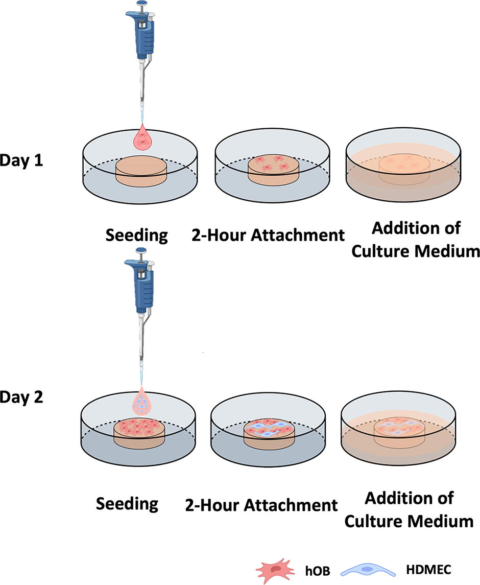
Sequential seeding technique for coculture with hOBs seeded on Day 1, using a small 30 µL droplet of culture medium containing cells to allow initial 2-h attachment only to the surface of the discs before adding hOB cell culture medium. On Day 2, endothelial cells were added to the discs allowing initial attachment for 2 h before adding HDMEC cell culture medium, which is the medium used during the rest of the experiment.
Cells in coculture were seeded on discs sintered at 1250°C to assess the effect of physicochemical surface properties on angiogenesis using high-density samples. Experimental protocols were established to deconvolute the effect of topography and electrochemical properties on protein and cell attachment, involving reduction of the differences in topography through polishing and abrading (resulting in scratches on the surface) for one set of samples and removing the electrochemical differences using an 8-nm gold sputter coating for the others, as previously described in section Deconvoluting Nanoscale Variations in Potential and Surface Roughness. The 1.25 wt% Si-HA and HA samples were divided into four groups, as presented in Table 2.
Sample Acronyms to Denote Treated Discs Used to Deconvolute Topographical and Electrochemical Surface Properties
Cell fixation and staining
Immunofluorescence staining was used to visualize the cell attachment and behavior and angiogenic potential of cells on the various surfaces. After each experiment, cells in mono and coculture were fixed with 4% paraformaldehyde (PFA, Thermo Fisher Scientific, UK) in PBS for 2 h after which the PFA was removed and the discs were thoroughly washed three times for 5 min with PBS. Well plates were stored at 4°C before staining.
Staining
To enable staining of the samples, fixed cells were first permeabilized for 15 min using permeabilization solution [0.5% (v/v) Triton X-100 in PBS] followed by a washing step with PBS (three times 5 min). Wells were blocked by immersing discs in blocking buffer [3% filter-sterilized bovine serum albumin (BSA) (w/v) in PBS] for 1 hour and were washed three times for 5 min in wash buffer [0.1% Tween 20 (v/v), 0.1% filter-sterilized BSA (w/v) in PBS]. All solutions were freshly prepared on the day of the experiment.
In monoculture, hOBs were treated with an actin filament staining using rhodamine (Life Technologies, UK), to enable assessment of their behavior, followed by staining with 4’,6-diamidino-2-phenylindole (DAPI, Sigma-Aldrich, UK). In coculture, both hOBs and HDMECs were stained with DAPI following specific HDMECs staining, to enable distinction between the osteoblasts and endothelial cells. Cell–cell junctions of HDMECs were stained with the primary antibody called antiplatelet endothelial cell adhesion molecule-1 (PECAM-1, Santa Cruz Biotechnology, USA), also known as the clusters of differentiation 31 (CD31) antibody, in combination with the primary anti-von Willebrand factor (vWF, Abcam, UK). PECAM is a cell surface glycoprotein specifically expressed at the endothelial cell–cell junction and vWF is a blood glycoprotein produced by endothelial cells and stored in granules. The secondary antibody Cy5-conjugated goat anti-mouse CD31 (Abcam, UK) and goat anti-rabbit (Abcam, UK) were subsequently used to detect the bound anti-PECAM and vWF, respectively. Table 3 shows the dilution ratios used for each staining agent.
Dye/Antibody Dilution Ratios for Cell Staining (Staining Agent:Diluent), Using Distilled Water as a Diluent for DAPI and Wash Buffer for All Other Staining Agents
Samples were incubated with anti-PECAM and vWF (coculture) or rhodamine (monoculture) for 1 hour after which the samples were thoroughly washed with wash buffer before adding the secondary antibodies anti-mouse CD31 and goat anti-rabbit, required only for coculture, which was left to attach for 1 hour in the dark. For both mono and coculture, discs were then rinsed thoroughly with wash buffer (three times) and one time with distilled water before being incubated with DAPI for 10 min at room temperature in the dark. After staining, the wells were rinsed three times with distilled water and stored at 4°C in the dark until required for fluorescence microscopy. Microscopy was carried out using a Carl Zeiss fluorescence microscope (Carl Zeiss AG, Oberkochen, Germany). Stitched images were taken to obtain a zoomed overview of the entire disc and, based on the information provided by these images; various zoomed-in single images were taken of representative areas. All samples were analyzed in triplicate.
Cell culture image analysis
The images obtained were used to examine the cell proliferation and vessel formation in mono and coculture, respectively. Analysis was carried out with ImageJ (National Institute of Health, Bethesda, USA) using a macro program.
Vessel analysis
Another macro program for ImageJ containing a two-step pipeline was applied to quantify the vascular network formation on the samples. First of all, the image resolution values are included in the macro program. Upon running the script, initial preprocessing steps are initiated, including manual adjustment of the image size and automatic optimization of the image quality. Automatic local thresholding was performed using the Phansalkar autocontrast, which estimates the local contrast value and is used to deal with low contrast images. 11 The optimized images are reloaded into ImageJ for analysis of the vascular network using the “Angiogenesis Analyzer” plugin. 11 The output presents various parameters of the microvascular network, including the number of vessels, junctions, and nodes as well as total vessel length and analyzed area enabling quantification of the vessel density, defined as vessel length over the measured area.
Statistical analysis
All protein adsorption and cell culture experiments were performed in triplicate to allow statistical analysis and each set of experiments was performed in duplicate. Statistical significance was determined by performing one-way ANOVA for multiple comparisons with one independent variable in combination with a Tukey’s posthoc test using SPSS software (SPSS 24 for windows) with a significance level of α = 0.05. The required homogeneity of variance for the statistical analysis was assessed using Levene’s test and if data fail the assumption, a Welch’s ANOVA was carried out with a Games–Howell posthoc test. Data are expressed as the mean ± standard deviation (SD).
Results
Material characterization and cell attachment
A summary of the surface characteristics of the biomaterials assessed in this study is presented in Table 4. First, both HA and 1.25 wt% Si-HA were phase pure with very low impurity levels. The disc density at a sintering temperature of 1250°C was similar for both biomaterials, having sintered densities above 90% of the theoretical density. The grain size was one of the major differences found between the samples with the smallest grain size found on 1.25 wt% Si-HA. Another important difference between the samples was the wettability, with HA samples being associated with moderate hydrophilicity, while 1.25 wt% Si-HA was highly hydrophilic.
Summary of Physicochemical Properties of HA and 1.25 wt%Si-HA. All Values Were Measured Using the Current Samples
In addition, as seen in Figure 3, a nanoscale variation in surface potential can be distinguished on both surfaces, which follow the characteristic grain surface, grain boundary structure on the discs. As a result, 1.25 wt% Si-HA exhibited most variation across the surface due to the smaller grains and there appeared to be increased differences between the potential found at different locations compared with HA. Altogether, these variations between the surfaces may all, to some extent, play a role in their elicited biological response.

Potential and topography profile of calcium phosphate discs analyzed through KPFM.
Cell attachment on calcium phosphate surfaces in mono and coculture
The osteoblast attachment was measured on surfaces over a time span of 5 days with and without FN precoating. Figure 4a compares the proliferative capacity of hOBs on HA and 1.25 wt%Si-HA. First, it can be seen that a FN precoating significantly improves the cell attachment on both biomaterials and proliferation is promoted resulting in populated surfaces after 5 days of incubation. Overall, 1.25 wt% Si-HA samples induced the highest osteoblast attachment on both FN and non-FN surfaces but the most prominent difference compared with HA samples was seen on the non-FN materials.

Fluorescent images of hOBs mono and cocultured with- and without FN precoating on HA and 1.25 wt%Si-HA discs for 1 and 5 day(s). Cell nuclei were stained with DAPI (blue) and actin filaments were stained using rhodamine (red) for monoculture and to distinguish between the cell types in coculture-specific endothelial cell markers, CD31 (red) and vWF (green), were used. Overall, in monoculture, 1.25 wt% Si-HA samples induced the highest osteoblast cell attachment on both FN and non-FN surfaces and promoted the formation of more distinct microfilaments aligned along the long axis of the cell compared with the other samples. An FN coating was found to considerably improve the cell morphology on all samples, although for 1.25 wt% Si-HA samples cells were already found to spread relatively well without the FN coating. In coculture, evenly spread vessel-like structures were visible on both HA and 1.25 wt% Si-HA with the thickest vessels and highest network complexity found on the latter samples, as evidenced by the many junctions. Scale bar in monoculture images corresponds to 150 µm. Scale bar in coculture images indicates 250 µm.
In terms of cell morphology, osteoblasts seeded on FN- and non-FN-1.25 wt% Si-HA exhibited more distinct microfilaments aligned along the long axis of the cell compared with the other samples, already after one day of incubation, especially on FN samples. On Day 5, cells seeded on FN-HA samples had reached nearly the same morphology of the microfilaments as seen on FN-1.25 wt% Si-HA samples. The effect of 1.25 wt% Si-HA on the microfilaments was, therefore, most visible on non-FN samples. At an early time point, a FN precoating was found to improve the cell morphology on HA, showing considerably better spreading of the osteoblasts. This positive influence of a FN precoating was also seen on 1.25 wt% Si-HA samples, but the cells were already found to spread relatively well without the FN precoating. At a later time point, the cells were able to strengthen their attachment through cell spreading on all samples. A precoating seemed to induce alignment of the osteoblasts after 5 days of incubation on HA and 1.25 wt% Si-HA samples.
After the cell ratio optimization (Supplementary Appendix A), the coculture protocol was applied to the HA and 1.25 wt% Si-HA surfaces to compare their ability to induce vessel formation. A coculture was carried out on non-FN precoated samples to test the ability of the biomaterials to stimulate vessel growth without externally applied stimuli. On all samples, with and without protein precoating, a coculture was successful, but the vessel density was lower without a precoating compared with the surfaces precoated with FN (Table 5). Within the FN and non-FN groups, vessel formation varied strongly between the chemically different surfaces, with the highest vessel density found on 1.25 wt% Si-HA within both groups. A visual representation of the vascular network on the different surfaces with and without an FN precoating is presented in Figure 4b and shows the difference in complexity of the networks. Vessel-like structures found on both HA and 1.25 wt% Si-HA surfaces were found to be evenly spread throughout the surface with the thickest vessels and highest network complexity found on 1.25 wt% Si-HA surfaces as evidenced by the many junctions.
Vessel-Density on Calcium Phosphate Discs With and Without FN Pre-Coating, Incubated for 8 Days. Vessel-like Network Formation was Successfully Induced on all Samples, With and Without Externally Applied Stimuli. The Highest Vessel Density was Found on 1.25 wt%Si-HA both with- and Without FN. Upper case Letters Indicate Statistical Significance Between Columns and Lower case Letters Indicate Significant Differences Between Rows at p < 0.05.
Deconvoluted effect of surface properties on the biological response
As seen in the first part of this article, there is a clear difference in the biological response induced by HA and 1.25 wt% S-HA surfaces, but the reason for this distinct behavior remains unclear due to the many physicochemical differences between the materials. To assess the deconvoluted effect of topographical and electrochemical differences between HA and 1.25 wt% Si-HA on the biological response, part of the samples were gold coated to mask the electrochemical differences between the surfaces and another part was polished and abraded to reduce the differences in topography. The experimental protocol was first validated and subsequently applied to assess cell behavior in coculture with and without FN precoating.
Protocol validation
Removing Electrochemical Differences
Removing the electrochemical differences between the surfaces can be achieved by coating the calcium phosphate discs with the same material. However, the coating should be thin enough to leave the differences in topography unaffected. In this section, a gold sputter coating, generally used for topographical analysis using SEM was assessed to achieve this goal.
Various coating thicknesses were assessed for their ability to shield the nanoscale potential variations across surfaces ranging from 6 to 9 nm. Figure 5a shows KPFM images of the potential profile across the three different calcium phosphate surfaces. From here, it can be concluded that a 6 nm coating was unable to successfully eliminate the nanoscale variations in surface potential. An 8 nm or thicker coating was observed to be necessary to obtain the desired outcome showing a uniform surface potential across the materials. However, in terms of topography, thicker coatings can induce changes in surface topography. As seen in Figure 5b, a coating thickness up to 8 nm leaves the topography mostly unaffected apart from the slight rounding of the local minima compared with the uncoated samples. However, a gold coating of 9 nm considerably affects the topography due to an increased influence of the coating on the rounding of the local minima. An 8 nm coating was therefore taken forward for further analysis.

Validation of gold-coating method for eliminating nanoscale variations in surface potential across calcium phosphate discs, representative data are displayed taken from 1.25 wt% Si-HA samples sintered at 1250°C.
A quantitative assay was carried out using Gwyddion software, measuring the RMS surface roughness, R q , of the gold-coated vs uncoated sample surfaces used in the experimental set-up to assess if a gold coating affects the surface roughness. As seen in Table 6, there was no significant difference between the R q values of all surfaces.
Root Mean Square Surface Roughness, R q , of Gold-Coated vs Uncoated Calcium Phosphate Discs. On Both Materials, No Significant Difference in the R q Was Found between the Coated and Noncoated Samples
Reducing Topographical Differences
To assess the effect of electrochemical differences between the calcium phosphate surfaces on the biological response, it is important to eliminate the difference in topography that naturally occurs after the production of the discs. Polishing and abrading all surfaces using the same technique was assessed as a method to reduce the differences in surface topography to such an extent that it will not induce significant changes in the biological response.
As seen in Figure 6, morphologically, both surfaces exhibited a similar appearance with clearly visible scratches. Further biological analysis will be carried out in the next section to confirm if the differences in topography were reduced enough to induce a similar biological response.
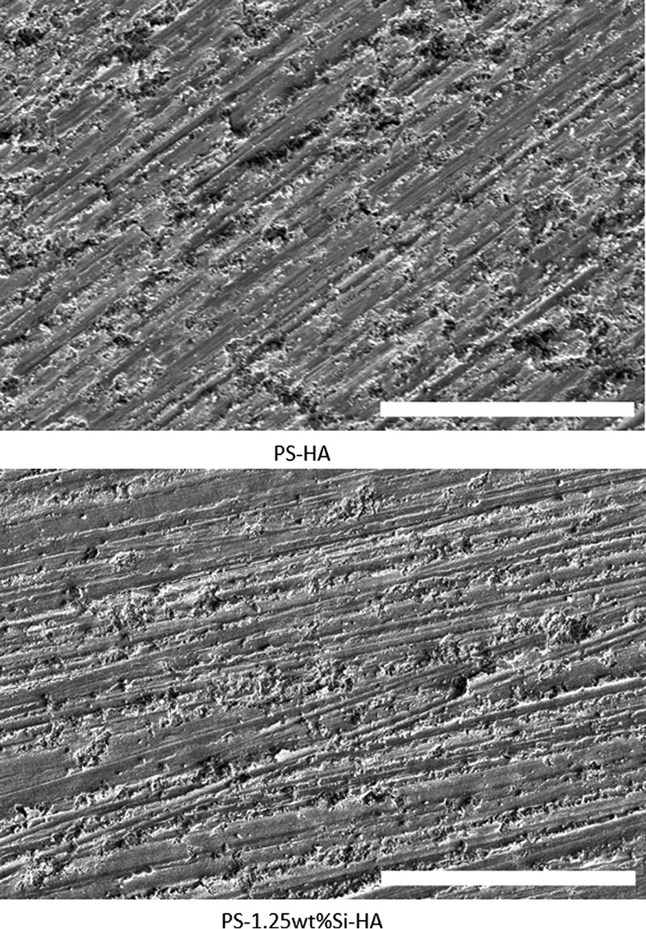
The effect of polishing and abrading on the surface morphology of calcium phosphate surfaces. All PS samples exhibit clear scratches. Both surfaces appeared similar after the PS treatment. Scale bar indicates 50 µm.
Effect of electrochemical and topographical surface properties on the biological response
Protein Adsorption
Using the experimental methodology, a protein adsorption assay was carried out and results are presented in Figure 7. As a final validation, a control group with PS-C discs was included, which induced a similar protein adsorption on both HA and 1.25 wt% Si-HA surfaces, indicating that the coating and polishing/abrading was effective enough in reducing the electrochemical and topographical differences between the surfaces. On the untreated samples, there was a clear difference in protein adsorption, with the highest adsorption on 1.25 wt% Si-HA. As these surfaces differ both in topographical and electrochemical features, the PS- and C samples were used to assess which of these two features contributed to this biological response. It was clear that with the unpolished but gold-coated surfaces, for all samples, there was a decrease in protein adsorption compared with the uncoated samples, but, despite having similar electrochemical properties due to the gold coating, there was still a difference in adsorption visible between the surfaces. The highest fluorescence was measured on the 1.25 wt% Si-HA discs and very little protein adsorption was detected on the HA surface. On the PS samples, similar adsorption capacities were found between 1.25 wt% Si-HA and HA, indicating that the electrochemical differences between the surfaces were not responsible for the distinct protein adsorption found on the untreated samples. For HA samples, within the coated and uncoated sample groups, the PS treatment slightly increased the protein adsorption compared with their unpolished/unabraded counterparts but reduced the adsorption on 1.25 wt% Si-HA surfaces, although not significantly for the uncoated sample set.

The effect of electrochemical and topographical surface features of calcium phosphates on protein adsoprtion. The highest protein adsorption was observed on UT-1.25 wt% Si-HA surfaces. Significant differences in protein adsorption capacities were observed on the C samples. PS treatment was found to improve protein adsorption for the HA and reduce the adsorption on 1.25 wt% Si-HA compared with both the uncoated and gold-coated counterparts. A comparable FN response was observed on PS-HA and PS-1.25 wt% Si-HA. A double asterisk indicates statistical significance at p < 0.01, respectively.
Cell Behavior in Coculture
To measure the cell behavior in response to surface properties a coculture of HDMECs and hOBs was applied to the untreated and treated surfaces with and without FN precoating, and the vessel densities are presented in Figure 8. Data were taken from duplicate independent experiments and similar trends were detected. Overall, it can be observed that the formation of a vessel-like network was more efficient on FN-precoated surfaces compared with non-FN samples. The vessel density on untreated surfaces was highest on 1.25 wt% Si-HA samples both for the FN and non-FN groups.
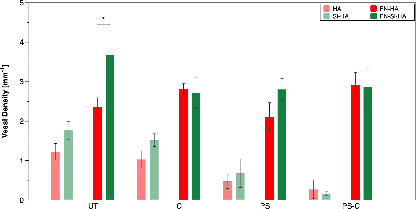
The effect of electrochemical and topographical surface features of calcium phosphates on cell behavior in coculture. While a functional coculture was achieved without a FN precoating, the addition of such a precoating increased the number of vessels obtained on nearly all samples. Overall, the densest vascular network was obtained on FN-UT-1.25 wt% Si-HA samples. While PS discs induced distinct responses on FN samples, no differences could be seen on FN-C samples. On non-FN samples, differences were found in both C- and PS-sample groups, but these differences were not statistically significant. However, a replicate experiment showed similar trends. A single asterisk indicates statistical significance at p < 0.05, respectively.
As seen in Figure 8, no difference in vessel density was found on the FN-C samples. Even when comparing outcomes of FN-C samples with the FN-PS-C samples, a similar vessel density was detected, indicating that the variability in surface topography did not affect the formation of vessel-like structures by cells in coculture when applying a FN precoating. Conversely, without a FN precoating, the difference in angiogenic potential for 1.25 wt% Si-HA, HA on coated samples was noticeable, revealing a higher vessel formation on 1.25 wt% Si-HA. While the differences observed for the non-FN samples were not statistically significant, a replicate experiment showed similar trends. On FN precoated samples without gold coating, the polishing and abrading treatment induced a strong enough change in the surface topography reducing the formation of a vascular network for 1.25 wt% Si-HA. This treatment did not affect vessel density for HA as measured on FN-PS-HA compared with FN-UT-HA. Without a precoating, on the contrary, PS treatment reduced the vessel formation on all samples considerably with and without gold coating.
In terms of both the FN- and non-FN-PS samples, the electrochemically diverse surfaces induced a distinct cell response, with the highest vessel density found on PS-1.25 wt% Si-HA samples, indicating an important impact of electrochemical surface features on cell behavior in coculture regardless of the FN precoating. The difference in vessel density between FN-PS-1.25 wt% Si-HA and FN-PS-HA was not found to be statistically significant. Surprisingly, the vessel formation slightly increased on the FN-C-HA and FN-PS-C-HA samples compared with their uncoated counterparts. This increase was not observed on samples without a FN treatment. FN-UT-1.25 wt% Si-HA surfaces did perform significantly better without a gold coating, compared with their coated counterpart.
Figure 9 displays a schematic representation of the results on the deconvoluted effect of topographical and electrochemical differences between HA and 1.25 wt% Si-HA on the vessel formation in a coculture with and without FN.
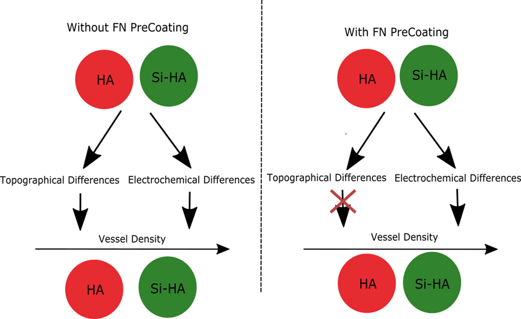
Schematic representation summarizing the results on the deconvoluted effect of topographical and electrochemical differences between HA and 1.25 wt% Si-HA on the vessel formation in a coculture with and without FN. Without FN precoating both topographical and electrochemical differences induced a distinct angiogenic response on 1.25 wt%Si-HA as opposed to HA. With a FN precoating, it appeared to be only the electrochemical differences that induced distinct vessel formation on the bioceramics.
Figure 10 shows fluorescence images of the vascular networks found on all sample groups treated with a FN precoating. First of all, it is clear that the network on FN-UT-1.25 wt% Si-HA surfaces was the most complex showing an increased number of branches and junctions compared with FN-UT-HA, resulting in an interconnected network. Similarly, within the FN-PS-group, 1.25 wt% Si-HA surfaces also appeared to induce increased self-assembly of endothelial cells compared with the other sample. Overall, the FN-C-HA samples exhibited more vessel-like structures compared with the FN-UT-HA discs but were unable to improve vessel formation compared with FN-UT-1.25 wt% Si-HA surfaces. Although for both calcium phosphates, their gold-coated counterpart appeared to promote the formation of thicker vessels, FN-UT-1.25 wt% Si-HA samples exhibited the most interconnected and complex vessel-like network. The vessels obtained on both FN-C- and FN-PS-C samples for both calcium phosphates exhibited a similar vessel count, but a clear difference can be seen in terms of morphology, with FN-PS-C samples exhibiting aligned vessels with fewer junctions and reduced interconnectivity. The FN-PS treatment, in general, was found to induce vessel alignment on both calcium phosphates both on coated and uncoated samples, resulting in a lower complexity compared with unpolished and nonabraded samples as evidenced by the fewer number of junctions and branches.

The effect of electrochemical and topographical features on vessel morphology. Cell nuclei of both hOBs and HDMECs were stained with DAPI (blue) and to distinguish between the cell type-specific endothelial cell markers, CD31 (red) and vWF (green), were used. The 1.25 wt% Si-HA surfaces accommodated the most complex vessel-like networks both on FN-UT- and FN-PS samples. A polishing and abrading treatment (PS) induced clear alignment on both coated and uncoated samples resulting in fewer vessel junctions and less interconnectivity. A gold coating without polishing and abrading appeared to promote formation of thicker vessels on all calcium phosphates, nonetheless, FN-UT-1.25 wt% Si-HA surfaces induced the most complex and interconnected network compared with all other coated or uncoated discs. Scale bar indicates 250 µm.
Discussion
This article aimed to systematically evaluate and compare the biological response to HA and 1.25 wt% Si-HA by gradually increasing the complexity of the system. First, osteoblasts in monoculture were added to the system and their interaction with the material both with and without FN precoating was assessed. After deconvolution of the surface properties, the effect of topographical and electrochemical surface features on FN adsorption alone was assessed. Finally, an additional cell type, HDMECs, was introduced into the in vitro system, further increasing the complexity of the methodology. To date, no such system has previously been applied to Si-HA and may, therefore, provide crucial new insights on their behavior in vivo compared with HA, using a more controlled in vitro environment.
Cell attachment
Following protein adsorption, cells arrive at the biomaterial and interact with the surface depending on the composition of the ion/water and protein layer. 12 Initially, attachment of the main cell type, osteoblasts, was assessed with and without FN precoating. Overall, an FN precoating was found to significantly improve the cell attachment on both biomaterials. The highest osteoblast attachment was observed on 1.25 wt% Si-HA surfaces compared with HA in both non-FN and FN groups. These findings were in agreement with the reduced affinity of HA surface to FN adsorption compared with 1.25 wt% Si-HA.
A further increase in complexity to the system was added by introducing an additional cell type that plays an important role in bone regeneration. Blood vessels provide the necessary nutrient and waste exchange for osteoblasts to prevent core degradation of the implant, and biomaterials should, therefore, successfully accommodate both cell types. In vitro assessment in coculture can help to improve the material properties to enable optimal bone cell and blood vessel growth in a more controlled environment and could provide increasingly valuable information on the behavior of biomaterials upon implantation. While cocultures have been widely applied to HA, in vitro coculture research on Si-HA is lacking.13–15
When establishing a coculture, several critical factors come into play. These include the choice of culture medium, the seeding technique, the cell ratio, and the potential inclusion of external stimuli, such as protein precoating. Generally, when studying angiogenic interactions rather than mineralization, researchers often use the coculture media specifically tailored for the endothelial cells.16,17 In the current study, we followed this approach. Furthermore, our laboratory’s previous investigations revealed that sequential seeding enhances coculture outcomes. By allowing osteoblasts (which exhibit a slower proliferation rate) to attach and initiate matrix protein production before introducing endothelial cells, more optimal coculture system was achieved. 10 The optimal cell ratio was determined based on cell ratio optimization assessment, which showed vessel formation on both hOB:HDMEC ratios of 50:50 and 70:30 with better performance on a 70:30 cell ratio. Using these parameters it was possible to establish successful cocultures on all biomaterials without further modification, such as using a coating of extracellular matrix proteins.
The results found in coculture on the precoated samples reflected the findings made in monoculture showing the highest vessel density on 1.25 wt% Si-HA. Both the angiogenic potential of HA and 1.25 wt% Si-HA increased on FN samples. Overall, 1.25 wt% Si-HA performed best both with and without FN precoating as indicated not only by the vessel density but also by morphological features, such as the complexity of the network, its interconnectivity, and the highest vessel thickness. However, without an FN precoating, the observed differences were not statistically significant. While, to the best of the author’s knowledge, no coculture studies have been published on Si-HA surfaces, our findings did correspond to previous observations made in an endothelial monoculture using leaching liquor, which showed that silicon doping can improve the angiogenic potential of HA-coated titanium implants. 18 Differences in morphology can occur in response to differences in surface chemistry, structure, and porosity of biomaterials either as a direct consequence or by altering the cytokine and growth factor production of cells, but it remains unclear how, and to what extent, each property influenced the detected biological response. 14 Taken together, 1.25 wt% Si-HA performed best in terms of FN adsorption and cell attachment in mono and coculture.
Over the years, many hypotheses and theories have been developed to explain the distinct biological responses occurring on different biomaterials. While it is clear that material properties such as topography, wettability, chemical composition, and the surface potential affect the protein and cell behavior, it remains unclear which of these interrelated properties dictates the biological response. To assess this, a methodology was established, allowing deconvolution between topographical and electrochemical effects on the biological response to gain more insight into the interaction between a biomaterial and a biological system.
Cell behavior in response to physicochemically different surfaces
Without FN precoating, a gold coating was found to slightly reduce the vessel formation on all calcium phosphates, but this difference was not significant despite the marked decrease in FN adsorption to these surfaces. Interestingly, with a FN precoating, a gold coating was found to improve the vessel formation on HA. A previous study found an increased osteoblast adhesion on gold-coated surfaces compared with uncoated HA after 4 h of incubation, which was ascribed to the increased hydrophobicity of these surfaces. 19 While generally hydrophobic surfaces are unfavorable for cell attachment, Kennedy et al. hypothesized that the increased proliferation on hydrophobic surfaces was a result of increased FN adsorption, nevertheless, as seen in this study, the protein quantity was actually lower on the less hydrophilic surfaces. 20 It may be possible that at longer incubation times the protein layer composition changes or that a more favorable protein conformation was achieved on the hydrophobic materials. Additionally, regardless of the more hydrophobic properties of gold-coated surfaces, the overall SE was significantly lower compared with the untreated calcium phosphates (Supplementary Appendix B). Although much remains unclear about how the SE affects cell attachment, a recent study did suggest that more moderate SEs of approximately 70 mJ.m−2 are associated with increased cell adhesion, growth, and proliferation. 21 It could, therefore, be the case that the lower SE measured on gold-coated surfaces induced the beneficial cell attachment. Nevertheless, despite these findings, this material is not suitable for tissue engineering due to the associated costs and its inertness preventing the necessary biodegradation.
Furthermore, in all cases, UT-1.25 wt% Si-HA performed best, making this material particularly interesting for the purpose of BTE. For HA, a FN precoating improved the cell response but not as much as it did for UT-1.25 wt% Si-HA. Taken together, all three electrochemically different materials (including the gold-coated surfaces) induced a strongly distinct cell response, and adding a FN precoating was found to increase the number of vessel-like structures on all samples. The current experimental methodology aimed to assess to what extent the physical and electrochemical properties contribute to these findings and how this may or may not be modulated by protein adsorption.
Relative topographical effects on the biological response
To assess the topographical effects on cell behavior, the calcium phosphates were gold coated to mask the electrochemical differences and allow the measurements to focus solely on the variation in surface topography. In terms of FN adsorption, on UT samples the highest FN adsorption was obtained on 1.25 wt% Si-HA samples. Guth et al. believed that the increased negative surface charge on Si-HA more strongly attracts the positively charged FN as opposed to HA, overcoming the stronger energy barrier induced by the necessary water displacement stemming from its higher hydrophilicity. 22 However, according to the current findings, the electrochemical differences between HA and 1.25 wt% Si-HA did not seem to significantly alter the protein adsorption with respect to its quantity as a similar fluorescence was detected on the PS samples. The significant difference in wettability of these samples as well as the surface potential distribution, therefore, does not seem to have caused the distinct FN adsorption on the UT surfaces. It is important to highlight that, in this case, the focus was on relative electrochemical effects and that these findings do not implicate that electrostatic interactions do not influence protein adsorption. In fact, when comparing PS- and PS-C samples, a marked electrochemical effect can be seen with the uncoated bioceramics inducing significantly higher protein adsorption. These findings are in agreement with literature stating that for HA surfaces’ protein adsorption is mediated by electrostatic interactions. 23 Nevertheless, the differences in electrochemical properties between 1.25 wt% Si-HA and HA were not found to be strong enough to induce more or less FN adsorption.12,24 With regard to the topographical effects on FN adsorption, it was clear that the protein strongly responds to differences in topography with the highest adsorption found on 1.25 wt% Si-HA with a grain size of 673 nm, followed by HA with a grain size of 1299 nm. FN adsorption, therefore, appeared to increase with reducing grain size, which is often associated with increased protein adsorption due to the entrapment of proteins at the grain boundaries as well as increased specific surface area for adsorption. 25 Nevertheless, both Webster et al. and Ribeiro et al. found a reduction in FN adsorption with reducing grain size but their assessment was carried out at a different scale with grain sizes in the range of 67–179 nm, whereas the minimum grain size assessed in this study was as high as 673 nm.26,27 Increasing protein adsorption with reducing grain size may, therefore, occur up to a certain grain size, after which a reversed relationship initiates, as schematically represented in Figure 11. Webster et al. also found different trends for different proteins in response to grain size, with laminin, a larger protein, attaching preferentially to larger grains and vitronectin, a smaller protein, attaching preferentially to smaller grains. 26 The trends observed may also be directly related to the number as well as the nature of grain boundaries on different surfaces, as the increased number of grain boundary-induced grooves with smaller grain sizes can promote the entrapment of proteins, but, can, at the same time, be unfavorable for larger proteins due to the stronger bending of the boundaries demanding stronger unfolding of the proteins to fit inside the grooves. Rather than stating that protein adsorption increased with reducing grain size and increased density of grain boundaries, it might be more accurate to state that, depending on the size of the protein, an optimal grain size and grain boundary density range exists with smaller proteins attaching preferentially to smaller grains than the larger proteins. Another trend showing that protein adsorption was strongly governed by the surface topography was the improved protein adsorption to PS-HA with 30 µm scratches as opposed to the UT counterparts.
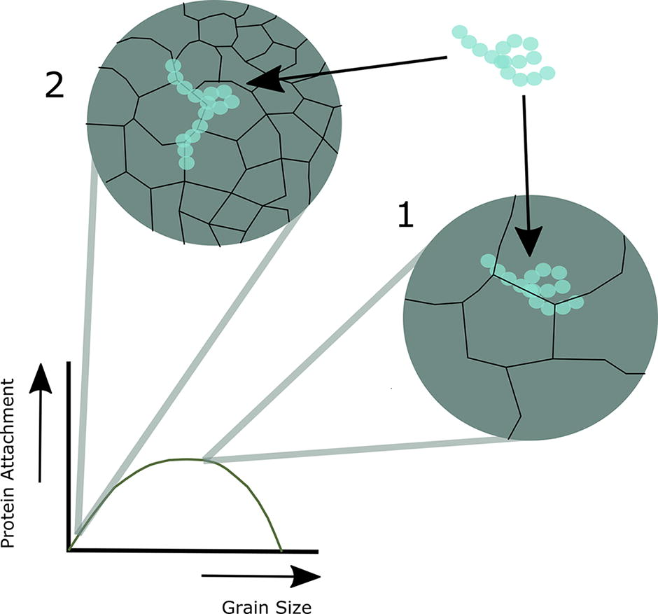
Schematic representation of the proposed relationship between grain size and protein adsorption. Smaller grains increase the number of grain boundary induced grooves that promote entrapment of proteins (
In a coculture, without a FN precoating, the cells seemed to respond to the differences in topography, with the lowest grain size observed for 1.25 wt% Si-HA inducing the highest vessel density. The nature of the grain boundaries may have played an important role in the observed enhanced cell behavior on samples with smaller grain sizes, as the disordered structure at these regions are associated with enhanced dissolution inducing voids and rougher regions on the sample surfaces, which may have had a beneficial influence on the cell attachment. 28 However, as the gold coating is believed to reduce differences in ion dissolution between the samples, but a similar trend was observed on the non-FN-C samples, factors other than dissolution-induced surface roughness are believed to also have played a role in the distinct biological response observed on UT samples without FN precoating. Overall, since the trend between the different surfaces seen for non-FN-C samples was comparable to that found for non-FN-UT samples, it can be concluded that topography plays a strong part in enhancing the vessel formation on 1.25 wt% Si-HA compared with HA when no FN precoating was applied.
Surprisingly, despite topography playing a prominent role in modulating FN adsorption and vessel formation on surfaces without FN precoating, all C samples treated with a 2-h FN precoating exhibited a comparable angiogenic potential. This indicates that with an FN precoating, the relative effect of the topographical differences between the samples on vessel formation was nearly eliminated. This finding was further validated by the similar vessel density found on the PS-C and C samples, with PS-C discs having a surface roughness introduced through abrading that deviated markedly from the original surface topographies seen on C-samples. It could be possible that without a FN precoating, the FN found in the cell culture medium, containing multiple proteins, was unable to compete for enough surface area on HA due to the unfavorable grain size compared with 1.25 wt% Si-HA resulting in a protein layer composition that was not as suitable for inducing vessel formation in coculture. Conversely, with a FN precoating, there may have been just enough FN attached to C-HA without interference from other proteins to make the topographical differences irrelevant, despite the significantly lower affinity to FN adsorption on C-HA compared with C-1.25 wt% Si-HA. It may be possible that the level of proteins attached to the surfaces with a FN precoating essentially smoothens the surfaces by preferentially depositing in the valleys induced by the grain boundaries reducing the differences in topography all together. The “smoothing” of the surfaces was suggested by a study carried out by Lamers et al. where cells were unable to respond to ridges with depths lower than approximately 34 nm. 29 The proteins created a uniform adhesive layer that they believe may have masked the nanopatterns on their surfaces. 29
Relative electrochemical effects on the biological response
PS samples were used to reduce the differences in topography between HA and 1.25 wt% Si-HA to assess the effect of electrochemical differences between the bioceramics on the angiogenic potential.
While there were no differences in protein quantity attached to PS-HA and PS-1.25 wt% Si-HA surfaces, there was a distinct cell response in coculture on both non-FN- and FN samples. Cells, therefore, appeared to respond to the electrochemical differences between HA and 1.25 wt% Si-HA regardless of the quantity of FN attached to its surfaces. This relative effect was especially visible on FN precoated samples, for which the topographical differences were previously discussed to be irrelevant for the cell behavior. The improved angiogenic potential on FN-UT-1.25 wt% Si-HA as opposed to FN-UT-HA was therefore believed to be a direct result of the differences in electrochemical properties, which was not affected by the quantity of protein adsorption. Multiple electrochemical differences were observed between the surfaces, including a higher wettability, a more negative surface charge, and enhanced nanoscale variations in surface potential on 1.25 wt% Si-HA as opposed to HA, which could all play a prominent role in modulating the biological response.
From the literature, it can be deduced that increased wettability is associated with increased cell attachment, but this is often attributed to its effect on protein adsorption.30,31 As it was found that electrochemical features did not alter the quantity of FN adsorption, it may be the case that the protein conformation on 1.25 wt% Si-HA surfaces was more favorable for cells in coculture. The increased hydrophobicity on HA samples may also have resulted in a stronger protein surface association, consequently preventing progressive enrichment of the surface or reorganization of FN into ECM-like structures, which is an important process in tissue healing.30,32 Regarding surface potential, it is often suggested that this feature has an indirect influence on cell attachment through protein adsorption. 33 As FN exhibits a positive charge at physiological pH, it may be possible that the increased negative surface charge on 1.25 wt% Si-HA beneficially influenced its adsorption. 22 However, as no increase in protein adsorption was found to be induced by the differences in electrochemical properties between HA and 1.25 wt% Si-HA, it may have more likely been the result of a more favorable protein orientation and/or conformation. A computational study modeling the orientation of the 7–10 th type III modules of FN (FN-III7-10) upon adsorption to electrostatically distinct HA surfaces found a preferred end-on orientation of FN on moderately negative HA surfaces (−0.025C/m2), having a charge similar to that observed for Si-HA by Vandiver et al. (≈−0.024C/m2), revealing an unfavorable positioning of the RGD cell-binding sequence and the synergy site (a sequence that enhances cell binding to the RGD site) at physiological ionic strength, as represented schematically in Figure 12a.34,35 The most favorable positioning of the cell-binding sites at physiological ionic strength was observed on weakly negatively (−0.005C/m2) and positively charged (0.008C/m2) surfaces as well as moderately positively charged surfaces (0.025C/m2). HA was observed to have a surface charge of approximately −0.011C/m2 and is, therefore, expected to induce an unfavorable orientation of the FN upon adsorption. However, the more negative surface charge of Si-HA did, in this case, also does not appear to be favorable for the FN orientation, but other characteristics of this surface affected by the silicate substitution may counteract the effect of the surface charge on the orientation of FN. Furthermore, since it is not only the orientation, but also the conformation of a protein that can be affected by the surface biochemistry, these surfaces may have induced a more favorable conformation.

Alternatively, the significantly less negative surface charge found at the grain boundaries of HA and 1.25 wt% Si-HA might locally induce a more favorable orientation of FN, as represented schematically in Figure 12b. Consequently, as Si-HA, in general, exhibits significantly more grain boundaries, more proteins will adsorb to this biomaterial with their cell-binding sites exposed. Furthermore, regardless of the orientation, due to the more negative net surface charge of Si-HA, more FN is being attracted to the surface increasing the probability for entrapment of the proteins inside the grain boundary-induced grooves, which induces a more favorable orientation of the protein (Fig. 12b).
Moreover, Thian et al. hypothesized that the favorable biological response on Si-HA is a consequence of ion dissolution promoting the formation of a bound silicate network, but they believed this to be associated with increased protein adsorption. 36 As the formation of a silicate network happens over time, there might be a change in the relative FN adsorption between the surfaces after long-term incubation. Alternatively, active mechanisms of silicate ions may be at the core of the bioactivity of Si-HA, as silicate has been found to inhibit the RANK/RANKL/OPG-signaling pathways related to osteoclast activation suppressing bone resorption and has been suggested to stimulate osteoblast development.37,38 Porter et al. observed preferential ion dissolution at the grain boundaries and triple junctions. 39 As Si-HA exhibits considerably smaller grains, resulting in more grain boundaries, this material displays a faster degradation rate, releasing a significant amount of silicate ions. 39 Due to this faster degradation rate, however, other passive mechanisms stemming from silicate substitution, such as increased Ca2+ and PO34 ion release enabling faster formation of a Ca-P layer, which is known to stimulate bone cells and their mineralization process, may also have been responsible for the induced biological response and its influence should not be overlooked in future dissolution studies.28,40
Conclusion
Taken together, while the differences in surface topography between the calcium phosphates were found to significantly affect protein affinity to the surfaces, they did not appear to play a role in the angiogenic potential of FN precoated surfaces. With a precoating, it was found that it was specifically the electrochemical differences between the surfaces that induced the distinct angiogenic responses. Allowing adsorption of FN through precoating without competition from other proteins appeared to provide the minimally necessary amount of FN to all samples regardless of the topography, inducing a similar cell response on the FN-C samples. As the electrochemical differences between the HA and 1.25 wt% Si-HA were not found to affect the protein affinity, it is believed that either the protein conformation was affected or that passive or active mechanisms related to the silicate substitution caused the improved cell behavior on 1.25 wt% Si-HA. Without a FN precoating, the differences in topography between HA and 1.25 wt% Si-HA began to play a more important role in determining the cell behavior, which can be associated with the strong influence of topography on protein adsorption, which may become more relevant when proteins inside the culture media have to compete for surface area. Without a precoating that allows FN to attach without competition from other proteins, the limiting factor of the larger grain sizes on HA for FN adsorption may have caused other proteins that are less important for osteoblast attachment to outcompete FN adsorption. The work has allowed us to draw a number of conclusions about biological response to the substrate materials and, overall, the electrochemical differences were found to be the most prominent factor influencing the cell behavior, while topographical differences dictated the quantity of protein attached to the surfaces. Furthermore, based on exploratory work using coculture that has been pioneered by Professor Kirkpatrick and his coworkers, we have gained significant new knowledge that will inform future optimization of scaffolds and substrates for tissue engineering.
Footnotes
Authors’ Contributions
EE: Conceptualization (lead); writing—original draft (lead); formal analysis (lead); and writing—editing (equal). RC: Writing—review (lead). SB: Conceptualization (supporting); Writing—review (lead) and editing (equal).
Disclosure Statement
There are no conflicts to declare.
Funding Information
This research was financially supported by the Engineering and Physical Sciences Research Council (EPSRC) and Geistlich Pharma AG. REC and SMB also acknowledge funding from EPSRC for an Established Career Fellowship, Grant No EP/N019938/1.
References
Supplementary Material
Please find the following supplemental material available below.
For Open Access articles published under a Creative Commons License, all supplemental material carries the same license as the article it is associated with.
For non-Open Access articles published, all supplemental material carries a non-exclusive license, and permission requests for re-use of supplemental material or any part of supplemental material shall be sent directly to the copyright owner as specified in the copyright notice associated with the article.