
Abstract
This study evaluated the efficacy of synthetic bone blocks, composed of hydroxyapatite (HA) or β-tricalcium phosphate (B-TCP), which were produced by additive manufacturing and used for the repair of critical size bone defects (CSDs) in rat calvaria. Sixty rats were divided into five groups (n = 12): blood clot (CONTROL), 3D-printed HA (HA), 3D-printed β-TCP (B-TCP), 3D-printed HA + autologous micrograft (HA+RIG), and 3D-printed β-TCP + autologous micrograft (B-TCP+RIG). CSDs were surgically created in the parietal bone and treated with the respective biomaterials. The animals were euthanized at 30 and 60 days postsurgery for microcomputed tomography (micro-CT) histomorphometric, and immunohistochemical analysis to assess new bone formation. Micro-CT analysis showed that both biomaterials were incorporated into the animals’ calvaria. The HA+RIG group, especially at 60 days, exhibited a significant increase in bone formation compared with the control. The use of 3D-printed bioceramics resulted in thinner trabeculae but a higher number of trabeculae compared with the control. Histomorphometric analysis showed bone islands in close contact with the B-TCP and HA blocks at 30 days. The HA blocks (HA and HA+RIG groups) showed statistically higher new bone formation values with further improvement when autologous micrografts were included. Immunohistochemical analysis showed the expression of bone repair proteins. At 30 days, the HA+RIG group had moderate Osteopontin (OPN) staining, indicating that the repair process had started, whereas other groups showed no staining. At 60 days, the HA+RIG group showed slight staining, similar to that of the control. Osteocalcin (OCN) staining, indicating osteoblastic activity, showed moderate expression in the HA and HA+RIG groups at 30 days, with slight expression in the B-TCP and B-TCP+RIG groups. The combination of HA blocks with autologous micrografts significantly enhanced bone repair, suggesting that the presence of progenitor cells and growth factors in the micrografts contributed to the improved outcomes. It was concluded that 3D-printed bone substitute blocks, associated with autologous micrografts, are highly effective in promoting bone repair in CSDs in rat calvaria.
Impact Statement
This study evaluated the effectiveness of two additive-synthetic bone block biomaterials, manufactured either alone or combined with autologous micrografts, used in the bone repair of surgically induced critical defects in rat calvaria. By examining the effects of these biomaterials on various parameters, including microtomography, histomorphometry, and immunohistochemistry, we sought to increase understanding of their biological behavior in the context of bone repair. The findings will contribute to the advancement of regenerative dentistry and could have significant clinical implications for improving outcomes of bone reconstructive procedures in humans.
Introduction
Dentists are often faced with issues related to low bone volume when considering the installation of dental implants. Various bone substitutes, including autogenous, allogeneic, alloplastic, and xenogeneic grafts, have frequently been used in different surgical techniques for reconstructing bone defects.1,2 Thes grafts are usually isolated from soft tissue by covering them with biological membranes, thus allowing osteogenic cell populations to repopulate the bone defect space.3,4
The ideal bone graft material should be osteoinductive, osteoconductive, osteogenic, biocompatible, and bioabsorbable and should have mechanical strength similar to that of natural bone, in addition to being cost effective. 5 Autogenous grafts are the gold standard owing to their osteogenic, osteoinductive, and osteoconductive properties.6–8 However, they are subject to limitations such as donor site morbidity and limited availability.9,10 In contrast, allografts and xenografts are easily obtained but can provoke immune responses and transmit infectious diseases.11,12
Calcium phosphate-based bioceramics, such as hydroxyapatite (HA) and tricalcium phosphate (TCP), are extensively studied owing to their chemical similarity to human bone. 13 TCP, particularly β-TCP, is highly osteoconductive and rapidly absorbed, facilitating bone replacement,14,15 whereas HA is osteophilic and osteoconductive and acts as a support for cell growth and bone neoformation. 16
Thanks to the advancements in tissue engineering treatments brought about by 3D printing, patient-specific bone substitutes can now be fabricated.17,18 Promising outcomes in bone regeneration have been observed in preclinical trials using printed bone substitutes.19,20
Recently, a synthetic absorbable membrane made of polydioxanone (PDO) was developed and showed promising results in bone regeneration without detectable systemic toxicity. 21 Combining this membrane with synthetic bone blocks showed synergy in promoting new bone formation, comparable with that of autologous grafts. 22
Cellular therapy, such as autologous micrografts, can improve biomaterials for tissue regeneration to produce biologically enhanced bone substitutes. 23 These micrografts exhibit cellular viability and growth factors essential for tissue regeneration.24,25
Considering the benefits of additive manufacturing of biomaterials and a biological cell type of therapy, it is reasonable to evaluate novel synthetic bone block biomaterials and their association with autologous micrografts to gain a better understanding of their biological behavior in bone repair, by analyzing microtomographic, histomorphometric, and immunohistochemical parameters in critical size defects (CSDs) in rat calvaria.
Material and Methods
Experimental design
Animals
The present study was approved by the Animal Experimentation Ethics Committee of the School of Dentistry of Ribeirão Preto—University of São Paulo (Process No. 2020.1.418.58.9) and was conducted in compliance with the Animal Research N3CR guidelines for Reporting In Vivo Experiments (ARRIVE). 26
Sixty male rats (Rattus norvegicus, Sprague-Dawley), 3 months old, weighing 250–300 g, were divided into five groups (n = 12): blood clot (CONTROL), 3D-printed HA (HA), 3D-printed β-TCP (B-TCP), 3D-printed HA + autologous micrograft (HA+RIG), and 3D-printed β-TCP + autologous micrograft (B-TCP+RIG). The animals were kept in cages maintained at a stable temperature and environment (24 ± 0.5°C, light control cycle = 12 h light, 12 h dark) and were fed a balanced diet (NUVILAB, 1.4% Ca, and 0.8% P + filtered water ad libitum).
The sample size calculation was performed using Statulator, an online statistical calculator, 27 to compare the results between two averages of a selected variable. Previous studies28,29 have shown data for selecting the variable—area of newly formed bone (NFB), with a difference of 1 unit between groups and a standard deviation of 0.5 units. A power of 90%, with a significance level of 5%, a bicaudal test, and groups with the same number of animals were the conditions established. The sample size calculated was six animals per group. Thus, to obtain a significant result, a total sample size of 60 animals was reached. Numbers identified the animals, and they were randomly separated by Microsoft Office Excel software (Microsoft, Redmond, WA), respecting a 1:1 allocation rate for each group.
Biomaterials (ceramic bone blocks and membranes)
The additive manufacturing procedure was used to produce the synthetic bone blocks, as previously described. 30 In summary, disks with a diameter of 5 mm were designed with a dense layer on top, a gyroid cell structure below (cell size 2.4 mm), and a wall thickness of 0.2 mm, mimicking the cortical and cancellous portions of bone. They were fabricated using lithography-based ceramics manufacturing (LCM) technology (Cerafab7500, Lithoz GmBH, Wien, Dallas, Austria) with a layer thickness of 25 μm. LithaBone TCP 300 (Lithoz GmBH, Wien, Austria) was used as the feedstock form for β-TCP scaffolds, whereas LithaBone HA 400 (Lithoz GmBH, Wien, Austria) was used for HA scaffolds. The feedstock was a slurry-based polymer mixture with ceramic loading. To eliminate any uncured slurry, the parts were manually removed from the build stage and cleansed in an ultrasonic bath. Subsequently, the discs were immersed in LithaSol 20 (Lithoz GmBH, Wien, Austria), a cleaning medium specifically designed for ceramic green parts. Finally, they were pressurized with air at approximately 6 bar. The green body samples were subjected to a 100-h sintering cycle in a muffle furnace with a catalytic converter, which included procedures of dehydrating (up to 205°C), debonding (up to 600°C), and sintering (hold temperature of 1,200°C for β-TCP and 1,300°C for HA).
Both the polydiaxonone (PDO, Plenum®Guide) absorbable-synthetic membranes and ceramic blocks were provided by Plenum® (M3 Health Ind. Com. de Prod. Med. Odont. e Correlatos S.A., Jundiaí, SP, Brazil).
Surgical procedure
Two intramuscular injections of xylazine hydrochloride (5 mg/kg; Rompum®–Bayer Animal Health, São Paulo, SP, Brazil) and 10% ketamine hydrochloride (50 mg/kg; Dopalen®–Ceva Animal Health Ltda., Paulínia, SP, Brazil) were used to sedate the animals. Trichotomy and asepsis of the dorsal region of the skull were performed using 2% chlorhexidine gluconate gel (Riohex®–Rioquimica, São José do Rio Preto, SP, Brazil). For each animal, a linear incision was made to allow access to the bone, and the tissue was detached. Once the animal’s left parietal bone was located, a CSD was created using a 5-mm external-diameter trephine (Harte Surgical Instruments, Ribeirão Preto, São Paulo, Brazil) adapted to a counter-angle at 1,000 rpm, with abundant irrigation with 0.9% NaCl. The integrity of the dura mater was preserved by gently removing the section of parietal bone from within the defect.
To facilitate the identification of the central portion of a CSD, two 1-mm marks, one anterior and one posterior to CSD, were made and filled with MTA–Fillapex (Angelus®, Londrina/PR, Brazil). Diamond drills (Diamond Tip 1015HL, KG Sorensen®, Cotia, São Paulo, Brazil) were used at high speed, under constant irrigation with 0.9% NaCl.
The defect was filled with blood clot, 3D-printed β-TCP scaffold, or 3D-printed HA in association with autologous micrograft, according to the groups previously described. In all groups, the defect was covered with a PDO membrane with 0.5 × 30 × 40, previously trimmed to the dimensions of a 10 mm square surface.
The membrane was fixed by absorbable sutures (Vicryl Ethicon 5-0, Johnson Prod., São José dos Campos, Brazil). Closure of the surgical wound was performed with nonabsorbable sutures (Silk Ethicon 4-0, Johnson Prod., São José dos Campos, Brazil) (Fig. 1).
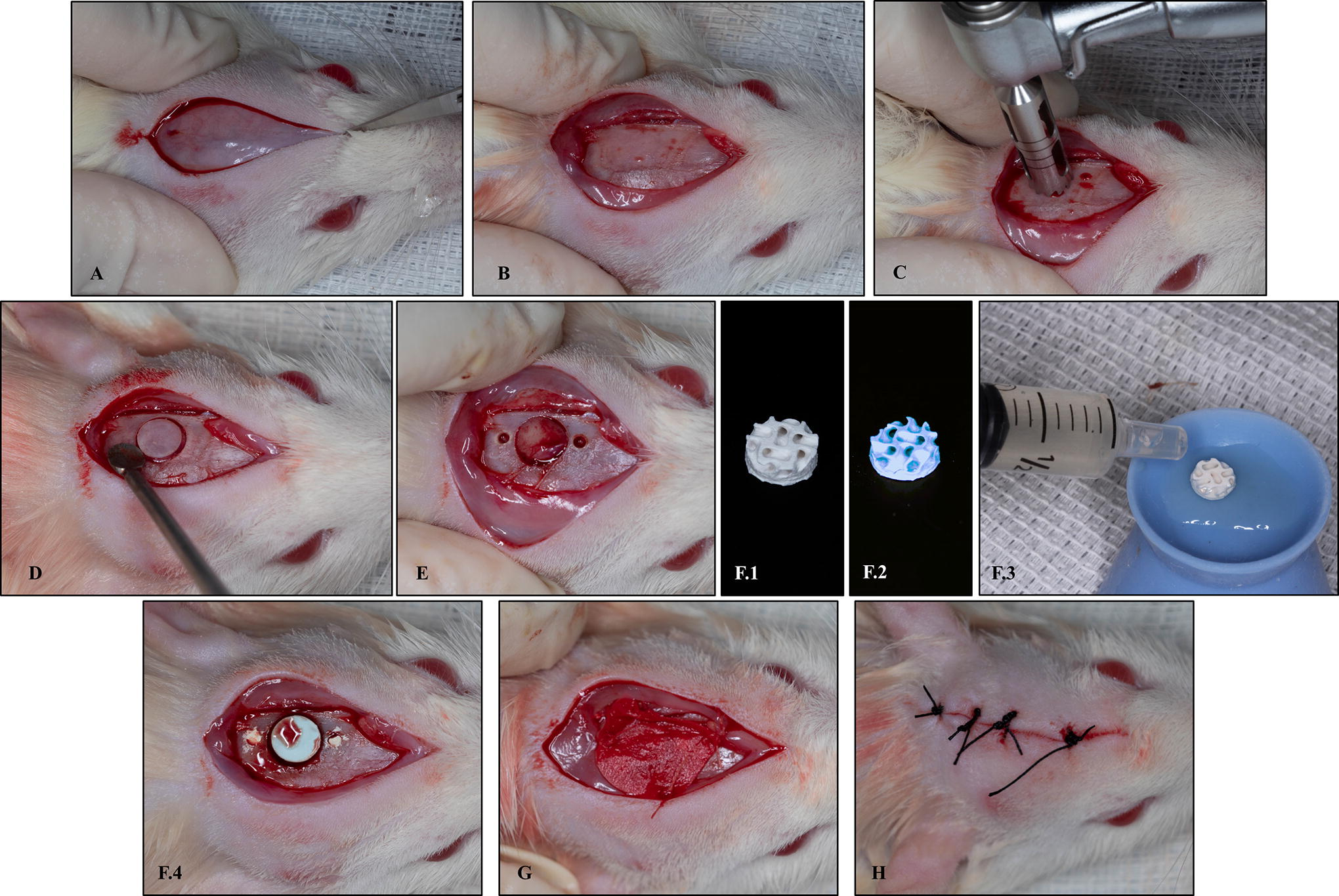
Surgical procedure.
Postoperatively, the animals received intramuscular injection of 24.000 IU/kg of penicillin G benzathine (single dose) at a dose of 0.2 mL (Small Animal Pentabiótico Veterinary, Fort Dodge® Animal Health Ltda., Campinas, SP), intramuscular anti-inflammatory Banamine® 0.2 mL/100g (Injectable Pet—Schering-Plough, Cotia, SP, Brazil) and 2 mg/kg of tramadol hydrochloride (Agner União®, Apucarana, PR, Brazil) every 12 h for 2 days. The animals were euthanized by anesthetic overdosage of xylazine and ketamine at time intervals of 30 and 60 days after surgery (n = 6 per group). Then, the tissues were collected for subsequent micro-CT, histomorphometric, and immunohistochemical analysis.
Autologous micrograft preparation
Preparation of the micrografts was carried out using the Rigeneracons® device (Human Brain Wave LLC, Turin, Italy) and according to the manufacturer’s manual as well as the protocol used by Araújo et al. 23 After the CSDs were created, in each animal of the B-TCP+RIG and HA+RIG groups, the calvarial periosteum removed was collected (5 × 5 mm square). Then, 0.8 mL of 0.9% NaCl was added to the Rigeneracons® device, using a syringe. Subsequently, the harvested tissue sample was placed on the grid inside the Rigeneracons®. After this, the helix of the device was manually adjusted to cover the sample. The adapter was used to connect the Rigeneracons® to the surgical motor, which operated at 80 rpm (15 Ncm) for 3 min. Once the mechanical disaggregation was completed, the micrograft suspension was collected from the device with a syringe, and the solution obtained was used to soak the customized bone block of the animals in the corresponding group.
Microcomputed tomographic analysis
After euthanasia, the calvaria were removed, reduced, and fixed in a 10% buffered formalin solution (Dinâmica®Química Contemporânea Ltda., Indaiatuba, SP, Brazil) for 48 h. Then, they were rinsed in running water for 24 h. After fixation, tissue was left in 70% alcohol (Labsynth® Produtos-Laboratórios Ltd.a., Diadema, SP, Brazil) to perform micro-CT. The specimens underwent scanning using the SkyScan 1272 microtomograph (SkyScan 1272 BrukerMicroCT, Belgium) with the following parameters: a slice thickness of 12 μm (90 kV and 111 μA), an Al 0.5 mm filter, a rotation step of 0.5 mm, a resolution of 2,016 × 1,344 μm, and an acquisition time of 47 min. X-ray projection images of the specimens were stored, reconstructed, and subsequently analyzed. The region of interest (ROI) was delineated by using NRecon software (SkyScan, 2011; Version 1.6.6.0). The bone defect filled with biomaterial blocks was evaluated with use of the CTAnalyser-CTAn software (2003-11SkyScan, 2012 BrukerMicroCT Version 1.12.4.0). Thirty slices were selected for analysis, focusing on the axial view of the ROI with a diameter of 5.0 mm at the center of the bone defect. Binary threshold values were set at grayscale values from 40 to 255 for total volume, including the biomaterial, and from 40 to 90 to select only the bone volume within the ROI, excluding the biomaterial volume. The 3D images were obtained using CTVox software (SkyScan, Leuven, Belgium, 2003; Version 3.3.1). This volume of interest designated was submitted to 3D analysis to determine the percentage values of bone volume (BV/TV) and bone surface density (BS/TV). 31
Laboratorial processing
The laboratory processing of the samples and subsequent analysis were performed at the Laboratory for the Study of Mineralized Tissues, Department of Basic Sciences, Araçatuba Dental School—São Paulo State University. Initially, the samples were fixed in 10% formaldehyde solution (Dinâmica®Química Contemporânea Ltd.a., Indaiatuba, SP, Brazil) for 48 h, followed by a 24-h wash in running water. Subsequently, they underwent decalcification in 10% EDTA (Exodo® Científica Química Fina Industria e Comércio Ltd.a., Sumaré, SP, Brazil) solution for a period of 6 weeks. Postdecalcification, the calvaria were dehydrated using a series of alcohol solutions, clearing with xylene, and then embedded in paraffin (Labsynth® Produtos-Laboratórios Ltd.a., Diadema, SP, Brazil). Microtomy was performed to obtain 5-µm-thick slices that were mounted on slides for subsequent histometric and immunolabeling analyses. 30
Histological and histomorphometrical analysis
The sections were deparaffinized using xylene and hydrated by means of a decreasing series of grades of ethanol, by staying in each concentration for 5 min. The sections were stained with hematoxylin (Labsynth® Produtos para Laboratórios Ltd.a., Diadema, SP, Brazil) for 8 min and with 1% eosin (Dinâmica® Química Contemporânea Ltd.a., Indaiatuba, SP, Brazil) for 20 s. The assembled slides were taken to an optical microscope (Leica DMLB, Heerbrugg, St. Gallen, Switzerland) to capture images using 4× and 10× magnifications. During image acquisition, the calibrated examiner performed the histological description in each of the groups.
The following histomorphometric parameters were evaluated: total area (TA) in mm2 of the defect originally created; area of NFB within the TA, calculated in mm2 and as a percentage value relative to the TA; area of remaining biomaterial (ARB) in the groups in which bone substitute was used, also measured as a percentage of TA; linear extent of the surgical created defect (ESD), with the boundaries delimited for the measurement of TA; and extension of newly formed bone (NBE), also calculated and expressed in mm and as a percentage relative to the ESD.
Immunohistochemical examination
Immunostaining was performed using the indirect immunoperoxidase technique, with polyclonal antibodies produced in goats (Santa Cruz Biotechnology, Inc., Dallas, TX) for the detection of target antigens (osteopontin—OPN [SC10593], osteocalcin—OCN [SC18319]), secondary antibody anti-goat produced in donkeys (Jackson ImmunosResearch Lab, West Grove, PA), using avidin-biotin (Vector Laboratories, Inc., Newark, CA) as a signal amplifier and diaminobenzidine (Agilent Dako Technologies, Inc., Santa Clara, CA) as the chromogen. At the end of the reactions, counterstaining was performed with Meyer’s hematoxylin. The analysis was performed at the center of the defect (ROI), where scores were assigned based on the extent of the positive staining area present in the ROI. Thus, mild score (with 25% of the area positively marked), moderate score (with 50% of the area positively marked), and intense score (with 75% of the area positively marked) were established. 31
Statistical analysis
All analyses were performed by a calibrated examiner without knowledge of the experimental groups. For examiner calibration, one-third of the sample was evaluated at two time periods with a 48-h interval. The intraclass correlation coefficient (ICC) was used to determine the examiner’s reproducibility in the two assessments. ICC values greater than 85% were considered to ensure examiner calibration. The data obtained were normalized and analyzed using GraphPad Prism 7.03 statistical software (GraphPad Software, La Jolla). Shapiro–Wilk test was used to assess data and confirmed their normal distribution. One-way ANOVA with the Tukey post hoc test were used to compare groups in the histomorphometric analyses. Two-way ANOVA with the Tukey post hoc test were performed to determine differences among the groups in the micro-CT analyses. The significance level of p was <0.05.
Clinical data
No animals were lost during the study, but 20 samples of 60-day follow-up groups had to be excluded (4 per group) owing to loss of integrity during histochemical processing affecting the histological and histomorphometrical analyses. Consequently, 60-day histological and histomorphometrical evaluations were not performed.
Results
Micro-CT analysis
Figure 2 shows representative three-dimensional reconstructions of each group analyzed. Figure 3 shows representative two-dimensional microtomographic cross-sections of each group analyzed. In the control group, minimal bone formation at the edges of the defect is observed at both 30 and 60 days. The bone formation inside the blocks can be observed in all test groups. In the HA group, new bone formation is observed inside the block and at the borders of the defect at 30 days, which increases at 60 days, indicating progressive integration of the material. The B-TCP group shows slight bone formation at 30 days, with visible improvement at 60 days, suggesting ongoing bone regeneration. In the HA+RIG group, bone formation was observed inside the bioceramic scaffold and at the borders of the defect. The bone formation is superior to the other groups at both 30 and 60 days, demonstrating even greater integration. The B-TCP+RIG group shows a similar result to HA+RIG group, with new bone formation observed at both 30 and 60 days. Among the biomaterials, the HA+RIG and B-TCP+RIG groups seem to exhibit more substantial bone formation, as they show increased new bone formation and integration with the surrounding bone tissue. It can be also noted that the adaptation of the biomaterial blocks was not entirely perfect in the created defects, since the defect was slightly larger than the biomaterial block (5 mm in diameter), so that it could be inserted into the cavity. However, this maladaptation was minimized by the fact that the defect was not created in the midline of the parietal bone but rather shifted to the left in a flatter portion of this bone, minimizing the negative effect that the bone curvature could have on the adaptation of the block.
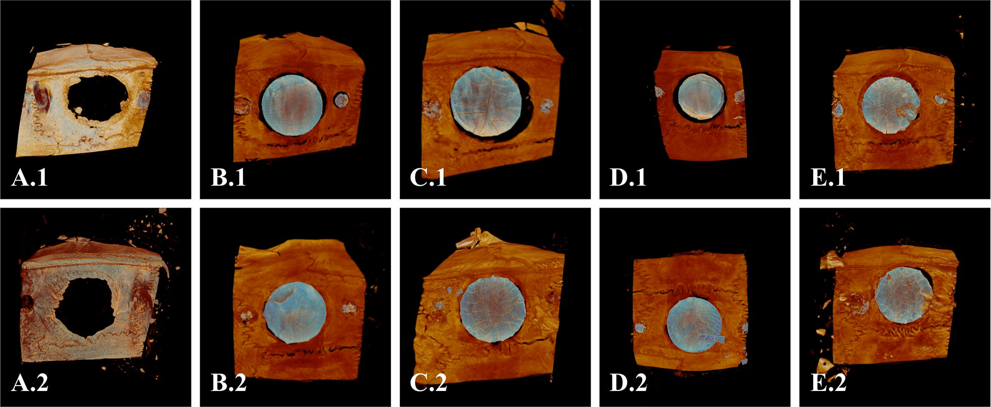
3D images obtained by CTVox software representing the bone formation of Control, HA, B-TCP, HA+RIG, and B-TCP+RIG groups.
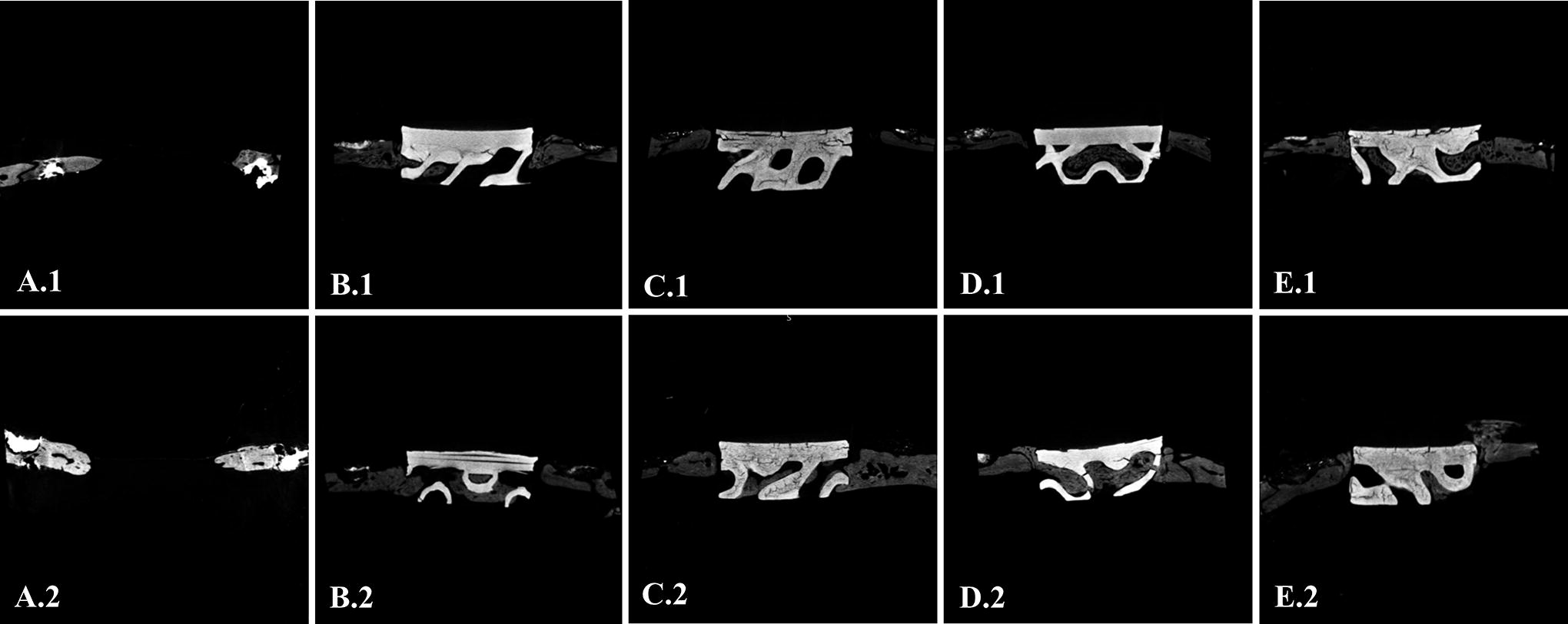
2D transversal images obtained by InVesalius software representing the bone formation of Control, HA, B-TCP, HA+RIG, and B-TCP+RIG groups.
Bone volume percentage (BV/TV)
In the analysis of BS/TV (Fig. 4A), at 30 days, no statistical differences were observed among the groups; however, higher values were present in HA+RIG and B-TCP+RIG groups. At 60 days, CONTROL (8.54 ± 5.06) and B-TCP (12.78 ± 5.20) groups had the lowest values, with significant differences compared with HA+RIG group (22.75 ± 5.43; p = 0.0002 and p = 0.0248, respectively).

Graphical representation of the results of microcomputed tomographic analysis:
Surface bone density (BS/TV)
In the analysis of BS/TV (Fig. 4B), CONTROL group had the lowest values at both 30 days (2.76 ± 1.68) and 60 days (3.57 ± 1.19), with significant differences compared with the other groups (p < 0.0001). No differences were found among the test groups at either evaluation time interval (p > 0.005).
Histological analysis
At 30 days postsurgery, the panoramic view (Fig. 5) shows the bone repair in progress, with NFB present in areas of the defect previously filled with biomaterial blocks. In the control group, only connective tissue is observed in the center of the defect, which is covered by the PDO membrane. In the tested groups, integration of the biomaterial blocks is evident. The grafts remained covered by the PDO membrane, and the blocks maintained their structural integrity. The edges of the defects showed organized connective tissue. Notably, in the HA, HA+RIG, and B-TCP+RIG blocks, bone formation was observed in proximity to the biomaterial remnants, with bone islands present in the center of the defect area. In contrast, the B-TCP group exhibited a higher prevalence of connective tissue than bone islands, when compared to the other groups. Overall, bone was consistently positioned adjacent to the biomaterial, with some connective tissue areas observed near the remnants of the biomaterial, mainly at the borders of the defect.

Representative photomicrographs of CONTROL, HA, B-TCP, HA+RIG, and B-TCP+RIG in the histological analysis at 30 days. Staining: Hematoxylin and eosin. Legend: *PDO Membrane; #3D-printed blocks; Arrow: Bone formation inside the block. Scale bar: 500 µm (magnification ×4).
Histomorphometrical analysis
Four central sections of each animal were assessed by histomorphometrical analysis, and the average of the four measurements was considered as the animal’s parameter value. The TA of all groups was analyzed (Fig. 6A). Groups with biomaterial had a larger TA because the biomaterial prevented the flap from collapsing into the defect. Significant differences were found between CONTROL (4.541 ± 0.057) and HA (6.087 ± 0.826; p < 0.0001), B-TCP (5.530 ± 0.095; p = 0.0012), and HA+RIG (5.926 ± 0.127; p < 0.0001) groups. Furthermore, significant differences were observed between B-TCP+RIG (5.137 ± 0.108) versus HA (p = 0.0018) and HA+RIG (p = 0.0111) groups.

Graphic representation of the results of the histomorphometrical analysis: (
In the analysis of the NFB in mm2 (Fig. 6B), the HA+RIG group (1.63 ± 0.54) had the best results, with significant difference from CONTROL (0.33 ± 0.18; p < 0.0001), HA (1.00 ± 0.44; p = 0.04), B-TCP (0.44 ± 0.20; p < 0.0001), and B-TCP+RIG (0.82 ± 0.28; p = 0.0047). The HA group also had significantly better results compared with CONTROL (p = 0.0236).
In analysis of the percentage of NFB (Fig. 6C), the HA group (19.24 ± 9.38) showed the best results, with significant difference from CONTROL (4.72 ± 2.24; p = 0.0069) and B-TCP (7.74 ± 3.5; p = 0.0434). The HA+RIG group (27.34 ± 9.29) had significantly better results than CONTROL (p < 0.0001), B-TCP (p = 0.0002), and B-TCP+RIG (15.8 ± 5.35; p = 0.0424).
In the analysis of ESD (Fig. 6D), no significant differences were found among CONTROL (5.402 ± 0.201), HA (5.251 ± 0.074), B-TCP (5.337 ± 0.015), HA+RIG (5.279 ± 0.17), and B-TCP+RIG (5.194 ± 0.059), indicating uniformity in the creation of the CSD (p = 0.0734).
Relative to the NBE (Fig. 6E), better results were observed in HA group (2.51 ± 0.94), with significant difference from CONTROL (0.713 ± 0.299; p = 0.0004) and B-TCP (1.403 ± 0.355; p = 0.0379). The HA+RIG group (2.895 ± 0.769) showed significant differences when compared with CONTROL (p < 0.0001), B-TCP (p = 0.003), and B-TCP+RIG (1.576 ± 0.518; p = 0.0097).
In the percentage analysis of the NBE relative to ESD (Fig. 6F), the HA group (50.08 ± 19.25) had better results, with significant difference from CONTROL (13.70 ± 6.05; p = 0.0003) and B-TCP (26.32 ± 6.73; p = 0.0233). HA+RIG (55.42 ± 14.81) also showed significant differences when compared with CONTROL (p < 0.0001), B-TCP (p = 0.004), and B-TCP+RIG (32.11 ± 10.78; p = 0.0268).
In ARB analysis (Fig. 6G), no significant differences were found among HA (43.37 ± 8.37), B-TCP (38.36 ± 6.07), HA+RIG (49.1 ± 8.71), and B-TCP+RIG (48.62 ± 7.59) (p = 0.0847).
Immunohistochemical examination
OPN is an extracellular matrix protein involved in initiating biomineralization responses, present in osteoblastic lineage cells and the extracellular matrix. At 30 days, moderate staining for OPN was observed in the cells and extracellular matrix of the HA+RIG group (Table 1 and Fig. 7). No positive staining was detected in the other experimental groups. At 60 days, slight staining was observed in the HA+RIG group, whereas the other groups showed no positive staining.
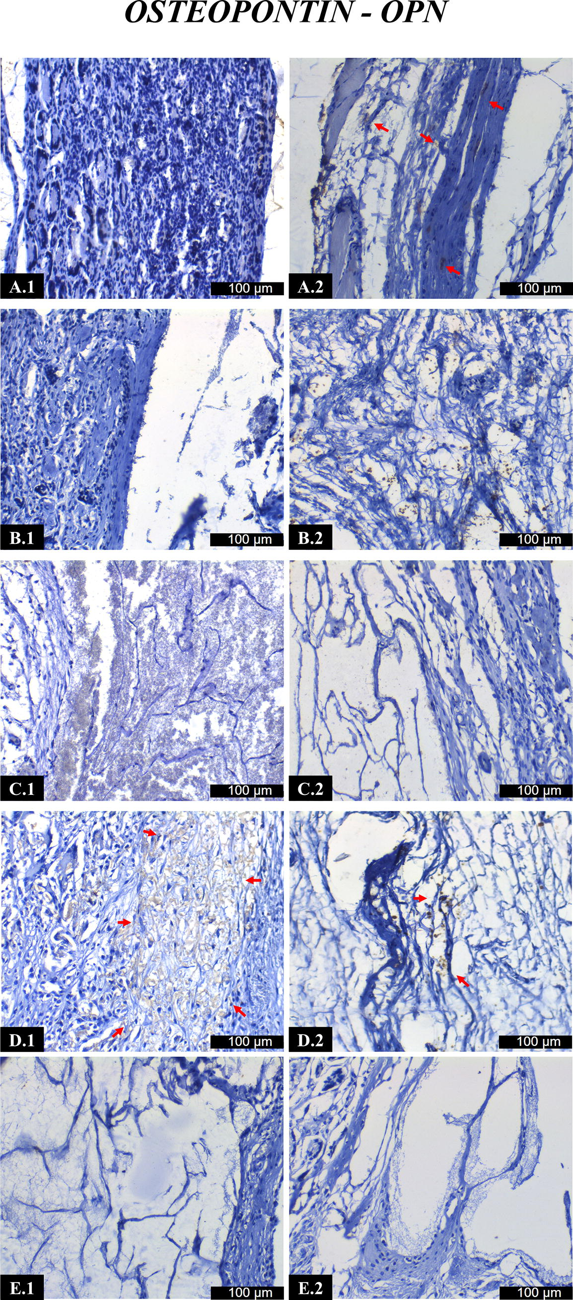
Representative photomicrographs of immunolabeling against osteopontin (OPN) antibodies in the different groups at 30 and 60 days of critical defect repair. (
Immunolabeling Scores in Control, HA, B-TCP, HA+RIG, and B-TCP+RIG Group at 30 and 60 Days of Bone Repair
Antibodies against OPN and OCN. Scores are evaluated at the border/center of the defect. Discrete labeling [+], moderate labeling [++], and intense labeling [+++].
B-TCP, β-tricalcium phosphate; HA, hydroxyapatite.
OCN is a key noncollagenous protein in the extracellular matrix, serving as a marker of bone mineralization, found in osteoblastic lineage cells and the extracellular matrix. At 30 days, slight staining for OCN was seen in the B-TCP and B-TCP+RIG groups, whereas moderate staining was observed in HA and HA+RIG groups (Table 1 and Fig. 8). At 60 days, slight staining was present in B-TCP, HA, and B-TCP+RIG groups, but no staining was observed in the HA+RIG group.

Representative photomicrographs of immunolabeling against osteocalcin (OCN) antibodies in the different groups at 30 and 60 days of critical defect repair. (
Discussion
The present study evaluated bone repair of two synthetic blocks for bone substitutes in association with autologous micrografts. A CSD model in rat calvaria was used to assess the regenerative process. The rat calvarial CSD model is considered one of the most predictable and widely used in vivo models in bone regeneration.32,33 Thus, studies on the efficacy of different biomaterials for the regeneration of calvarial defects are generally conducted in small animal models. 34
3D printing technology allows bone substitutes to be tailor-made35,36 for application in bone augmentation procedures.37–39 Despite the many advantages, one of the main disadvantages is the lower mechanical strength of 3D-printed alloplastic bone, which could be inferior to that of natural human bone. 40 In the present study, the blocks did not suffer damage pre- and postoperatively, as they were in areas without compression. Moreover, at 30 and 60 days of follow-up, the blocks were macroscopically fully incorporated into the animal’s calvaria.
The results of micro-CT imaging analysis showed that the blocks were incorporated into the animals’ calvaria. Both biomaterials exhibited new bone formation. There was a numerical increase in values for the groups associated with autologous micrografts (HA+RIG and B-TCP+RIG), especially the HA+RIG group at 60 days, which showed a statistically significant difference compared with the control group. Similar results were reported by Pitol-Palin et al. 30 at 60 days of repair, they observed similar results for BV/TV in the biomaterial groups (HA and B-TCP blocs) when compared with the autologous group.
In the histological analysis, the synthetic blocks were incorporated into the recipient site with new bone formation, without histological signs of necrosis, osteolysis, or foreign body reaction. The presence of the PDO membrane was noted in all samples. Organized connective tissue and formation of bone tissue were observed at the defect margins. At 30 days of follow-up, bone islands were observed in intimate contact with the B-TCP and HA blocks. These results were similar to those reported in previous studies, in which alloplastic biomaterials demonstrated excellent compatibility after implantation in bone defects or on bone surfaces.41,42 Pitol-Palin et al. 30 obtained similar results to those of the present study using HA and B-TCP blocks in conjunction with PDO membranes. In the present study, new bone formation values were statistically higher in the HA blocks (HA and HA+RIG groups), and there was also a tendency for improvement when the autologous micrograft was incorporated into the block.
The better performance of HA can, in part, be explained by its characteristics, such as the macroporosity and interconnected porosity of synthetic HA, which allowed the adhesion, proliferation, and differentiation of osteoprogenitor cells, as well as revascularization and subsequent new bone growth when implanted in vivo.43,44 In addition to being an osteoconductive material, HA can induce osteoinduction under certain conditions. The mechanism of osteoinduction by a synthetic material is still unclear, but several factors, such as microporosity, surface area, geometry, and topography, are important,45–48 with microporosity having a positive effect on increasing ectopic bone formation.49–51 The blocks used have a gyroid cell structure with a cell size of 2.4 mm (referring to the pore size) and a wall thickness of 0.2 mm (referring to the trabecular thickness of the biomaterial). Studies have shown that gyroid scaffolds demonstrated significant cell proliferation in vitro compared with cubic scaffolds.48,52
Immunohistochemical analysis allows visualization of tissue responses by means of expression of proteins related to the bone repair process.22,53,54 OPN is a marker of the initial point of biomineralization, showing active osteoblasts and cement lines (reversal lines), indicating the onset of the repair process. At 30 days, the HA+RIG group showed moderate OPN staining, whereas the other groups showed no staining; at 60 days, it showed slight staining, similar to CONTROL group, whereas the others remained unstained. These results suggested greater bone tissue formation in the HA+RIG group. In contrast, OCN staining indicates osteoblastic activity and the mineralization process, as this protein precipitates with Ca+ in the collagen matrix on the final stages of the mineralization process.22,53,54 After 30 days, the HA+RIG and HA groups showed moderate OCN expression, and the B-TCP and B-TCP+RIG groups showed slight OCN expression, suggesting that Ca+ deposition in the matrix was higher in the HA groups. Pitol-Palin et al. 30 obtained similar results, with the HA group showing slight OPN staining compared with the other groups. Both groups (HA and B-TCP) showed moderate OCN expression at 30 days. In contrast, other studies55,56 comparing HA/B-TCP scaffolds with autologous grafts showed that OPN staining was intense at 30 days in the autologous group and slight in the biomaterial group, indicating earlier osteoblastic activity in the former. At 60 days, the autologous group showed intense OCN expression, indicating bone maturation, whereas the biomaterial group showed slight expression of this protein, representing the formation of immature tissue during this period. In the present study, immunohistochemical analysis results showed that the repair process occurred in both biomaterials evaluated, with the HA groups outperforming the B-TCP groups.
Recent advances in tissue engineering have explored the potential of autologous micrografts when used to biofunctionalize bone substitutes. In the present study, different analyses have shown that the groups associated with micrografts presented better results irrespective of the type of synthetic block used. Several studies have confirmed the osteogenic and regenerative properties of micrografts produced by Rigenera® owing to their progenitor cell content.23,57,58 With the suspension of autologous micrografts, it is possible to imbibe the block, creating a bioactive biomaterial in a few minutes without significantly impacting postoperative morbidity. In this way, autologous micrografts are incorporated into the scaffolds, allowing the release of progenitor cells and growth factors over time. Autologous micrografts provide osteoinductive properties to the scaffold, promoting cell differentiation and proliferation. 23 This effect was probably in the present study, evidenced by the better results of the groups with the association of micrografts with the biomaterials.
Among the main issues and challenges found in the present study, we can mention the difficulties in histological processing, which led to the failure of many specimens from the 60-day follow-up group and to difficulties in immunohistochemical analysis. To optimize our results and reduce the number of animals, the micro-CT specimens were also used for histological examination. Consequently, the specimens were exposed and incubated for a long time in alcohol. This resulted in tissue impairment and failure of a part of the specimens, especially in the 60-day follow-up group, evidenced during the immunolabeling analysis, where the histological sections were too fragile (the protocol contains many steps and temperature changes) and often it was not possible to acquire images of the slides. Also, in the 60-day follow-up groups, there were great difficulties in decalcifying the specimens, preventing histological sectioning. Perhaps one of the reasons for this is the nature of the biomaterial blocks, i.e., their composition and three-dimensional arrangement, that after 60 days resulted in a tight integration with the surrounding bone tissue, which made the decalcification process extremely difficult. Even after twice the time used for the 30-day samples, it was not possible to make histological sections. These clinical data could be correlated with the increased mechanical capacity promoted by the HA present in the scaffolds, which is characterized by a slower resorption rate and a high osteoprogenitor potential.30,55,59,60 In addition, the macro-geometry of the scaffolds in association with the abovementioned characteristics increases the mechanical capacity of the bioceramics, as they act as a structure throughout the bone regeneration process.61,62
The results of the present study showed that the use of bone substitute blocks of synthetic origin, manufactured by 3D printing, associated with membranes also of synthetic origin and printed manufacture, led to an adequate bone regeneration process in CSDs in rat skulls. The results also pointed to the beneficial effect of using autologous micrografts to biofunctionalizer bone substitutes. Although this is a preclinical study with the inherent limitations of animal type and evaluation time, the former is a classical model used in the literature. It allows the extrapolation of the results found here, with reservations, to propose future clinical studies, to evaluate in patients, whether the effectiveness of this therapy, evidenced here, could be maintained in humans.
Conclusion
Synthetic bone substitutes produced by additive manufacturing, such as HA and B-TCP blocks, particularly in combination with autologous micrografts, demonstrated efficacy in bone regeneration in rat calvarial defects. These promising results suggest that such biomaterials may represent alternatives for reconstructive bone procedures, supporting future studies in patients.
Footnotes
Acknowledgments
The authors would like to acknowledge Adriana Luisa Gonçalves de Almeida and Juliana Moura for their technical assistance during the experiments.
Authors’ Contributions
E.M.: Methodology, investigation, formal analysis, and writing—original draft preparation. A.C.L.: Investigation (experimental work, histomorphometrical analysis), validation. L.P. and R.O.: Investigation (laboratorial processing). J.S.: Writing—review and editing, resources. M.M.: Software, data curation. A.N. Jr.: Formal analysis, visualization. S.S.S.: Conceptualization, methodology, project administration, supervision, and writing—review and editing. All authors have read and agreed to the published version of the article.
Disclosure Statement
Jamil Shibli has financial relationships (shareholder) with M3 Health Ind. Com. de Prod. Med. Odont. e Correlatos S.A., Jundiaí, Brazil, and declares conflicts of interest. The remaining authors (Eladio Muñoz, Ana Carolina Loyola, Letícia Pitol-Palin, Roberta Okamoto, Michel Messora, Arthur Novaes Jr., and Sergio Scombatti) declare no conflicts of interest.
Funding Information
This research was funded by Plenum Bioengenharia, M3 Health Indústria e Comércio de Produtos Médicos, Odontológicos e Correlatos S.A, Jundiaí, Brazil.