
Abstract
Plant-derived hydrocolloids offer promising prospects in biomedical applications. Among these, Flaxseed hydrocolloid (FSH) can form a soft, elastic, and biocompatible hydrocolloid with tunable viscosity and superior swelling capacity, making it an attractive scaffold. This study introduces a green extraction method for FSH, employing a single-step aqueous extraction process and fabrication of FSH scaffold. Despite growing interest, the pristine form of FSH has not been investigated for sustainable long-term three-dimensional (3D) cell culture. Here, FSH scaffolds were thoroughly characterized for their morphological, chemical, mechanical, and biological properties. 3D cell culture experiments were conducted using NIH-3T3 mouse fibroblast cells, and cell viability was assessed using live/dead and Alamar Blue assays. High cell viability was sustained for long term compared with 2D cell culture. Cell adhesion and 3D cellular morphology on FSH scaffold for 30 days were monitored by scanning electron microscopy analysis. Also, collagen type-I and F-actin expressions were analyzed by immunostaining after 30 days of culture, resulting in 5- and 4-fold increments of fluorescence intensity, respectively. Results indicate sustained cell viability in the long term and favorable cell–material interaction, demonstrating the potential of FSH as a scaffold. This study emphasizes the importance of the green extraction approach, improving the biocompatibility and functionality of FSH tissue engineering applications.
Impact Statement
Flaxseed hydrocolloid (FSH) is a promising scaffold for biomedical applications due to its biocompatibility and tunable properties. This study introduces a green extraction method for FSH and evaluates its use in 3D cell culture with NIH-3T3 mouse fibroblast cells. The findings indicate high cell viability and enhanced cell–material interactions over 30 days, highlighting the potential of FSH for tissue engineering.
Introduction
Polysaccharide-based polymers have gained significant interest in the biomedical field owing to their multifunctional features, such as biocompatibility, biodegradability, and hydrocolloid-forming ability.1–8 Plant-derived hydrocolloids are abundant in polysaccharides, uronic acids, glycoproteins, and bioactive compounds.9,10 Flaxseed or linseed (Linum usitatissimum) is comprised of heterogeneous polysaccharides, mainly neutral arabinoxylan (75%) and acidic rhamnogalacturonan (25%).11,12 Flaxseeds have bioactive units that offer multifunctional properties such as antibacterial, anti-inflammatory, and antioxidant. 11 Moreover, it can possess a soft and elastic hydrocolloid structure that is biocompatible and biodegradable, with high viscosity and remarkable water-holding capacity, thus gaining attention in biomedical applications, especially in tissue engineering and wound dressing applications.13,14 In literature, it is commonly utilized in combination with other biomaterials to form hybrid scaffolds such as cellulose, 12 collagen, 15 and silk fibroin 16 to improve specific properties for different purposes. However, despite the growing interest in FSH, the pristine form of FSH was not investigated thoroughly and there remains a need for a comprehensive investigation of its applicability in long-term three-dimensional (3D) cell culture. The extraction method used for polysaccharide-based hydrocolloids is crucial in biomedical applications. Some methods lead to loss of bioactive properties, while some methods cause cytotoxicity during cell culture due to the usage of toxic chemicals. Therefore, using green extraction method plays an important role in tissue engineering in terms of cell viability and biocompatibility.
This study aims to develop a green extraction method, which utilizes a single-step aqueous extraction to mitigate the aforementioned disadvantages. Furthermore, it provides rapid extraction by eliminating tedious purification and isolation steps. Herein, the applicability of green-extracted pristine FSH was investigated as a scaffold for 3D cell culture. Subsequent to the aqueous extraction of FSH, cross-linking was achieved through the utilization of 1-ethyl-3-(3-dimethylaminopropyl) carbodiimide–N-hydroxy succinimide (EDC–NHS). FSH scaffold was characterized using Fourier transform infrared-attenuated total reflectance (FTIR-ATR), compression testing, and scanning electron microscopy (SEM) for the determination of morphological, chemical, and mechanical characteristics. Swelling and protein adsorption analyses were performed as part of the characterization process. After characterization, 3D cell culture studies were carried out using NIH-3T3 fibroblast cells, then cell viability was evaluated by live/dead and Alamar Blue assays. Morphology and adhesion of cells on FSH scaffolds were visualized by SEM analysis, and immunostaining of F-actin and collagen type-I was performed to monitor their expressions during a 30-day period. The outcome of this study highlights the suitability of FSH scaffold in providing a proper microenvironment for 3D cell culture and tissue engineering applications.
Materials and Methods
Extraction of FSH
Extraction and gelation of FSH were performed using green extraction methodology based on previous reports with some modifications.17,18 Flaxseeds were purchased from the local market in Aegean region of Turkey. Throughout the process, a single-step aqueous extraction was utilized to avoid toxic solvents and maintain the bioactive properties of FSH. Briefly, 100, 150, and 200 mg/mL FSHs were stirred in ultra-pure water at 85°C for 3 h to allow solubilization of polysaccharides, uronic acids, and other bioactive compounds, forming a viscous hydrocolloid solution. Following the complete gelation, hydrocolloid was collected by filtration to remove insoluble impurities and cross-linked by EDC–NHS where 0.4 M EDC and 0.1 M NHS were mixed with FSH in proper ratios. For lightly cross-linked FSH 5:1 (L-XD), moderately cross-linked FSH 3:1 (M-XD), and heavily cross-linked 1:1 (H-XD) FSH:cross-linker ratios were utilized. Further, FSH was subjected to a freeze-drying process to fabricate sponge-like porous scaffolds to be used in 3D cell culture.
Characterization of FSH scaffold
Optimization and characterization of FSH scaffolds in terms of hydrocolloid concentration and cross-linking degree were performed by FTIR and SEM analyses. FTIR-ATR (PerkinElmer, USA) was used to analyze FSH, within the range of 400–4000 nm−1, and FTIR graphs were plotted using OriginPro 2016 software (Version 93E, Northampton, MA). Further characterizations of FSH scaffolds were done in terms of scaffold morphology, % porosity, mechanical properties, swelling, and protein adsorption capacities. Scaffold morphology was analyzed using SEM (FEI Quanta, 250 FEG), and % porosity was calculated using SEM images by ImageJ software (NIH). FSH scaffolds were also subjected to mechanical analysis by Texture Analyzer (Stable Micro Systems, TA, XT plus C). The mechanical analysis was conducted as a compression test using a 5 kg load cell and a 35 mm diameter probe at a 0.83 mm/s compression speed.14,19,20 Swelling capacity was analyzed by immersing the scaffolds in PBS; dry and wet weights of scaffolds were measured at specific time points (0–24 h) and calculated using the equation given elsewhere. 21 Protein adsorption capacity of FSH scaffolds was determined according to the bicinchoninic acid (BCA) assay protocol (Pierce, Thermo Scientific). Following the 2 h incubation in protein solutions at 37°C, absorbance was measured at 562 nm wavelength by Multiskan™ GO Microplate Spectrophotometer (Thermo Fischer Scientific); then, the amount of total adsorbed protein was calculated using a standard bovine serum albumin (BSA) curve.
3D cell culture studies
NIH-3T3 mouse fibroblast cell line (ATCC® CRL-1658™) was utilized as a model cell line in 3D cell culture. The cells were maintained in Dulbecco’s modified Eagle medium (DMEM) (Gibco) supplemented with 10% fetal bovine serum (Gibco) and 1% penicillin/streptomycin (P/S) under 5% CO2 at 37°C. 1 × 10⁶ cells were seeded on FSH scaffold after sterilization and 24-h conditioning in DMEM. Cell viability was assessed using live/dead and Alamar Blue assays. Live/dead analysis was conducted with CytoCalcein™ Green and propidium iodide (PI) dyes (AAT Bioquest) and observed under a fluorescence microscope (Zeiss Axio Observer). 22 Alamar Blue analysis was performed to evaluate cell viability quantitatively using a spectrophotometer at 570 and 600 nm.
Cell adhesion and 3D cellular morphology were monitored on FSH scaffold for 1, 7, 15, and 30 days using SEM analysis. For this, the scaffolds were fixed using 1% osmium tetroxide and then examined using SEM. Furthermore, immunostaining was done to visualize the expression of collagen type-I and F-actin on days 1, 7, 15, and 30. Hence, cells on FSH scaffolds were fixed, permeabilized, and blocked with 4% paraformaldehyde, 0.1% Triton X-100, and 1% BSA, respectively. The stained cells were visualized by fluorescence microscope and fluorescence intensity was quantified using Image J software (NIH).
Statistical analysis
Each experiment was repeated at least with three replicates, and the results were displayed as the mean value along with the standard deviation. Two-way analysis of variance (ANOVA) was utilized for statistical analysis of relative cell viability, while one-way ANOVA was utilized for statistical analysis of fluorescence intensity of collagen type-I and F-actin on days 1,7 and 15 in comparison with day 30 via GraphPad Prism 9 software (GraphPad Prism, Inc., San Diego, USA). The statistically significant differences between groups were represented as follows: *p ≤ 0.05, **p ≤ 0.01, ***p ≤ 0.001, ****p ≤ 0.0001.
Results
Extraction of FSH
FSH extraction was performed through green, single-step aqueous extraction, and hydrocolloid was collected at the end of 3 h incubation. This extraction method offers substantial advantages over previously reported extraction techniques.10,23,24 Unlike other methods that rely on harsh chemical usage, our approach excludes toxic solvents, which can negatively impact cell viability and introduce cytotoxicity risks in cell culture applications. The water-based extraction preserves the bioactive properties of FSH, as well its antibacterial, anti-inflammatory, and antioxidant characteristics, which are essential for biomedical applications, 23 and provides a biocompatible 3D microenvironment for cell culture studies.
Later, FSH was cross-linked with EDC–NHS and freeze-dried to obtain sponge-like FSH scaffolds as shown in Figure 1. FTIR-ATR analysis was used to optimize hydrocolloid concentration and hydrocolloid:cross-linker ratio. The FTIR spectrum of pristine FSH characteristically has distinct peaks correlated with functional groups found in the polysaccharide and protein components. O–H stretching band (3200–3500 cm−1) corresponds to hydroxyl groups majorly found in cellulose and hemicellulose content of FSH and is responsible for the gelation and water-holding behavior of hydrogel structure. 18 A slight C–H stretching band which refers to methyl groups in glycoprotein structure, was observed at 2800–3000 cm−1. 25 C–O stretching band was observed at 1650–1750 cm−1 and refers to carbonyl groups associated with carboxylic acid found in uronic acid subunits. 26 Following the cross-linking through EDC–NHS, the formation of expected peptide bonds owing to cross-linking, N–H stretching (3315 cm−1), C = O stretching (1641 cm−1), N–H bending (1563 cm−1), and C–N stretching (1260 cm−1) peaks were observed in the cross-linked FSH samples. The expected peaks corresponding to the peptide bond formation were observed at similar intensities for each cross-linked sample (L-XD, M-XD, and H-XD) of 100 mg/mL FSH, which indicates that high-density cross-linking occurred even at L-XD group (Fig. 2A). For 150 mg/mL FSH, these peaks started to be observed at L-XD sample, and compared with it higher intensities were observed at a similar level for M-XD and H-XD samples with the increase in cross-linker ratio (Fig. 2B). The cross-linking efficiency of EDC–NHS was lower in 200 mg/mL FSH samples due to excess polysaccharide content. 27 Expected peaks that indicate cross-linking were noted in L-XD sample of 200 mg/mL FSH, while a gradual increase was observed for increasing cross-linker ratio (Fig. 2C).

Green extraction of FSH and fabrication of scaffolds through freeze-drying. FSH, flaxseed hydrocolloid.
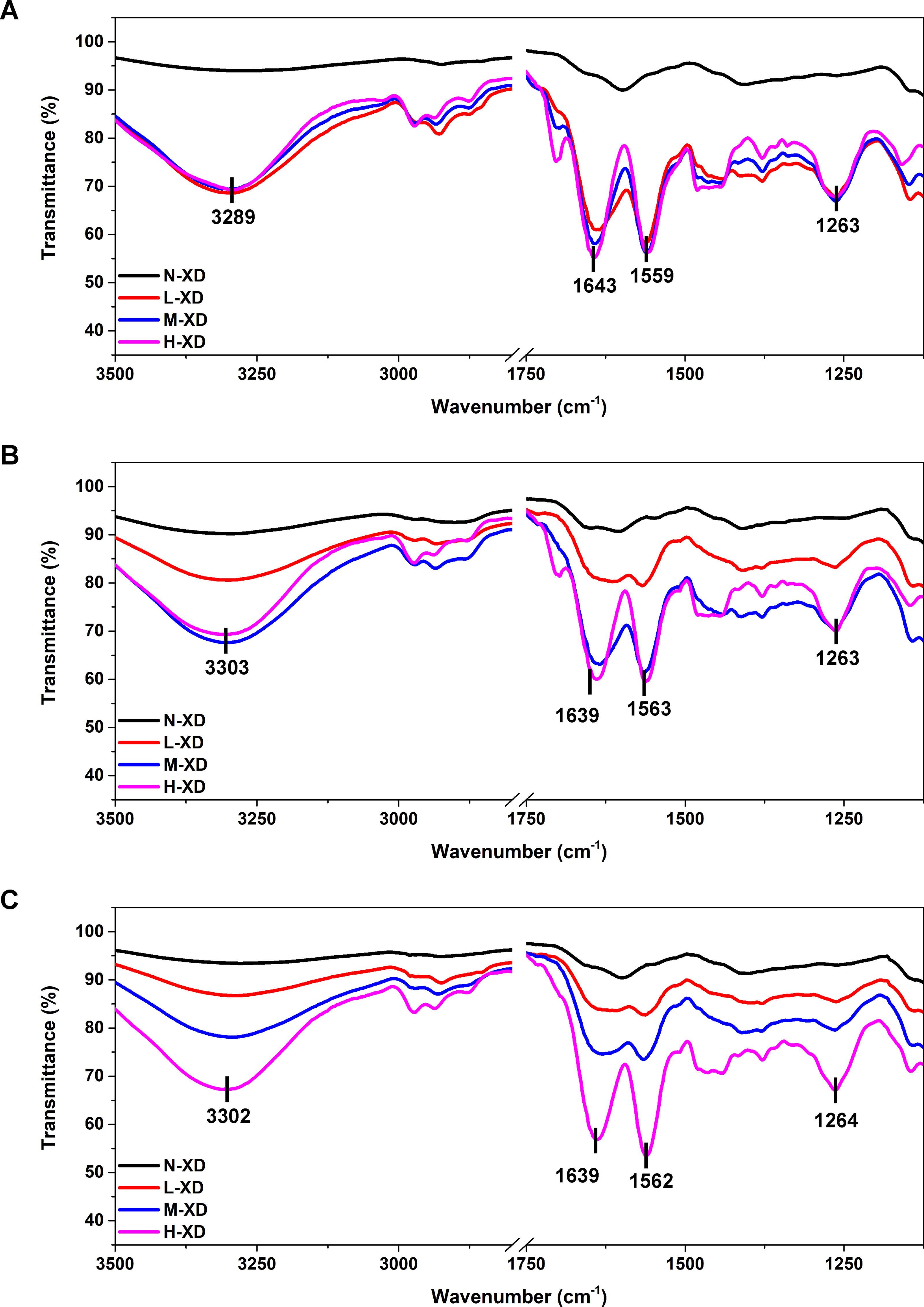
FTIR spectrum and fingerprint regions of FSH in varied cross-linking ratios (N-XD, L-XD, M-XD, H-XD):
Following FTIR analysis, texture, and viscosity of FSH samples were investigated, and morphological analysis was carried out by SEM. The increase in FSH concentration leads to a highly viscous and more heterogeneous hydrocolloid (Fig. 3A–C). Porosity of non/cross-linked FSH samples was investigated and it was observed that N-XD 100 mg/mL FSH has a highly porous structure (30.5%). As expected, an increase in cross-linker ratio resulted in decreased pore size, and very low porosity (0.4%) was observed especially for H-XD (Fig. 3D and G). The morphology of N-XD 150 mg/mL FSH was not significantly changed by cross-linking, where porous structures were obtained in all cross-linked samples (Fig. 3E), and % porosity of 150 mg/mL FSH was calculated in the range of 31.8%–27.8% (Fig. 3G). On the other hand, the most porous structure was obtained in N-XD 200 mg/mL FSH (31.9%) (Fig. 3F). The % porosity of 200 mg/mL FSH was obtained as 31.2%, 27.4%, 23.2% for L-XD, M-XD, and H-XD, respectively.
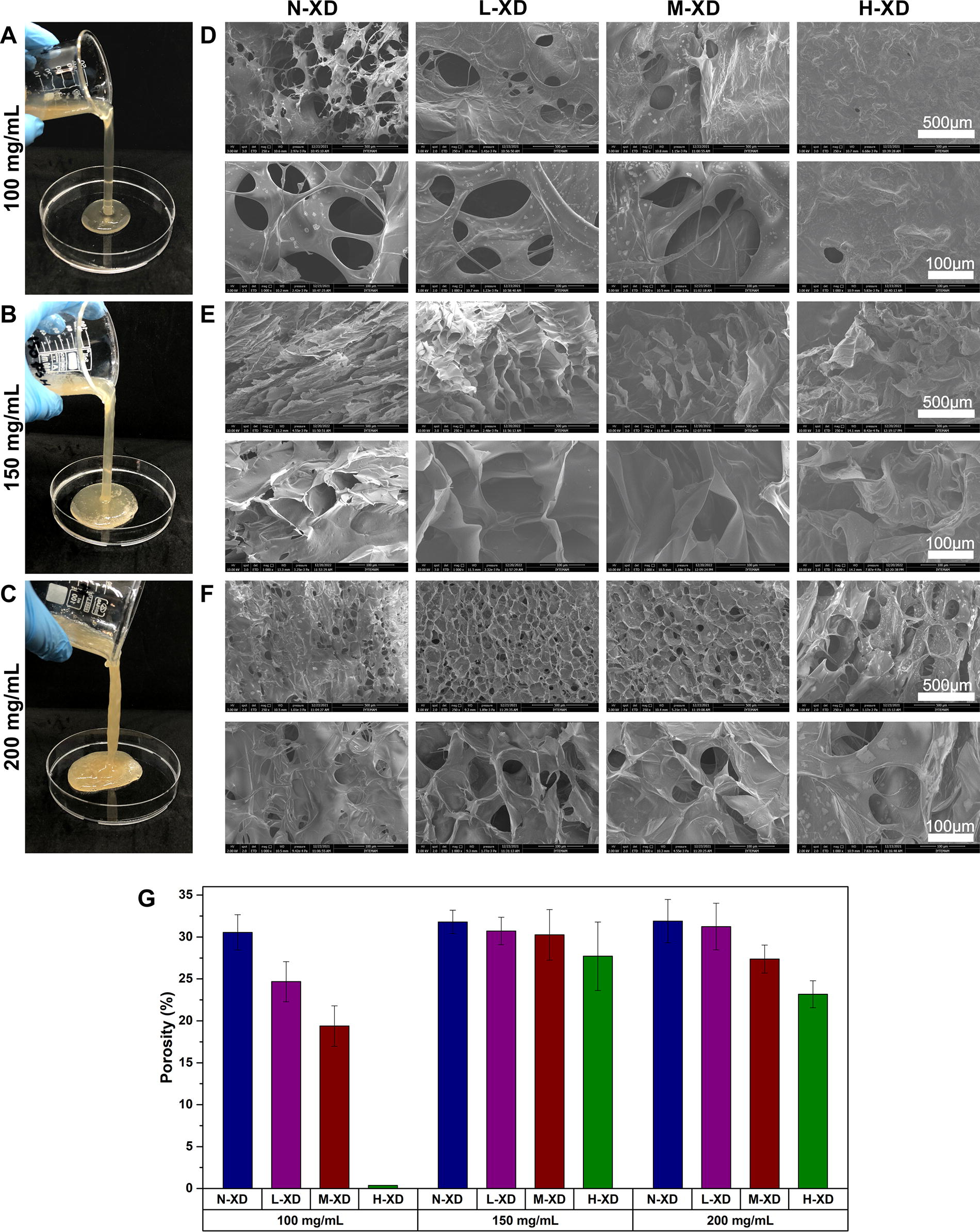
Images of N-XD FSH samples showing the consistency of hydrocolloid:
For further characterization and 3D cell culture studies 150 mg/mL FSH concentration was used due to the heterogeneous structure of 200 mg/mL FSH. The mechanical characterization of FSH scaffolds was performed by Texture Analyzer, and Young’s modulus (E) values were calculated using the linear region of the compression plots. Young’s modulus of L-XD FSH scaffold was obtained as 2 Pa, while increased cross-linker ratio improved the Young’s Modulus resulting in 3 and 30 Pa for M-XD and H-XD FSH scaffolds, respectively (Fig. 4A). As expected, an increased cross-linker ratio leads to formation of a denser and more tightly packed structure with less space between polymer chains; hence, the required pressure becomes higher for compression, which resulted in Young’s modulus increment. 28 As a result, cross-linking ratio is a key factor to tune mechanical characteristics of scaffold and enables fabrication of scaffold for varied tissue engineering applications.29,30 The swelling study showed that all samples reached maximum swelling around 3 h, which is followed by slight deswelling, especially for L-XD and M-XD samples, and then reached equilibrium (Fig. 4B). H-XD samples held 5.1 times more water than their own weight at the end of 24 h, while M-XD and L-XD samples had 16.1- and 22.8-fold higher swelling capacities, respectively. BCA assay was employed to determine the protein adsorption profile of FSH scaffolds before 3D cell culture studies. The amount of adsorbed protein increased with increasing concentration and then reached a plateau between 1500 and 2000 µg/mL (Fig. 4C). The maximum amount of adsorbed protein was 1111.6 µg/mL at equilibrium which is higher than alternative plant-derived hydrocolloids reported in the literature.14,15,21,31,32

Biocompatibility assessment of FSH scaffold
The cytocompatibility of the FSH scaffold was investigated by live/dead and Alamar Blue assays (Fig. 5) and results revealed that high cell viability was sustained through 15 days on 3D cell culture. Small spheroid formation was obtained on day 1, starting from day 7 cellular assemblies started to spread, and covered the entire scaffold on day 15 (Fig. 5A). In contrast, cells covered the entire surface of two-dimensional (2D) cell culture even on day 1; however, cell death increased on day 7 due to contact inhibition, and detachment of cell clusters was observed on day 15. On the other hand, overgrowth of cells in 2D cell cultures over long culture times can lead to the cells death due to contact inhibition. This can mislead the result, thus a comparison of cell viability between 2D and 3D cultures on day 30 was not conducted. These results highlighted the limitation of 2D cell culture maintaining high cell viability in the long term, likely due to a lack of spatial organization and insufficient surface area compared to 3D FSH scaffold.33,34 Furthermore, these findings were validated via Alamar Blue analysis, quantitatively where cell viability gradually increased in 3D cell culture, whereas it decreased in 2D cell culture through 15 days (Fig. 5B). In addition to cell viability studies, SEM analysis was employed to observe cell adhesion and 3D morphological changes for 30 days, as depicted in Figure 6. Initial assessments on day 1 revealed the formation of cellular aggregates, showing successful cell adaptation to the FSH scaffold. 35 These aggregates evolved into 3D spheroid structures (shown by yellow arrows) and started to cover the scaffold through 30 days of culture.
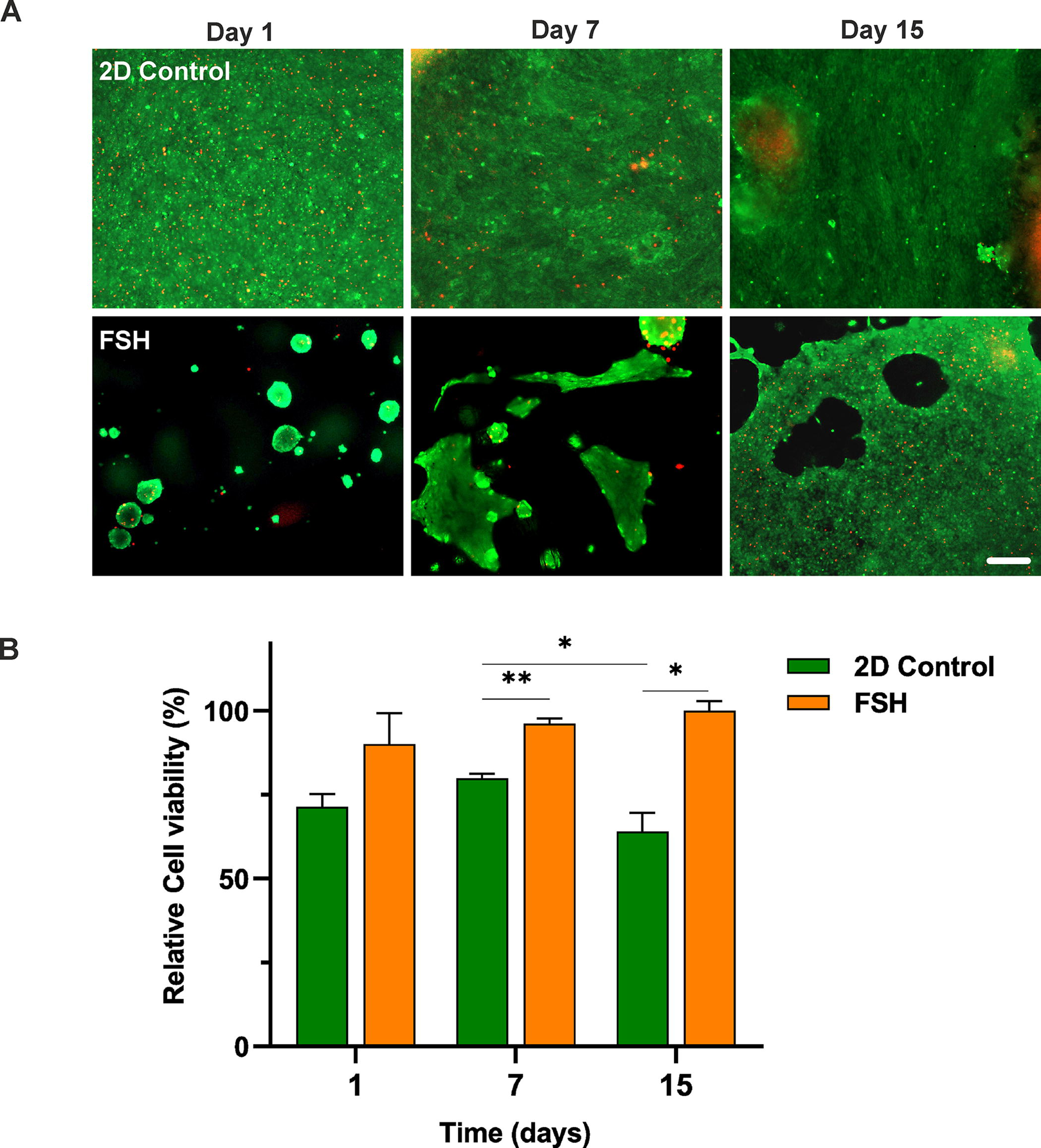
Cell viability analysis on FSH scaffold via

SEM analysis of NIH-3T3 cells on FSH scaffold for 3D cellular morphology monitoring on days 1, 7, 15, and 30 (scale bar: 50 and 20 µm). 3D, three-dimensional
The expression profiles of cellular and extracellular matrix (ECM) components were analyzed through immunostaining of collagen type-I and F-actin, along with DAPI staining for 3D cultured cells on FSH scaffold, as shown in Figure 7A. Fluorescence intensities of F-actin and collagen type-I were measured using immunostaining images (Fig. 7B and C). F-actin was expressed during 3D cell culture and there was a 4-fold increment of F-actin intensity from day 1 to day 30 (Fig. 7B). On the other hand, collagen type-I expression was also observed during 3D culture and its intensity increased through 30-day culture. There is a 5-fold increment of collagen type-I intensity from day 1 to day 30 (Fig. 7C).

Discussion
The results of this study demonstrate the successful extraction of FSH using an eco-friendly aqueous method. FTIR-ATR analysis confirmed the cross-linking process with EDC–NHS, revealing characteristic peaks associated with functional groups and peptide bonds. In addition, the findings highlighted that the hydrocolloid concentration is an important parameter for effective cross-linking which is in line with the literature.36,37 Further characterization of FSH scaffolds by texture, viscosity, and morphological analyses highlighted how increasing FSH concentration resulted in higher viscosity and decreased porosity as cross-linker ratios increased. Mechanical testing showed the porosity of low hydrocolloid concentration (100 mg/mL) was affected significantly by the increment of the cross-linking ratio since the number of polymer chains was limited. 27 On the other hand, in medium (150 mg/mL) and high concentration (200 mg/mL), samples were not affected remarkably owing to the high number of polymer chains to be cross-linked. 27 Decreased cross-linking degree resulted in high swelling capacity for M-XD and L-XD samples which correlates with the literature.38–40 On the other hand, H-XD sample has higher cross-linking degree and a stiff polymeric network, which limits the uptake of water molecules. 41 This indicates that the swelling capacity was inversely related to the degree of cross-linking, with lightly cross-linked scaffolds showing higher water retention. The FSH scaffold demonstrates good protein adsorption capacity, which indirectly reflects its ability to support cell adhesion and create an optimal microenvironment for cell culture in tissue engineering applications. In the literature, it was proved that hydrophilicity directly affects the adsorption of proteins and cells since cells interact with scaffold through cell adhesion proteins.42,43 Therefore, the hydrophilic nature of FSH is expected to enhance the protein adsorption capacity and favor cell adhesion. 15 These findings emphasize the importance of controlling FSH concentration and cross-linking degrees to optimize scaffold properties like porosity, stiffness, and swelling capacity. The ability to fine-tune these properties makes FSH a promising material for tissue engineering applications, aligning with previous studies that have shown the effect of cross-linking density on scaffold characteristics.
Moreover, the high cell viability observed in 3D cultures compared with 2D cultures underlines the advantage of using FSH scaffolds to support long-term cell growth. This outcome can be ascribed to the presence of polysaccharides like arabinoxylan in flaxseed, 44 that serves as a natural adhesive for cells like within the ECM, thus establishing a conducive microenvironment for cell adhesion and proliferation. 15 In addition, these results overlap with previously reported studies indicating polysaccharide-based scaffolds, such as quince seed hydrocolloid and psyllium seed hydrocolloid scaffolds, and promote cell adhesion and high viability.14,21,31,32,45–47 Furthermore, SEM analysis confirmed enhanced cell adhesion, with 3D spheroid structures tightly adhering to the FSH scaffold over a 15-day period. As the cells adapted to the scaffold, the 3D cellular structures progressively expanded to cover the entire scaffold during long-term culture. SEM result displayed that FSH scaffold provided better cell adhesion and spreading, 15 demonstrating the topography of the FSH scaffold promotes cell–material interaction, cell attachment, and spreading, thus promoting 3D cell culture formation. Increasing collagen type I and F-actin expression after day 15 can be attributed to the adaptation of cells to 3D microenvironment. This situation significantly depends on the type of scaffold and nature of the cells, where expression of ECM content might require more time in 3D cell culture and enhanced after cell adaptation.32,48,49 The enhanced expression of collagen type-I and F-actin further validates the scaffold’s ability to promote a suitable 3D microenvironment, consistent with the behavior of other polysaccharide-based scaffolds.14,21,31,32,50
Conclusion
This study presents a comprehensive investigation of extraction, gelation, and characterization of FSH scaffold. Here, a green and single-step aqueous extraction methodology was utilized, where FSH provided porous structures, high swelling capacity, and remarkable protein adsorption. High cell viability in 3D cell culture was sustained in FSH for long term, while it could not be maintained in 2D cell culture. Long-term culture results indicated successful cell–material interaction and increased collagen type-I and F-actin expression. Overall, this study underscores an eco-friendly alternative for the extraction of FSH and shows its potential as a biocompatible scaffold with a conducive microenvironment in further 3D cell cultures.
Footnotes
Acknowledgments
R.B.-K. gratefully acknowledges the Scientific and Technological Research Council of Turkey (TUBITAK) 2211-A National Graduate Scholarship program. The authors thank İzmir Institute of Technology Biotechnology and Bioengineering Research and Application Center and İzmir Institute of Technology Materials Research Center.
Authors’ Contributions
Ö.Y.-S.: Conceptualization, investigation, methodology, and writing—original draft. R.B.-K.: Conceptualization, investigation, methodology, and writing—original draft. A.A.-Y.: Conceptualization, supervision, and writing—review and editing.
Disclosure Statement
There are no conflicts to declare.
Funding Information
This study was supported by the Scientific and Technological Research Council of Turkey (TUBITAK) under grant number 120C155.