




Abstract
We established a model of hypothermic hypoxia/reoxygenation injury of fibroblasts, simulated the process of ischemia/reperfusion injury during cardiopulmonary bypass, and studied the effects of cardiac fibroblasts on cardiomyocyte activity, connexin 43 (Cx43), and calmodulin kinase II (CaMKII) expression. Furthermore, the effects of sevoflurane-treated fibroblast culture medium on cardiac activity, Cx43 protein, and CaMKII expression were observed. The results showed that the fibroblast culture medium damaged by hypothermic hypoxia/reoxygenation could reduce the beating frequency of cardiomyocytes, increase the mortality of cardiomyocytes, decrease the relative expression of Cx43, and increase the relative expression of CaMKII. However, sevoflurane containing hypothermic hypoxia/reoxygenation injury fibroblast culture medium can increase the beating frequency of cardiomyocytes, reduce the mortality of cardiomyocytes, increase the relative expression of Cx43 protein, and decrease the relative expression of CaMKII. The results suggest that the antiarrhythmic effects of sevoflurane on the expression of Cx43 and CaMKII are related to fibroblasts.
Introduction
Reperfusion arrhythmia is the main manifestation of myocardial ischemia/reperfusion injury, especially ventricular arrhythmia, which is the key factor affecting the success or failure of cardiopulmonary bypass (Talwar et al., 2015; Tse et al., 2016; Chin et al., 2017). In the past, the studies on ischemia/reperfusion injury mainly focused on cardiomyocytes, but there were few studies on the interaction between fibroblasts and cardiomyocytes.
Sevoflurane has antiarrhythmic effect (Gong et al., 2012; Wang et al., 2019), previous study of our group confirmed that the electrophysiological instability of isolated rat heart induced by hypothermia and global ischemia/reperfusion can be improved by sevoflurane, and this effect is related to sevoflurane improving the downregulation and distribution disorder of myocardial connexin 43 (Cx43) protein expression after hypothermic ischemia/reperfusion (Li et al., 2018).
Calmodulin kinase II (CaMKII) is a molecule that can lead to arrhythmia. A large amount of evidence shows that overactivation of CaMKII can cause ventricular remodeling, which makes the cardiac conduction system more likely to produce reentrant and cause fatal ventricular arrhythmias (Howard et al., 2018).
Therefore, in this study, normal cardiomyocytes were cultured in hypothermic hypoxia/reoxygenation fibroblast culture medium to verify the indirect effect of fibroblasts on cardiomyocytes during ischemia/reperfusion injury, and the effects of hypothermic hypoxia/reoxygenation fibroblast culture medium containing sevoflurane on the activity of primary cardiomyocytes and the expression of Cx43 and CaMKII, so as to provide a new idea for the clinical treatment of reperfusion arrhythmias, were also studied.
Methods
All experiments were approved by the Institutional Animal Care and Use Committee of Guizhou Medical University, and conducted in accordance with the Guide for the Care and Use of Laboratory (Animal use protocol No. 1900702).
Acquisition of primary fibroblasts and cardiomyocytes
Eighteen healthy sprague dawley suckling mice (newborn 2–3 days, weight 10–15 g, no limitation with sex) were provided by the Animal experiment Center of Guizhou Medical University. After the suckling rats were killed by cervical dislocation, the rats were disinfected with alcohol. Then open the chest to expose and squeeze out the heart, cut off the left ventricle, put it into a clean beaker, and rinse with phosphate-buffered saline (PBS) solution for three times. The heart tissue washed with PBS was washed once with Penicillin-Streptomycin solution, and then continued to be washed with PBS solution. The heart was cut up, and an appropriate amount of 0.125% trypsin was added, which was stored in the refrigerator at 4°C overnight. The next day, the heart tissue was digested with 0.08% trypsin 3–5 times. After each digestion, the supernatant (containing cells) was taken, and the same amount of Dulbecco's modified Eagle's medium containing 10% fetal bovine serum was added to the supernatant to terminate digestion. After digestion, the mixed liquid containing cells was filtered by cell filtration, and the filtered cell suspension was centrifuged (radius 15 cm, 1000 rpm, 5 minutes). The precipitates were collected and resuspended in the culture medium, inoculated in a Petri dish, and cultured in a culture chamber (37°C, 5% carbon dioxide [CO2]). After 1.5 hours, the fibroblasts were attached to the culture dish wall and the cell suspension was sucked out. Take a new Petri dish and put the cell suspension (including cardiomyocytes) to continue culture, and observe the growth of cardiomyocytes every day. The culture medium was changed for the first time after 24 hours. The cell concentration was adjusted and inoculated into the cell culture bottle at the density of 1 × 10⁵ cells/mL.
Experimental grouping and processing
Fibroblasts cultured for 4 days were randomly divided into three groups: control group (Group C), hypoxia reoxygenation group (Group I/R), and sevoflurane group (Group SF), with 5 × 10⁵ cells in each group. Cardiac fibroblasts in Group C were cultured (37°C, 5% CO2+95% air) for 5 hours after replacement of new culture medium. In Group I/R and Group SF, the original culture medium was replaced by the medium containing 95% N2+5% CO2. The mixture of 95% N2 and 5% CO2 was continuously blown into the anoxic device at a rate of 5 L/min for 15 minutes. The inlet and outlet were closed, and then, the cells were incubated in a refrigerator at 4°C for 1 hour under low temperature. After oxygen deficiency, the cell culture bottle was taken out from the anoxic device and put into a 37°C incubator for reoxygenation (5% CO2+95% air) for 4 hours. In Group SF, 2.5% sevoflurane was continuously injected into the culture bottle connected with vapor 2000 immediately after reoxygenation for 10 minutes. After culture, the culture medium of each group was collected, and the fibroblast conditioned medium was filtered through a 0.22 mm filter to remove any cell debris. Before use, the conditioned media were concentrated 10 × using a centrifugal filter device with a 3000 MW cutoff, and then diluted to 1 × with the control media, to exclude any possible influence of the previous consumption of media by cardiac fibroblasts. Three bottles of normal cardiomyocytes were randomly selected and added into the fibroblast culture medium after the above treatment for 16 hours. The experimental process is shown in Figure 1.

Experimental design drawing. CaMKII, calmodulin kinase II; Cx43, connexin 43.
Observation and recording of beating frequency of cardiomyocytes by tape recorder
Connecting the phase contrast microscope and the tape recorder, 10 cardiomyocytes with spontaneous beating were randomly selected under high-power microscope to observe and record the beating rate, repeated thrice.
Determination of cardiomyocyte survival rate by 3-(4,5-dimethylthiazol-2)-2,5-diphenyltetrazolium bromide method
Twenty microliters of 3-(4,5-dimethylthiazol-2)-2,5-diphenyltetrazolium bromide (MTT) solution (5 mg/mL) was added to each well and cultured at 37°C for 4 hours. After the termination of culture, the supernatant was absorbed and discarded, and 150 μL dimethyl sulfoxide was added to each well. The absorbance of each well at 490 nm was measured by enzyme-linked immunosorbent assay (ELISA). The results were expressed as A value. The survival rate of cardiomyocytes was calculated according to the calculation formula, and the survival rate (%) = A490 of the experimental group/A490 × 100% of the control group.
Determination of relative expression of Cx43, p-Cx43, and CaMKII by Western blot
The relative expression of the three groups of proteins was determined according to the instructions of the total protein extraction kit. Cardiomyocytes were scraped off and total protein was obtained by centrifugation with radius 15 cm and 12,000 R/min at 4°C for 10 minutes. Take each group of myocardial cells, add appropriate amount of cell lysate and protease inhibitor according to the kit instructions, operate on ice, mix well, and use Bicinchoninic acid (BCA) protein concentration determination kit to quantify. The separation gel and concentrated gel were prepared according to the molecular weight of protein. The upper layer voltage was 80 V and the lower layer voltage was 120 V. The 200 mA constant current was used to transfer the film for 2 hours, and the sealing solution was sealed for 2 hours. The rabbit anti-rat first antibody (Cx43 antibody, 1:800; Cx43 ser368, 1:1000; CaMKII, 1:1000) was incubated overnight at 4°C, and sheep anti-rabbit second antibody (1:10,000, which has been labeled with horseradish peroxidase) was added for 1 hour at room temperature. After electrochemiluminescence, exposure and development were made. Taking the band gray value of the internal reference glyceraldehyde-3-phosphate dehydrogenase protein as the standard, the relative expression of the corresponding protein was calculated by ImageJ software.
Statistical method
SPSS 22.0 software was used for statistical analysis. The measurement data of normal distribution were expressed by mean ± standard deviation (x ± s). One-way analysis of variance was used for comparison between groups. Least significant difference and student-newman-keuls methods were used for pairwise comparison between groups, and values of p < 0.05 were considered statistically significant.
Results
Beating frequency of myocardial cells
Spontaneous pulsatility is an important sign of good state and good electrophysiological characteristics of primary myocardial cells of neonatal rats. Therefore, we counted the beating frequency of cardiac muscle cells. Compared with Group C, the beating frequency of cardiomyocytes in Group I/R was lower (p < 0.05), but there was no significant difference between Group SF and Group C (p > 0.05) (Fig. 2).
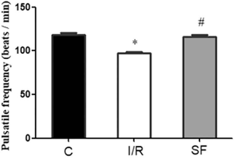
Comparison of beating frequency of myocardial cells in three groups. Compared with Group C, *p < 0.05; compared with Group I/R, #p < 0.05. C, control group; I/R, hypothermic hypoxia/reoxygenation injury group; SF, sevoflurane+hypothermic hypoxia/reoxygenation injury group.
The survival rate of cardiomyocytes
Compared with Group C, the survival rate of myocardial cells in Group I/R and Group SF was lower than that in Group C (p < 0.05), while the survival rate of myocardial cells in Group SF was higher than that in Group I/R (p < 0.05) (Fig. 3).
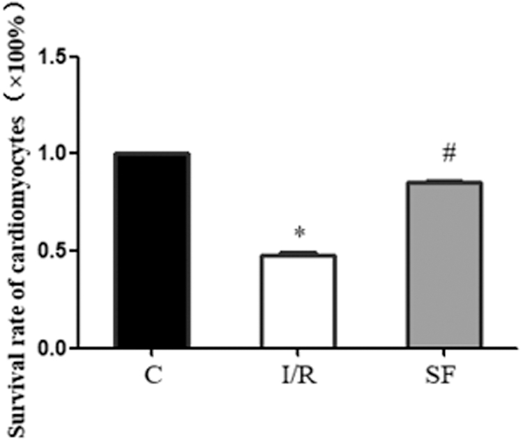
Comparison of myocardial cell survival rate among three groups. Compared with Group C, *p < 0.05; compared with Group I/R, #p < 0.05. C, control group; I/R, hypothermic hypoxia/reoxygenation injury group; SF, sevoflurane+hypothermic hypoxia/reoxygenation injury group.
Relative expression of Cx43 in cardiomyocytes
Compared with Group C, the relative content of Cx43 in Group I/R and Group SF was decreased (p < 0.05), and the relative content of Cx43 in Group SF was increased compared with Group I/R (p < 0.05) (Fig. 4A).
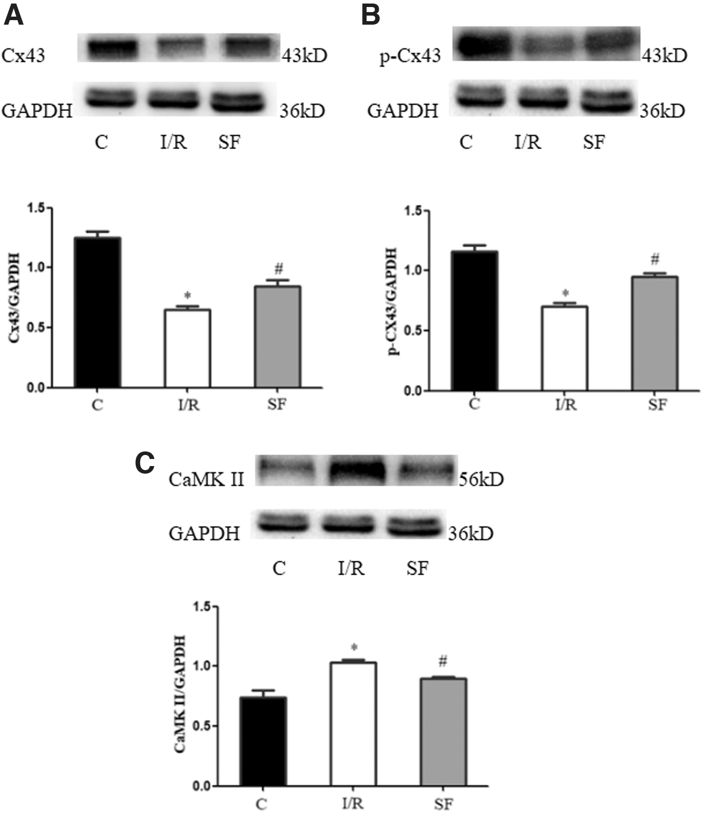
Relative expression of proteins in cardiomyocytes.
Relative expression of p-Cx43 in cardiomyocytes
Compared with Group C, the relative content of p-Cx43 protein decreased in Group I/R and Group SF (p < 0.05). The relative content of p-Cx43 protein in Group SF was higher than that in Group I/R (p < 0.05) (Fig. 4B).
Relative expression of CaMKII in cardiomyocytes
Compared with Group C, the relative content of CaMKII in Group I/R and Group SF increased (p < 0.05); compared with Group I/R, the relative content of CaMKII was decreased in Group SF (p < 0.05) (Fig. 4C).
Discussion
More and more attention has been paid to the role of cardiac fibroblasts in pathological changes such as arrhythmia and cardiac fibrosis (Pellma et al., 2016; Ma et al., 2018). It can affect cardiac electrophysiology through a variety of mechanisms, including the release of chemical mediators, extracellular matrix deposition, and direct electrical coupling with cardiomyocytes (Brown et al., 2005; Dostal et al., 2015; Chacar et al., 2017). Studies have confirmed that the medium derived from infarcted cardiac fibroblasts can significantly slow down the electrical conduction velocity of normal myocardial cells and shorten the action potential time (Vasquez et al., 2010). Hypoxia fibroblasts from the heart failure can reduce the beating frequency of cardiomyocytes in an arachidonic acid lipoxygenase-dependent manner by paracrine (Sandstedt et al., 2018).
In this study, we established a model of hypoxia/reoxygenation of fibroblasts, simulated the process of ischemia/reperfusion injury in vivo, excluded the direct effect of hypoxia/reoxygenation injury on cardiomyocytes, and verified the indirect effect of fibroblasts on cardiomyocytes in the process of hypoxia/reoxygenation injury. It is suggested that fibroblasts may participate in the process of myocardial injury by releasing chemical mediators.
Gap junction is an important structure of electrical conduction between fibroblasts and cardiomyocytes and adjacent cardiomyocytes (Michela et al., 2015; Tse and Yeo, 2015). Connexin is mainly composed of Cx43, which is mainly phosphorylated form works (Xue et al., 2019). Végh et al. (2013) showed that the increase of phosphorylation level of Cx43 can reduce the occurrence of arrhythmia. The change of Cx43 during myocardial ischemia/reperfusion injury can increase the risk of arrhythmia (Schulz et al., 2015; Zhou et al., 2020). Previous studies of our group also showed that the antiarrhythmic effect of custodiol HTK-Solution containing sevoflurane on hypothermic ischemia/reperfusion arrhythmia is related to the phosphorylation of Cx43 serine 368 (Li et al., 2018). Inhibiting the corresponding change of Cx43 is a new target for the treatment of myocardial ischemia/reperfusion injury (Boengler and Schulz, 2017).
CaMKII is a molecule that can cause arrhythmia (Wu et al., 2002). It has been found that the phosphorylation of serine at position 571 in Nav1.5 mediated by CaMKII can lead to the prolongation of monophasic action potential duration (MAPD), the increase of MAPD dispersion, and the increase of arrhythmia susceptibility after ischemia/reperfusion in isolated mouse heart (Howard et al. 2018). In addition, CaMKII overactivation can phosphorylate Ca2+ release channel, that is, RyR2,to increase abnormal Ca2+ release from sarcoplasmic reticulum, which further prolongs the duration of Ca2+ transient, while Ca2+-MAPD coupling prolongs MAPD (Neef et al., 2010; Park et al., 2014; Wei et al., 2017). Sevoflurane could alleviate reperfusion arrhythmia by ameliorating transmural dispersion of repolarization (TDR) and 90% time history of action potential repolarization (MAPD90) in isolated rat hearts after ischemia/reperfusion. Previous studies of our team also have shown that cold ischemia injury results in upregulation of CaMKII, prolongation of epicardial MAPD90, increase of TDR, and increase of fatal ventricular arrhythmias (Li et al., 2019).
Based on the previous research, the relative expression of Cx43, p-Cx43, and CaMKII in cardiac myocytes was detected in the culture medium of low-temperature hypoxia reoxygenation fibroblasts. It was found that the relative expression of Cx43 and p-Cx43 in the fibroblast culture medium of hypothermia hypoxia reoxygenation was decreased, and CaMKII was increased. While the relative expression of Cx43 and p-Cx43 was increased and CaMKII was decreased in cultured fibroblasts containing sevoflurane. It is suggested that sevoflurane can improve the expression of Cx43 and CaMKII in cardiomyocytes during global ischemia/reperfusion injury by affecting fibroblasts. Sevoflurane has anti-inflammatory effects, and reduces oxidative stress and mitochondrial damage (Yu et al., 2015). It is concluded that the effect of sevoflurane on fibroblasts is related to the inhibition of inflammatory reaction of fibroblasts after hypothermic hypoxia/reoxygenation injury.
However, there are still some limitations in this study. First, the specific mechanism of fibroblasts on cardiomyocytes needs further study; the possible media in this mechanism also need to be further extracted and verified. Second, in comparison with adult cardiomyocytes, neonatal cells have the advantage of being easily cultured; therefore, the mice we selected were 2–3 days old, which could not completely represent the age range of patients undergoing cardiopulmonary bypass. Finally, because the cardiomyocytes we used were normal cardiomyocytes, we did not distinguish the sex of suckling mice. Whether there are differences in the effects of different sex suckling mice on cardiomyocytes need to be further studied.
In conclusion, hypothermic hypoxia/reoxygenation fibroblast culture medium containing sevoflurane can improve cardiomyocyte activity and regulate the expression of Cx43, p-Cx43, and CaMKII in cardiomyocytes; the specific mechanism needs further study.
Footnotes
Author Disclosure Statement
No competing financial interests exist.
Funding Information
No funding was received for this article.