




Abstract
Lung injury occurring in the early stage of heat stroke (HS) leads to hypoxia and further aggravation of other organic damage. Lactoferrin (LF) is an iron binding protein with anti-inflammatory and antioxidant effects. This study focuses on the protection of preadministration of bovine lactoferrin (BLF) against lung injury in rats with HS. Sixty-four Sprague-Dawley male rats were divided into four groups randomly: control (CON)+phosphate-buffered saline (PBS) (n = 16), HS+PBS (n = 16), HS+low-dose BLF (LBLF) (n = 16), and HS+high-dose BLF (HBLF) (n = 16). CON+PBS and HS+PBS were preadministered 10 mL/kg PBS for 1 week. HS+LBLF and HS+HBLF were preadministered 100 and 200 mg/kg BLF for 1 week, respectively. The HS onset time and the survival rate were recorded, and bronchoalveolar lavage fluid was obtained to measure protein concentration. Lung was obtained for pathological analysis and wet/dry weight ratio measurement; later, the content of malondialdehyde (MDA), activity of myeloperoxidase (MPO), and superoxide dismutase (SOD) were measured in lung tissue homogenate. The results indicated that BLF preadministration could delay the HS onset time, enhance the survival rate, the levels of serum inflammatory cytokine and MDA content in HS+LBLF and HS+HBLF showed significant reduction compared with HS+PBS, while a significant elevation of SOD activity and reduction of MPO activity in HS+HBLF. Our results demonstrate that BLF preadministration could relieve lung injury in HS rats by enhancing thermal endurance, and alleviating serum inflammatory response and pulmonary oxidative stress damage.
Introduction
Heat stroke (HS) is a disease characterized by core body temperature >40 ± 0.5°C, central system lesions (delirium, convulsion, coma, etc.), systemic inflammation, and multiple organ dysfunction (Epstein and Yanovich, 2019). Even with aggressive treatment, 75% of the HS patients developed multiple organ degeneration, leading to high mortality (Varghese et al., 2005).
As the organ for gas exchange and heat dissipation of the body, the lung is one of the principal organs suffering damage after heat stress (Liu et al., 2020), of which lung injury will lead to hypoxia and further aggravation of other organic damage (Chang et al., 2013). However, the underlying mechanism on lung injury caused by HS remains unclear.
Lactoferrin (LF) is an 80 kDa iron-binding glycoprotein existing widely in mammalian tissues and body fluids, which has been proved to make an effect of protection against lung injury caused by sepsis and coronavirus infection (Han et al., 2019; Campione et al., 2020). LF is highly homologous among diverse species (Fernandes and Carter, 2017). In this study, we focused on the preservation of lung in HS rats by preadministration of bovine lactoferrin (BLF) to provide a prevention and treatment method of HS.
Materials and Methods
Animals and materials
Male Sprague-Dawley rats (weight, 250–300 g) obtained from Sippr B&K Laboratory Animal Ltd. (Shanghai, China) were all raised in the Specific Pathogen Free Animal Experiment Center of the Navy Medical University in China. BLF was purchased from Weideli Chemical Science and Technology Co., Ltd. (Hubei, China) (Purity: 98%). All experimental protocols were approved by the Animal Ethics Committee of Naval Medical University according to the Guide for the Care and Use of Laboratory Animals of the National Institutes of Health.
Animal grouping and BLF administration
The rats were raised at an ambient temperature of 24 ± 1°C and a humidity of 50 ± 5% with a 12-hour day/night circadian rhythm. Food and water were acquired ad libitum. A total of 64 rats were randomly divided into four groups as follows: control+phosphate-buffered saline (PBS) (CON+PBS) group, HS+PBS (HS+PBS) group, HS+low-dose BLF (LBLF) (HS+LBLF) group, and HS+high-dose BLF (HBLF) (HS+HBLF) group. Rats in CON+PBS and HS+PBS were administered intragastrically with 10 mL/kg PBS once a day for 1 week. Rats in HS+LBLF and HS+HBLF were administered intragastrically with 100 and 200 mg/kg BLF once a day for 1 week, respectively.
HS protocol
A constant temperature and humidity chamber was utilized to stabilize the experimental temperature at 40 ± 0.5°C and relative humidity at 60 ± 5%. Core temperature (Tc) of rats was monitored by a temperature capsule (Flamingo Technology Co., Ltd., Shenzhen, China) implanted in the abdomen. The HS protocol was referred to the previous one reported (Li et al., 2021). Rats in the HS groups were placed in the chamber, with Tc of rats being continuously monitored. A stable Tc of 42°C was conducted as the criterion for HS onset (Lin et al., 2020). Rats in the CON+PBS group were placed in a 25 ± 0.5°C, humidity 50 ± 5% environment for 1 hour without food and water.
Survival time investigation and sample collection
Rats that met the criterion of HS onset were transferred to a 25 ± 0.5°C, humidity 50 ± 5% environment for 3 hours with food and water, where the survival time of HS groups was recorded. Additional experiments were required if the number of alive rats was less than the minimum requirement of each group (n = 8). Eight rats in each group were anesthetized with isoflurane at 3 hours after HS onset. Blood samples were collected through the abdominal aorta, then centrifugation at 3000 rpm for 10 minutes at 4°C. The supernatant was collected and stored at −80°C.
The thoracic cavity of the rat was opened, with the main bronchus fully exposed, and then, the right main bronchus and right pulmonary vessels were ligated with sutures below the forked trachea. An incision was made in the lower one-third of the trachea, and a polyethylene tube was inserted into the left lung. Lavage was performed with 2 mL of cold PBS infused into the left lung three times, and each lavage was extracted three times. The bronchoalveolar lavage fluid (BALF) was obtained in centrifuge tubes (on ice), and then centrifugated at 1500 rpm for 10 minutes at 4°C.
The supernatant was collected and stored at −20°C for further measurement. The left lung was immediately immersed in 4% paraformaldehyde solution and treated with a previous method for pathological analysis (Li et al., 2021). The right upper leaf and right middle leaf were used to measure the wet/dry weight ratio. The right lower lobe and right posterior lobe were obtained to make lung tissue homogenate for further determination.
Measurement of inflammatory cytokine levels
Tumor necrosis factor-α (TNF-α), interleukin-1β (IL-1β), and IL-10 are common proinflammatory cytokines, whose levels were determined with enzyme-linked immunosorbent assay kits [Multi Sciences (Lianke) Biotech Co., Ltd., Hangzhou, China] by a previously reported method (Xia et al., 2017).
Measurement of lung wet/dry weight ratio and BALF protein concentration
The right upper leaf and right middle leaf were harvested and weighted, then placed in an oven at 65°C for 48 hours at constant weight. The dried leaves were weighted and the wet/dry weight ratio was calculated. The protein concentration of BALF was measured with a bicinchoninic acid protein assay kit (Jiancheng Bioengineering Institute, Nanjing, China) according to the instructions.
Measurement of myeloperoxidase activity
Myeloperoxidase (MPO) activity of lung tissue homogenate was determined with an MPO assay kit (Jiancheng Bioengineering Institute) according to Liu et al. (2010).
Measurement of lipid peroxidation level and superoxide dismutase activity
The level of lipid peroxidation was determined by measurement of malondialdehyde (MDA) content. The content of MDA and superoxide dismutase (SOD) activity in lung tissue homogenate were determined by the MDA kit (Jiancheng Bioengineering Institute) and SOD kit (Jiancheng Bioengineering Institute), respectively, according to Deng et al. (2013).
Pathological analysis and scoring
Eight regions of each lung section were randomly selected by a pathologist for pathological scores blindly, and scrutinized at × 200 magnification (Leica DM 2000, Wetzlar, Germany). Each region was scored from 0 to 3 based on factors of alveolar wall thickness, lung inflammatory infiltration, and congestion. The average score of each section was calculated, according to which statistical analysis was conducted.
Statistical analysis
All experimental data are described as mean ± standard deviation. GraphPad Prism (Version 8.3.0; GraphPad Software, La Jolla, CA) was used for statistical analysis and plotting. The survival rate was analyzed by the log-rank test. Data from multiple groups were processed with one-way analysis of variance test followed by the Tukey method. p-Value of 0.05 or less was considered to be statistically significant.
Results
BLF preadministration attenuated hyperthermia and reduced mortality
Rats were removed out of the chamber when the Tc reached 42°C. Compared with HS+PBS group, the duration of Tc reaching the onset of HS appeared significantly longer in HS+LBLF group and HS+HBLF group (57.50 ± 4.57 minutes vs. 69.25 ± 6.71 minutes vs. 90.13 ± 4.45 minutes, p < 0.001, n = 8) (Fig. 1). The number and survival time of death rats were recorded within 3 hours after modeling, with no significant difference in survival rate between HS+PBS group and HS+LBLF group (50.00% vs. 68.75%, p = 0.231), but a significant reduction in HS+HBLF group (50.00% vs. 87.50%, p = 0.014) (Fig. 1).

BLF preadministration attenuated hyperthermia and reduced mortality.
BLF did not affect wet/dry weight ratio of lung and protein concentration of BALF
Our research indicated no significant difference in lung wet/dry weight ratio between HS groups (4.79 ± 0.41 vs. 5.07 ± 0.44 vs. 4.88 ± 0.50 vs. 5.10 ± 0.20, p = 0.105) (Fig. 2). Consistently, no significant difference in BALF protein concentration (g/L) was founded (1.00 ± 0.18 vs. 1.23 ± 0.25 vs. 1.27 ± 0.29 vs. 1.06 ± 0.20, p = 0.074) (Fig. 2).
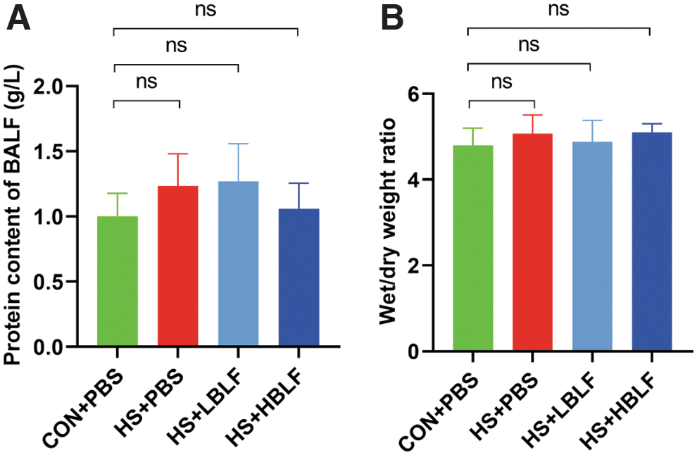
BLF did not affect the wet/dry weight ratio of lung and protein concentration of BALF.
BLF reduced levels of serum proinflammatory cytokines
The elevation of IL-1β (pg/mL), IL-6 (pg/mL), and TNF-α (pg/mL) in serum indicates the aggravation of inflammatory reaction. Inflammatory cytokines in HS+PBS group showed a significant increase compared with CON+PBS group (761.40 ± 56.66 vs. 351.50 ± 54.49, p < 0.01; 55.26 ± 1.67 vs. 26.09 ± 3.83, p < 0.01; 418.80 ± 21.14 vs. 207.90 ± 18.01, p < 0.01). While preadministration with LBLF and HBLF notably decreased the levels of IL-1β, IL-6, and TNF-α (614.30 ± 40.44 vs. 522.40 ± 54.17 vs. 761.40 ± 56.66, p < 0.001; 44.25 ± 3.21 vs. 40.54 ± 3.69 vs. 55.26 ± 1.67, p < 0.001; 356.50 ± 26.87 vs. 317.10 ± 25.97 vs. 418.80 ± 21.14, p < 0.001), as shown in Figure 3.
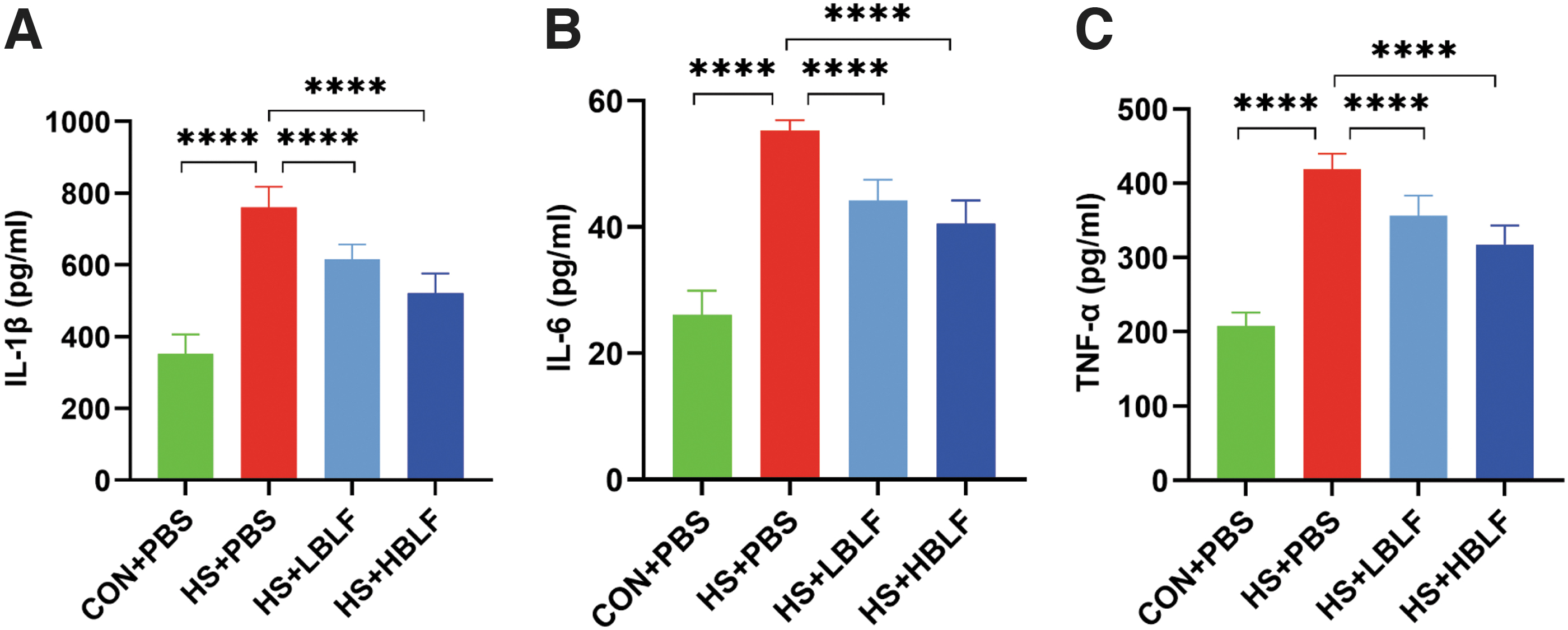
BLF reduced levels of serum proinflammatory cytokines. IL-1β (pg/mL)
BLF decreased pulmonary MPO activity
The MPO activity (U/g) in lung tissue homogenate of rats in HS+PBS group was significantly higher than that in CON+PBS group (12.66 ± 2.20 vs. 4.09 ± 0.82, p < 0.001). While no significant improvement was found in the HS group of HS+LBLF group (11.17 ± 1.58 vs. 12.66 ± 2.20, p = 0.21). The preadministration of HBLF significantly alleviated the MPO activity in the lungs of HS rats (6.49 ± 0.92 vs. 12.66 ± 2.20, p < 0.001) (Fig. 4).
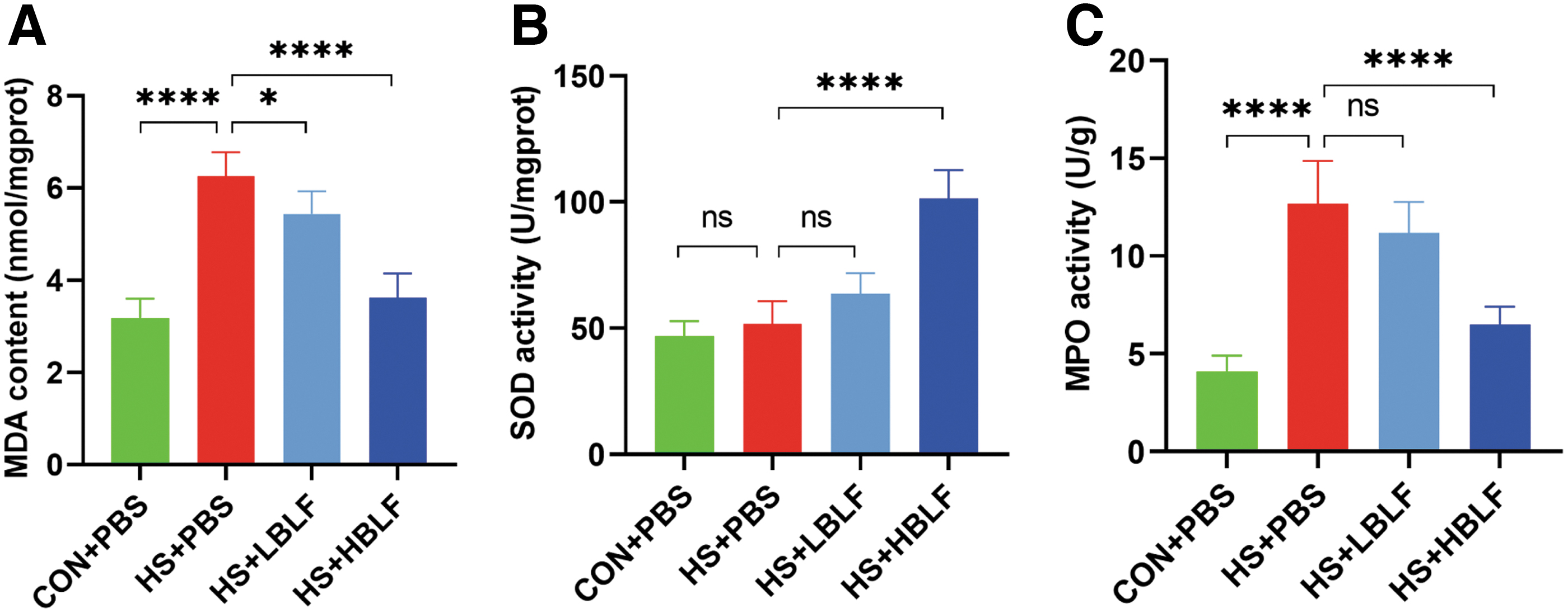
BLF increased SOD activity and reduced MPO activity and lipid peroxidation level of lung. MDA content
BLF increased SOD activity and reduced lipid peroxidation level of lung
No statistical difference in SOD activity (U/mgprot) between the CON+PBS and HS+PBS groups (51.59 ± 9.11 vs. 46.97 ± 5.87, p = 0.728). SOD activity in HS+LBLF group slightly increased compared with HS+PBS group with no statistically significant (63.66 ± 8.10 vs. 51.59 ± 9.11, p = 0.051), and was significantly higher in HS+HBLF group than that in the other three groups (101.30 ± 11.47, p < 0.001) (Fig. 4). MDA content (nmol/mgprot) of lung tissue homogenate in HS+PBS group appeared significantly higher than that in CON+PBS group (6.26 ± 0.52 vs. 3.18 ± 0.43, p < 0.001). Preadministration with LBLF and HBLF could significantly reduce the concentration of MDA (5.44 ± 0.49 vs. 3.62 ± 0.52 vs. 6.26 ± 0.52, p < 0.001), respectively (Fig. 4).
BLF attenuated pulmonary pathological injury
Lung biopsy of rats in HS+PBS group presented significant thickening of alveolar wall, severe infiltration of inflammatory cells in lung interstitium, and severe hyperemia in lung interstitium and alveolar cavity, which were effectively reversed by preadministration with BLF, with the pathological scores being reduced, as shown in Figure 5.
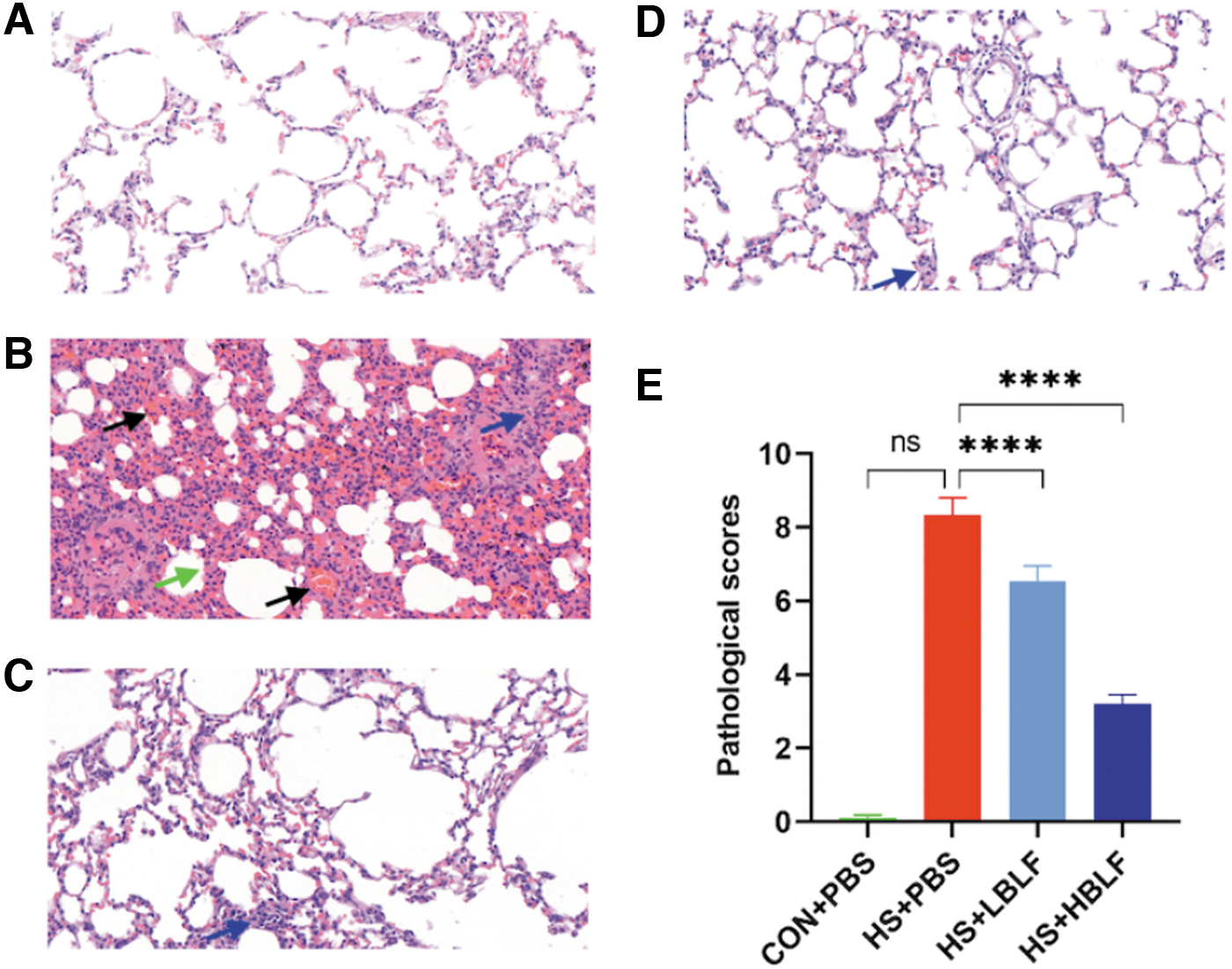
BLF attenuated pulmonary pathological injury. The left lung was harvested 3 hours after HS and stained with H&E. Representative pathological sections of CON+PBS group
Discussion
The incidence of HS shows a growing trend as reported (Leyk et al., 2019), the fatality of which remains high even after appropriate treatment. Lung is an important organ for heat dissipation, and the early protection of its function can effectively promote body heat dissipation while avoiding aggravating hypoxemia. That the heat stress can cause damage to lung vascular endothelial cell is the trigger factor of lung injury in HS (Al and Bouchama, 2018), of which the potential mechanism of injury remains unclear. LF can inhibit proinflammatory cytokines and promote the generation of anti-inflammatory cytokines (Kanwar et al., 2015). However, no studies have explored the protective effect of pre-oral LF on HS lung injury. In this study, the HS rat model was established after preadministration of PBS and BLF at low and high doses.
The preadministration of BLF significantly prolonged the time of Tc for rats to reach onset of HS, and the increase in oral concentration significantly improved the survival rate. In conclusion, both high and low doses of BLF can effectively enhance the thermal endurance of rats. However, the LBLF group did not show significant improvement in the survival rate of HS rats, while increasing the oral dose reversed this phenomenon.
BALF protein concentration can robustly reflect the severity of alveolar exudation. In this study, we compared the protein concentration of BALF in HS groups with CON+PBS groups, and found no significant difference between them, which may be related to the mild exudation 3 hours after HS occurred.
LF regulates cellular immunity at the cellular level through nuclear factor-kappa B and mitogen-activated protein kinase signaling pathway, decreasing the proinflammatory cytokines (Kanwar et al., 2015). HS promotes the leakage of gastrointestinal ischemia and endotoxin into the systemic circulation, aggravating the occurrence of lung injury (Selkirk et al., 2008; Tong et al., 2016). MPO is highly expressed in various inflammatory cells, the activity of which will be increased by the aggregation of inflammatory cells stimulated by related factors (Chen et al., 2020). In the present study, BLF preadministration could effectively inhibit the levels of IL-1β, IL-6, and TNF-α in the serum of HS rats. MPO activity in lung tissue homogenate was significantly increased after HS, suggesting the aggravation of inflammation, while BLF preadministration could reduce MPO activity. Our result showed that the remission of lung injury may be influenced by the remission of serum inflammation.
Heat shock protein can alleviate oxidative stress in the lung, so as to relieve lung injury (Chang et al., 2013). Multiple oxidative stress-related genes (Aqp3, Cygb, SOD2, Hmox1) were confirmed in lung injury caused by HS (Liu et al., 2020). SOD2, also known as manganese SOD, is a metal enzyme widely existing in organisms, which catalyzes the decomposition of superoxide ions into H2O2 and oxygen in mitochondria, removing superoxide ion free radicals generated by the mitochondrial respiratory chain electron transport system to reduce oxidative stress damage (Wang et al., 2018). MDA is the terminal product of lipid peroxidation, the concentration of which can reflect the severity of lipid peroxidation.
LF exerts antioxidant activity by scavenging nitric oxide and 1,1-diphenyl-2-trinitrophenylhydrazine free radicals (Habib et al., 2013). Oral administration of LF can significantly elevate the activity of SOD (Farid et al., 2021). Our experiment suggested that SOD in lung tissue homogenate of rats with BLF preadministration increased significantly and the MDA content decreased after HS. According to previous reports (Minzhou Zhang et al., 2017; Snipe et al., 2018; Lin et al., 2019), we hypothesize that multiple factors are involved in lung injury after HS (hyperthermal cytotoxicity, disseminated intravascular coagulation, intestinal endotoxemia, oxidative stress injury, etc.), evoking the occurrence of acute lung injury and even acute respiratory distress symptoms.
Conclusions
Our study demonstrated that BLF preadministration could relieve lung injury in rats with HS by enhancing thermal endurance and alleviating serum inflammatory response and pulmonary oxidative stress damage. It suggests that LF preoral may play an effective role in the prevention and treatment of HS. In addition, we found that BLF also had a special protection on other organs of HS. Further studies will be designed to explore the protective effect and specific mechanism of BLF on HS.
Availability of Data and Material
The data sets analyzed during the current study are available in the ScienceDB repository (https://www.scidb.cn/en/s/feyaIj).
Footnotes
Acknowledgment
The authors sincerely thank Yang Guang for English language revisions.
Author Disclosure Statement
No competing financial interests exist.
Funding Information
This study was supported by the Shanghai Military Civilian Integration Industry Development Project (Grant No. 2019-jmrh1-kj52) and National Science Foundation of China (grant 81570073 to M. T.).