




Abstract
The aim of this study is to address the limited research on skin frostbite models and the gaps in pathological identification of time-series injuries in frostbitten skin, which hinder comprehensive assessment of injury severity. A deep second-degree frostbite model was developed in BALB/c nude mice, and staining identification was performed at various stages, from the onset of frostbite to the healing process. Continuous observations at multiple time points provided a more accurate and comprehensive standard for comparison in frostbite treatment experiments. A deep second-degree frostbite model was developed using BALB/c nude mice. Histopathological examination was performed with hematoxylin-eosin (HE) staining, while Masson’s Trichrome (MT) staining was used to observe collagen recovery. Additionally, immunofluorescence staining was conducted to analyze epidermal cells and dermal structures. A deep second-degree frostbite model was successfully developed in BALB/c nude mice. Histopathological characteristics of mouse skin tissue were examined through HE staining at various time points. MT staining highlighted changes in the morphology and thickness of the original fibers. Immunofluorescence staining offered a detailed evaluation of the damage and recovery of appendages, including hair follicles and sweat glands. The deep second-degree frostbite model in BALB/c nude mice establishes a standard for studying skin frostbite injuries and developing related treatments.
Introduction
Frostbite occurs when ice crystals form in intercellular spaces and within cells due to a drop in external temperature or prolonged exposure to cold, damp environments, resulting in endothelial damage to blood vessels (Hu and Zou, 2022). In recent decades, globally, there has been an increase in the frequency and intensity of extreme temperature events, including heatwaves and cold snaps (Xie et al., 2024). Epidemiological studies indicate that the incidence of frostbite has risen, particularly in cold northern regions and among military personnel (Van et al., 2025). As a prevalent winter condition among soldiers, frostbite manifests in forms such as “trench foot,” “peripheral frostbite,” and “chilblains” (Turner et al., 2025). Advancing research on frostbite treatment is essential to safeguard the health of residents in low-temperature environments and soldiers stationed under extreme conditions.
Second-degree frostbite is the most common type of skin frostbite, characterized by blister formation that affects the entire skin layer, accompanied by significant pain. Within 2–4 days of frostbite occurrence, skin symptoms worsen, marked by an increase in inflammatory cells, leading to epidermal crusting, erythema, swelling, and pain. Prompt implementation of effective early treatment measures is critical (Raleigh et al., 2022). Research on frostbite treatment spans several areas, including drug development, physical therapy, and cell therapy, with particular focus on treating common second-degree frostbite (Tu et al., 2020; Jellestad and Holm, 2023; Zhang et al., 2023). Among these, cell therapy has shown distinct advantages in promoting wound healing by reducing cell apoptosis, enhancing angiogenesis, and accelerating collagen synthesis. In recent years, its application in frostbite treatment has garnered increased attention. Thus, establishing animal models of frostbite that reflect clinical phenotypes is essential for studying the mechanisms of frostbite and improving clinical diagnosis and treatment. Currently, frostbite animal models are primarily based on larger organisms such as rats and rabbits, but there are no reports of immune-deficient mouse models for skin frostbite (Jiao et al., 2016; Shen et al., 2016; Rothenberger et al., 2014; Sun et al., 2020). Researchers in this study established a deep second-degree frostbite model in immune-deficient BALB/c nude mice by refining the freezing treatment method, thereby creating a reliable animal model for cellular therapy research in frostbitten skin. Additionally, staining methods for identifying frostbite skin tissues were optimized, and continuous sampling at multiple time points enabled detailed analysis of physiological and pathological changes at different stages of frostbite in mouse skin. The findings from these studies not only improve the accuracy of frostbite skin identification but also provide valuable reference data for frostbite treatment research.
Materials and Methods
Experimental animals
The animal experiments in this study were approved by the Institutional Animal Care and Use Committee of the National Center for Protein Science (Ethical Approval Number: NCPSB-20230419-23MO), and all experiments were conducted at this facility. A total of 25 male BALB/c nude mice (purchased from Beijing Vital River Laboratory Animal Technology Co., Ltd.), aged 6–8 weeks and weighing 17–23 g, were used. The mice were housed in groups of five per cage and acclimated in a stable environment for 5 days prior to the experiment.
Mice were randomly assigned to five groups, with five mice in each group: the control group, day 1 post-frostbite (D1 group), day 3 post-frostbite (D3 group), day 7 post-frostbite (D7 group), and day 14 post-frostbite (D14 group). At the respective time points, mice were euthanized, and skin tissues were collected for fixation.
Major reagents
The main reagents used in the experiment included 1.25% ready-to-use tribromoethanol solution (Aibei Biotech), Carprofen (Selleck), Harris Hematoxylin Staining Solution (Biotopped), Modified Masson Trichrome Staining Kit (SolaiBio), CEA (Abcam), and Ck14 (Abcam).
Equipment
The primary instruments and equipment used in the study included a paraffin embedding station (Thermo Fisher Scientific), a paraffin microtome (Thermo Fisher Scientific), a refrigerator (Haier Group), a digital slide scanning system (Hamamatsu Photonics K.K.), and a super-resolution laser scanning confocal microscope imaging system (Nikon Corporation).
Methods
Establishment of frostbite model
BALB/c nude mice were anesthetized via intraperitoneal injection of tribromoethanol solution (Aibei Biotech, 0.2 mL/10 g). Post-anesthesia, the back skin of the mice was disinfected with 75% ethanol. A circular paper disc with a diameter of 2.5 mm was placed on the back skin, and the area was marked with an animal-safe marking pen. The skin on both sides (head and tail) of the marked area was gently grasped to divide the region into two semicircular sections. Using forceps, two thick copper discs that had been placed in dry ice (−78°C) for 15 minutes were applied to the marked areas, ensuring the copper discs adhered tightly to the skin without applying excessive pressure. The discs were manually held in place to maintain contact. This study adopted a cold-heat-cold alternating frostbite method (Auerbach et al., 2013). The mouse skin was frozen for 1 minute with the copper discs, removed, and rewarmed for 3 minutes. The procedure was repeated by replacing the copper discs, freezing for another minute, and rewarmed for 3 minutes. This cycle was repeated three times. During the frostbite procedure, a constant temperature heating pad was used to prevent a decrease in the core body temperature of the mice. After the frostbite procedure, carprofen (Selleck, 5 mg/kg) was administered subcutaneously for pain relief, and the mice were monitored before being returned to their cages for continued breeding. For the control group, the same procedures of anesthesia, skin disinfection, and skin marking were followed. Instead of the frostbite procedure, a sham frostbite operation was performed using thick copper discs at room temperature, followed by carprofen injection. Post-procedure, the mice were observed and then returned to their cages for continued breeding.
Tissue embedding and paraffin sectioning
Five mice were selected from each group: control, D1, D3, D7, and D14. These mice were euthanized using a mixture of carbon dioxide gas. They were placed in a sealed container and exposed to the carbon dioxide mixture until cessation of movement and respiration. The gas supply was then turned off, and the mice were observed for an additional two minutes to confirm fatality. The skin from the frostbite area on the back of the mice was excised, with a portion placed in 4% paraformaldehyde solution for fixation for approximately 48 hours. After 48 hours, the skin tissue was transferred to an embedding cassette and immersed in 75% ethanol overnight (approximately 12 hours). The following day, the tissue was moved to 80% ethanol for 60 minutes, followed by immersion in 90% ethanol, 95% ethanol, and 100% ethanol for 15 minutes each. The tissue was then placed in 100% ethanol for 45 minutes, followed by immersion in xylene for 30 minutes. Finally, the tissue was transferred to the paraffin cylinder for two immersion cycles, each lasting 30 minutes. After the final paraffin immersion, the skin was removed and a small amount of paraffin was added to the mold. The skin was positioned with the cross-section facing down in the mold and placed on the cold point of the embedding machine to solidify the paraffin while keeping the tissue upright. A significant amount of paraffin was then added, and the mold was placed on a cold plate to allow the paraffin to solidify, completing the embedding process. The embedded blocks were then sectioned into slices, each 4 μm thick, and placed in a 60°C oven for more than 6 hours.
Hematoxylin-eosin staining
After the sections were deparaffinized, they were immersed in distilled water for 5 minutes, stained with hematoxylin for 3 minutes, and then rinsed in water to stop the staining process. The sections were briefly differentiated in hydrochloric acid and then rinsed under running water for 20 minutes. Next, the sections underwent dehydration in 80% ethanol, 90% ethanol, and 95% ethanol, each for 5 minutes, followed by staining with eosin for 1 minute. The sections were then further dehydrated in 100% ethanol and cleared in xylene. Finally, they were mounted using a resin mounting medium. Once the resin had solidified, the sections were scanned with a digital slide scanning system to save the experimental results.
Masson’s Trichrome staining
Following the protocol outlined in the Modified Masson Trichrome Staining Kit (SolaiBio, Catalog No.: G1346), tissue sections were deparaffinized and then immersed in distilled water for 3 minutes prior to staining. Histology pens were used to outline the tissue, and the mordant solution was applied to the sections. The sections were incubated in a 60°C oven for 1 hour for mordanting, followed by a 10-minute wash under running water. The sections were then stained with Celestine Blue solution for 3 minutes, followed by two washes with water, each lasting 15 seconds. Mayer’s hematoxylin staining solution was applied for 3 minutes, followed by two additional 15-second washes with water. The sections were differentiated in an acidic differentiation solution for several seconds, washed with water to halt differentiation, and then rinsed under running water for 10 minutes. The sections were stained with Ponceau Fuchsin solution for 10 minutes, followed by two 15-second washes with water. Phosphomolybdic acid solution was applied for approximately 10 minutes, after which the excess solution was discarded. Without washing the sections, the toluidine blue staining solution was applied directly for 5 minutes. After rinsing off the toluidine blue solution with a weak acid solution, the sections were treated with the weak acid solution for an additional 2 minutes. The sections were then dehydrated in 95% ethanol for 30 seconds, followed by two further dehydration steps in absolute ethanol: the first for 30 seconds and the second for 1 minute. The sections were cleared in xylene twice, each for 1–2 minutes, and then mounted using a resin mounting medium. Once the resin mounting medium had solidified, the sections were scanned using a digital slide scanning system to document the experimental results.
Immunofluorescence staining
The sections were deparaffinized and placed in distilled water for 5 minutes. They were then incubated in an antigen retrieval solution for tissue repair and allowed to cool naturally to room temperature after the repair process. The sections were washed three times in phosphate buffered saline (PBS) buffer solution, each for 3 minutes. The tissue on the sections was outlined using a histology pen, and goat serum blocking solution was applied to cover the tissue, followed by incubation at room temperature for 1 hour. After the blocking step, the blocking solution was discarded, and the primary antibody, diluted in goat serum blocking solution, was applied to cover the tissue and incubated overnight at 4°C.
Post-incubation overnight, the sections were allowed to reach room temperature and washed three times with PBS buffer solution, each for 3 minutes. The appropriate secondary antibodies, diluted in PBS buffer solution and protected from light, were added to the sections, which were then incubated at room temperature for 1 hour in the dark. Post-incubation, the sections were washed again with PBS buffer solution. 4′,6-diamidino-2-phenylindole was subsequently added to stain the nuclei for 5 minutes. After another wash, an antifade mounting medium was applied, followed by the placement of a coverslip. The edges were sealed with nail polish, and the sections were left to dry. Once the nail polish had dried, the sections were imaged using a confocal imaging microscopy system.
Results
Establishment of a deep second-degree frostbite model in mouse skin
In this study, a deep second-degree frostbite model in mouse skin using a modified cold-heat-cold alternating frostbite method was successfully established, which improved the contact between the frozen copper discs and the mouse skin. The skin conditions of normal mice and those at 1-, 3-, 7-, and 14-days post-frostbite were observed, and changes in the frostbitten skin were recorded. One day post-frostbite, the skin on the back of the mice appeared grayish-white, as shown in Figure 1. Three days post-frostbite, red eschars appeared on the skin surface, and the skin lost its elasticity. Seven days post-frostbite, large areas of eschars formed on the skin, and pus appeared subcutaneously, completely covering the skin surface. Fourteen days post-frostbite, eschars remained on the skin surface, with edges showing signs of imminent sloughing, and little pus remained subcutaneously.
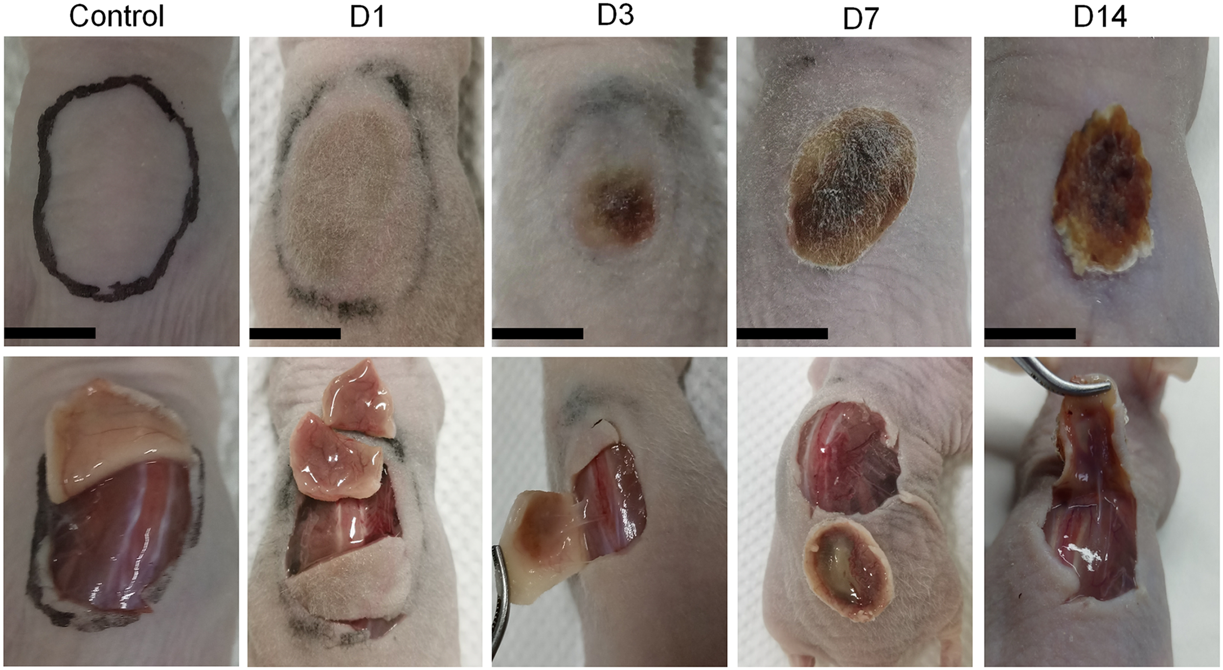
Observation of the skin tissues derived from deep second-degree frostbite model of BALB/c Nude mice.
Histopathological characteristics of deep second-degree frostbite model in mouse skin
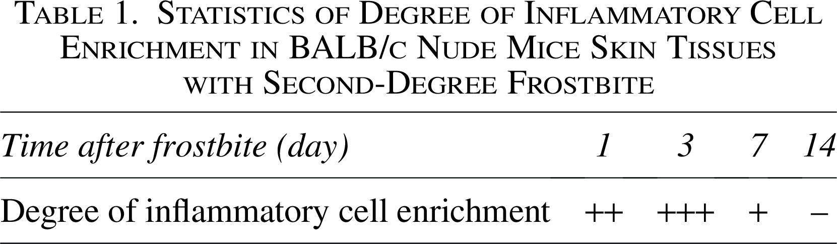
To provide a comprehensive assessment of the histopathological features of mouse skin following frostbite, HE staining was performed on frostbitten skin at 1-, 3-, 7-, and 14-days post-injury. One day post-frostbite, the epidermal cells in the mouse skin were lost, some adipocytes were ruptured, and neutrophils were present in the dermis, as shown in Figure 2. Three days post-frostbite, some adipocytes remained ruptured, and both neutrophils and lymphocytes were observed in the dermis, with additional inflammatory cells present in the muscle layer. Seven days post-frostbite, the adipocytes were ruptured, and numerous neutrophils and lymphocytes were visible in the dermis, along with partial rupture of blood vessels and proliferation of new cells in the subcutaneous tissue. Fourteen days post-frostbite, the original dermal tissue was completely necrotic, and newly proliferated cells began to re-differentiate, forming skin structures such as hair follicles and other appendages. Random fields from the HE-stained images were selected, and inflammatory cells were counted based on nuclear morphology. The number of inflammatory cells in the dermis was highest at 3 days post-frostbite, accompanied by extensive infiltration of inflammatory cells, as shown in Figure 3. At 14 days post-frostbite, the original dermal layer of the mouse skin was completely necrotic, with nearly no inflammatory cells detected in the newly formed tissue. The qualitative statistical analysis of inflammatory cells in the mouse skin at the four time points is detailed in Table 1.

HE staining of BALB/c Nude mice skin tissues from normal controls and deep second-degree frostbite model groups (bar: 500 μm, 100 μm). HE, hematoxylin-eosin.
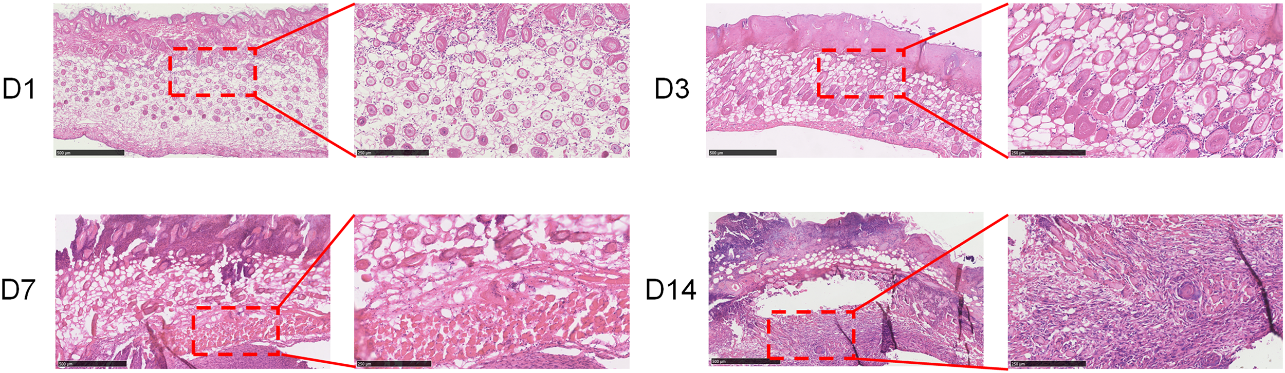
Qualitative comparison of inflammatory cell in BALB/c Nude mice skin tissues with second-degree frostbite based on HE staining (bar: 500 μm, 250 μm). HE, hematoxylin-eosin.
Statistics of Degree of Inflammatory Cell Enrichment in BALB/c Nude Mice Skin Tissues with Second-Degree Frostbite
Observation of collagen and muscle fibers in deep second-degree frostbite model using Masson staining
The status of collagen and muscle fibers in skin tissue serves as a crucial indicator for assessing whether the skin retains its basic functions, with collagen fibers playing a significant role in the generation of new cells. The damage and recovery of collagen fibers in the frostbitten skin of mice using Masson staining was observed next. One day post-frostbite, the collagen fibers in the mouse skin were disrupted, while the muscle fibers temporarily remained intact, with no significant change in the thickness of the collagen fiber layer, as shown in Figure 4. Three days post-frostbite, the collagen fibers were disrupted, and the morphology of the muscle fibers changed, accompanied by a noticeable reduction in the thickness of the collagen fiber layer. Seven days post-frostbite, the collagen fibers in the mouse skin were broken, and the muscle fibers were extensively ruptured, compromising their structural integrity. The collagen fiber layer was the thinnest at this time point. Fourteen days after the frostbite procedure, the original dermal layer of the mouse skin had undergone complete necrosis, with the collagen fibers entirely absent. New tissue cells began to produce collagen fibers.
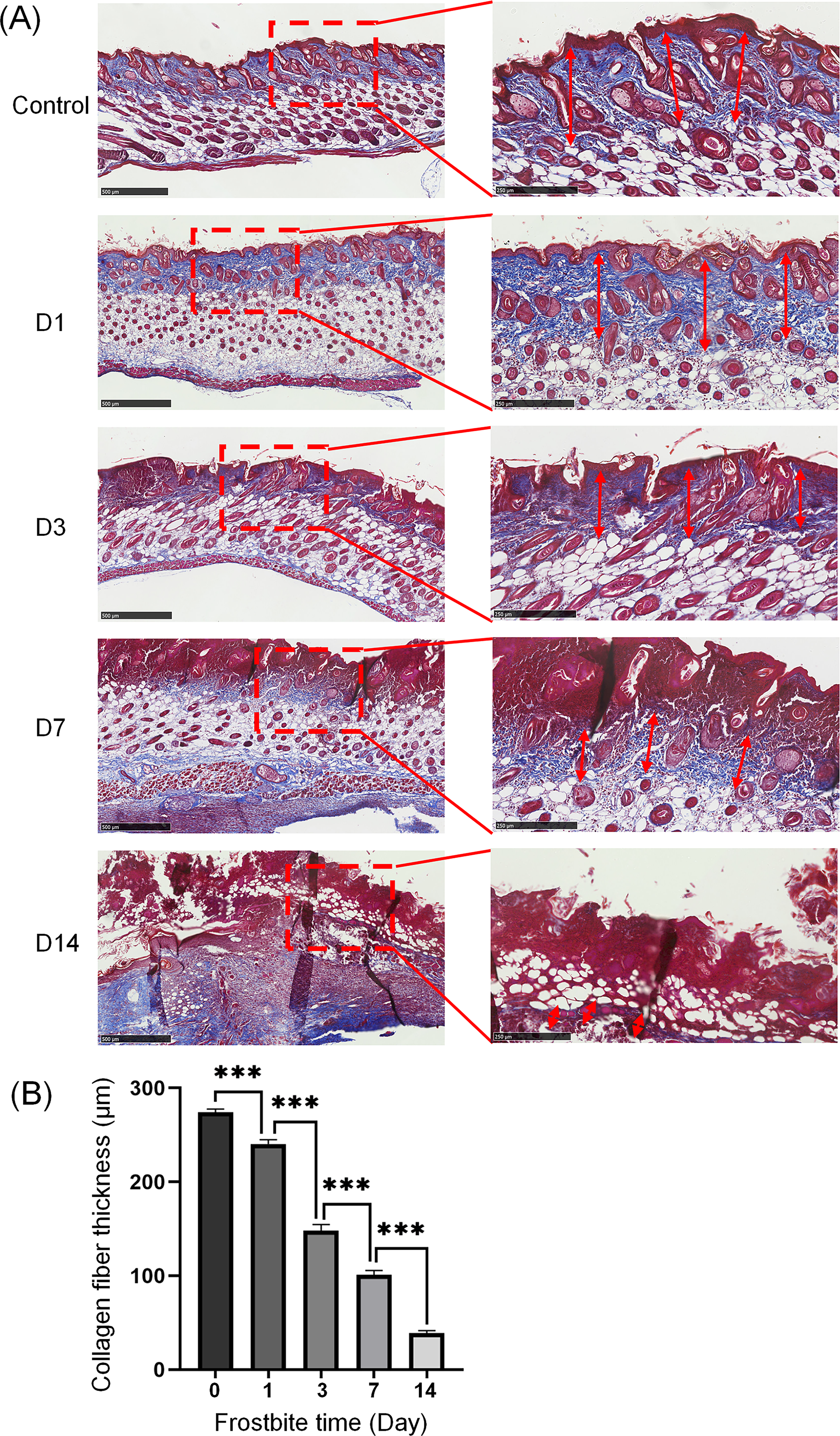
Masson staining of BALB/c Nude mice skin tissues from normal controls and deep second-degree frostbite model groups (bar: 500 μm, 250μm).
Immunofluorescence staining of deep second-degree frostbite model in mouse skin
Although BALB/c nude mice exhibit minimal hair growth on their skin surface, they still possess hair follicles within the skin, with sebaceous glands accompanying the growth of hair follicles in the dermis. Immunofluorescence staining on mouse skin tissue using antibodies against the epidermal cell marker CK14 and the glandular marker CEA was performed to observe the growth of skin appendages. Seven days post-frostbite, the original dermal layer of the mouse skin was completely necrotic, as shown in Figure 5. Fourteen days post-frostbite, newly proliferated epidermal cells, hair follicles, and sebaceous glands were observed in the mouse skin tissue. Their morphology was clearly visible under a 40× microscope.

Immunofluorescence staining of BALB/c Nude mice skin tissues from normal controls and deep second-degree frostbite model groups (bar: 100 μm).
Discussion
Clinically, the treatment of tissue frostbite primarily involves the use of medications for rewarming, anti-inflammatory effects, infection prevention, anticoagulation, thrombolysis, and circulation improvement (Musi et al., 2022; Crooks et al., 2022; Liskutin et al., 2020; Drinane et al., 2022; Ta et al., 2000; Swartz, 2020). Additionally, methods that promote angiogenesis and collagen synthesis play a crucial role in accelerating the recovery of frostbitten tissues (Öner et al., 2023; Wang et al., 2022; Zhang et al., 2023). Studies have shown that, compared with traditional treatments, cell therapy has a more beneficial effect on promoting angiogenesis and collagen synthesis during the recovery process of frostbite (Isozaki et al., 2022; Li et al., 2024). The use of combined therapies for frostbite has increased in recent years, with the application of cell therapy alongside drug therapy emerging as one of the most promising strategies for treatment (Volkova et al., 2023; Sun et al., 2022). A skin frostbite model using immune-deficient mice, reducing interference from immune cells and avoiding rejection reactions, has been established in this study. This provides an ideal animal model for studying cell therapy from various sources for frostbite, allowing the research to focus on the positive impacts of cell therapy.
Most existing frostbite models and treatments have been developed using animals such as rats and rabbits, with fewer reports on models established using mice. Mice offer distinct advantages over other animals, including lower costs and greater ease of manipulation in experiments. BALB/c nude mice, a commonly used immunodeficient model, not only facilitate research on cell therapy but also eliminate the need for depilation during frostbite modeling, thereby minimizing potential skin damage that could occur during depilation. However, the skin of BALB/c nude mice is more fragile than that of regular mice. In this study, a frostbite model using 5-mm-thick copper discs, replacing the commonly used magnets, has been approved. The copper discs were applied to the mouse skin with forceps for frostbite modeling. The use of copper discs ensures a consistent low temperature during the injury process and prevents pressure damage from the magnetic force of magnets. This modification significantly reduces non-freezing pressure injuries, protects subcutaneous blood vessels, and eliminates other potential factors affecting the frostbite experiment.
In conclusion, this study successfully developed a deep second-degree frostbite model in the skin of BALB/c nude mice using the cold-heat-cold alternating frostbite method. By replacing magnets with thick copper discs and refining the method for applying the frozen copper discs to the skin, the model was effectively established. Additionally, through the use of immunohistochemistry, MT staining, and immunofluorescence staining, the pathological features, collagen fiber structure and thickness, as well as the damage and recovery of skin appendages, including hair follicles and sweat glands, at various time points in the frostbite model were thoroughly assessed. This approach sets a standard for future research on frostbite injury and potential treatments.
Authors’ Contributions
Conception and design of the research: T.L., Y.Z., and J.M. Acquisition of data: T.L., W.W., X.L., and M.H. Analysis and interpretation of the data: T.L., D.G., W.W., X.L., and M.H. Statistical analysis: D.G. and Y.Z. Obtaining financing: Y.Z. and J.M. Writing of the article: T.L. Critical revision of the article for intellectual content: D.G., Y.Z., and J.M. All authors read and approved the final draft.
Footnotes
Acknowledgments
The authors would like to acknowledge the hard and dedicated work of all the staff who implemented the intervention and evaluation components of the study.
Author Disclosure Statement
The authors declare that they have no competing interests.
Funding Information
This work was supported by National Natural Science Foundation of China (No. 32270697) and National Key Research and Development Program of China (No. 2021YFA1301603), and the Open Fund of State Key Laboratory of Medical Proteomics (SKLP-O202204).
Ethics Approval and Consent to Participate
The experiment was approved by the Animal Ethics Committee of Beijing Institute of Lifeomics (No. NCPSB-20230419-23MO). Animal models of disease are subject to the 3Rs of Responsible Research, that is: Replace animal experiments with alternatives where possible, Reduce the number of animals used to a scientifically justified minimum, and Refine the procedure to minimize animal harm.
Availability of Data and Materials
The datasets used and/or analyzed during the current study available from the corresponding author on reasonable request.