
Abstract
Background:
Thyroid nodules are a common clinical problem, and differentiated thyroid cancer is becoming increasingly prevalent. Since the publication of the American Thyroid Association's guidelines for the management of these disorders was published in 2006, a large amount of new information has become available, prompting a revision of the guidelines.
Methods:
Relevant articles through December 2008 were reviewed by the task force and categorized by topic and level of evidence according to a modified schema used by the United States Preventative Services Task Force.
Results:
The revised guidelines for the management of thyroid nodules include recommendations regarding initial evaluation, clinical and ultrasound criteria for fine-needle aspiration biopsy, interpretation of fine-needle aspiration biopsy results, and management of benign thyroid nodules. Recommendations regarding the initial management of thyroid cancer include those relating to optimal surgical management, radioiodine remnant ablation, and suppression therapy using levothyroxine. Recommendations related to long-term management of differentiated thyroid cancer include those related to surveillance for recurrent disease using ultrasound and serum thyroglobulin as well as those related to management of recurrent and metastatic disease.
Conclusions:
We created evidence-based recommendations in response to our appointment as an independent task force by the American Thyroid Association to assist in the clinical management of patients with thyroid nodules and differentiated thyroid cancer. They represent, in our opinion, contemporary optimal care for patients with these disorders.
In 1996, the American Thyroid Association (ATA) published treatment guidelines for patients with thyroid nodules and DTC (10). Over the last decade, there have been many advances in the diagnosis and therapy of both thyroid nodules and DTC. Controversy exists in many areas, including the most cost-effective approach in the diagnostic evaluation of a thyroid nodule, the extent of surgery for small thyroid cancers, the use of radioactive iodine to ablate remnant tissue following thyroidectomy, the appropriate use of thyroxine suppression therapy, and the role of human recombinant thyrotropin (rhTSH). In recognition of the changes that have taken place in the overall management of these clinically important problems, the ATA appointed a task force to re-examine the current strategies that are used to diagnose and treat thyroid nodules and DTC, and to develop clinical guidelines using principles of evidence-based medicine. Members of the taskforce included experts in thyroid nodule and thyroid cancer management with representation from the fields of endocrinology, surgery, and nuclear medicine. The medical opinions expressed here are those of the authors; none were dictated by the ATA. The final document was approved by the ATA Board of Directors and endorsed (in alphabetical order) by the American Association of Clinical Endocrinologists (AACE), American College of Endocrinology, British Association of Head and Neck Oncologists (BAHNO), The Endocrine Society, European Association for Cranio-Maxillo-Facial Surgery (EACMFS), European Association of Nuclear Medicine (EANM), European Society of Endocrine Surgeons (ESES), European Society for Paediatric Endocrinology (ESPE), International Association of Endocrine Surgeons (IAES), and Latin American Thyroid Society (LATS).
Other groups have previously developed guidelines, including the American Association of Clinical Endocrinologists and the American Association of Endocrine Surgeons (11), the British Thyroid Association and The Royal College of Physicians (12), and the National Comprehensive Cancer Network (13) that have provided somewhat conflicting recommendations due to the lack of high quality evidence from randomized controlled trials. The European Thyroid Association has published consensus guidelines for the management of DTC (14). The European Association of Nuclear Medicine has also recently published consensus guidelines for radioiodine (RAI) therapy of DTC (15).
The ATA guidelines taskforce used a strategy similar to that employed by the National Institutes of Health for its Consensus Development Conferences (
What is the appropriate evaluation of clinically or incidentally discovered thyroid nodule(s)? What laboratory tests and imaging modalities are indicated? What is the role of fine-needle aspiration (FNA)? What is the best method of long-term follow up of patients with thyroid nodules? What is the role of medical therapy of patients with benign thyroid nodules? How should thyroid nodules in children and pregnant women be managed?
What is the role of preoperative staging with diagnostic imaging and laboratory tests? What is the appropriate operation for indeterminate thyroid nodules and DTC? What is the role of postoperative staging systems and which should be used? What is the role of postoperative RAI remnant ablation? What is the role of thyrotropin (TSH) suppression therapy? Is there a role for adjunctive external beam irradiation or chemotherapy?
What are the appropriate features of long-term management? What is the role of serum thyroglobulin (Tg) assays? What is the role of US and other imaging techniques during follow-up? What is the role of TSH suppression in long-term follow-up? What is the most appropriate management of patients with metastatic disease? How should Tg-positive, scan-negative patients be managed? What is the role of external radiation therapy? What is the role of chemotherapy?
The initial ATA guidelines were published in 2006 (16). Because of the rapid growth of the literature on this topic, plans for revising the guidelines within 24–36 months of publication were made at the inception of the project. Relevant articles on thyroid cancer were identified using the same search criteria employed for the original guidelines (16). Individual task force members submitted suggestions for clarification of prior recommendations, as well as new information derived from studies published since 2004. Relevant literature continued to be reviewed through December 2008. To begin the revision process, a half-day meeting was held on June 2, 2007. The Task Force was broadened to include European experts and a head and neck surgeon. Three subsequent half-day meetings were held on October 5, 2007; July 13, 2008; and October 5, 2008, to review these suggestions and for additional comments to be considered. The meeting in July 2008 also included a meeting with six additional surgeons in an effort to produce guidelines related to central neck dissection that would be as authoritative as possible. The organization of management guideline recommendations is shown in Table 1. It was agreed to continue to categorize the published data and strength of recommendations using a modified schema proposed by the U.S. Preventive Services Task Force (17) (Table 2).
If viewing these guidelines on the Web, or in a File, copy the Location Key to the Find or Search Function to navigate rapidly to the desired section.
R, recommendation; T, table; F, figure.
Adapted from the U.S. Preventive Services Task Force, Agency for Healthcare Research and Quality (17).
[A1] Thyroid Nodule Guidelines
A thyroid nodule is a discrete lesion within the thyroid gland that is radiologically distinct from the surrounding thyroid parenchyma. Some palpable lesions may not correspond to distinct radiologic abnormalities (18). Such abnormalities do not meet the strict definition for thyroid nodules. Nonpalpable nodules detected on US or other anatomic imaging studies are termed incidentally discovered nodules or “incidentalomas.” Nonpalpable nodules have the same risk of malignancy as palpable nodules with the same size (19). Generally, only nodules >1 cm should be evaluated, since they have a greater potential to be clinically significant cancers. Occasionally, there may be nodules <1 cm that require evaluation because of suspicious US findings, associated lymphadenopathy, a history of head and neck irradiation, or a history of thyroid cancer in one or more first-degree relatives. However, some nodules <1 cm lack these warning signs yet eventually cause morbidity and mortality. These are rare and, given unfavorable cost/benefit considerations, attempts to diagnose and treat all small thyroid cancers in an effort to prevent these rare outcomes would likely cause more harm than good. Approximately 1–2% of people undergoing 2-deoxy-2[18F]fluoro-
[A2] What is the appropriate evaluation of clinically or incidentally discovered thyroid nodule(s)? (See Fig. 1 for algorithm)
With the discovery of a thyroid nodule, a complete history and physical examination focusing on the thyroid gland and adjacent cervical lymph nodes should be performed. Pertinent historical factors predicting malignancy include a history of childhood head and neck irradiation, total body irradiation for bone marrow transplantation (24), family history of thyroid carcinoma, or thyroid cancer syndrome (e.g., Cowden's syndrome, familial polyposis, Carney complex, multiple endocrine neoplasia [MEN] 2, Werner syndrome) in a first-degree relative, exposure to ionizing radiation from fallout in childhood or adolescence (25), and rapid growth and hoarseness. Pertinent physical findings suggesting possible malignancy include vocal cord paralysis, lateral cervical lymphadenopathy, and fixation of the nodule to surrounding tissues.
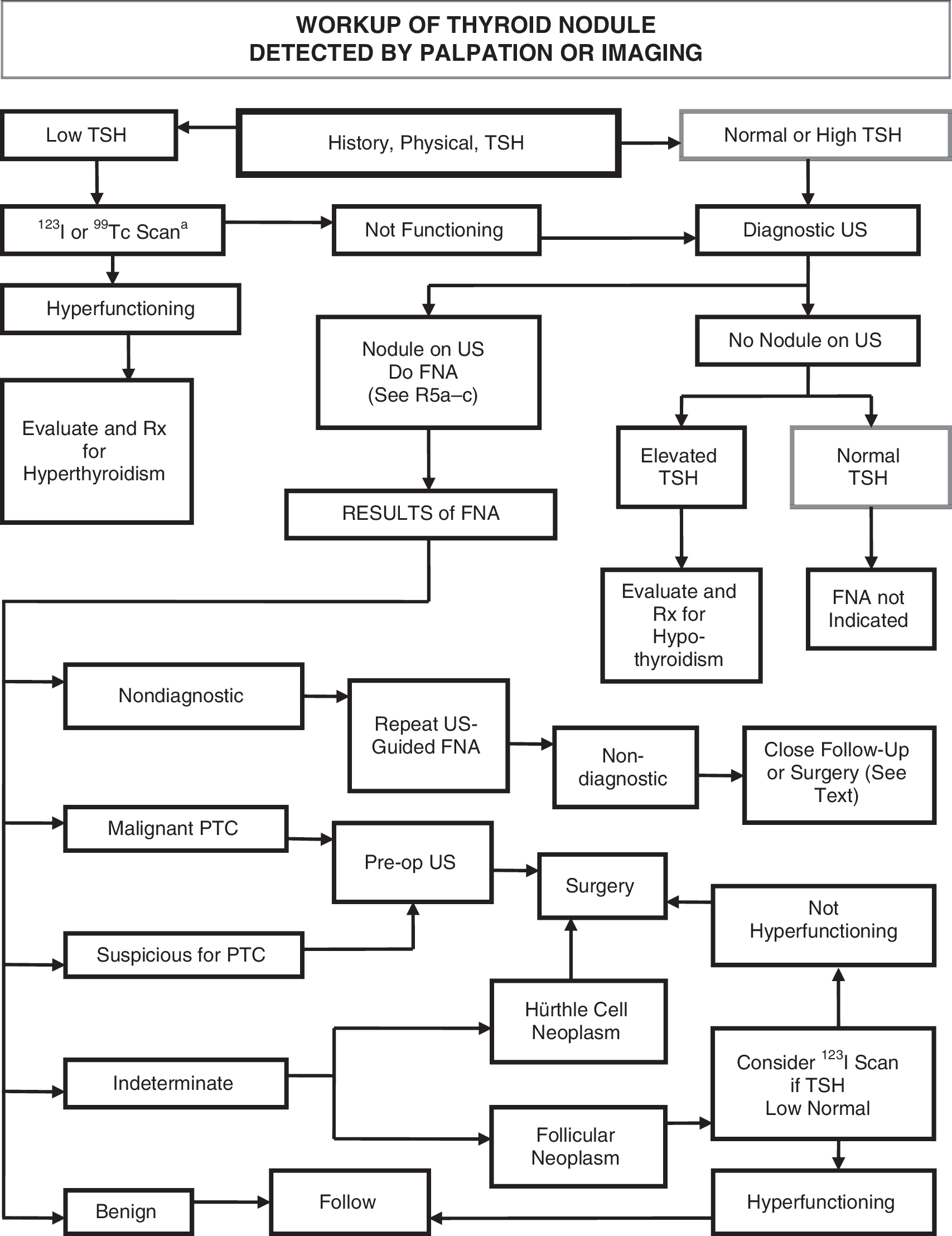
Algorithm for the evaluation of patients with one or more thyroid nodules.
[A3] What laboratory tests and imaging modalities are indicated?
[A4] Serum TSH with US and with or without scan
With the discovery of a thyroid nodule >1 cm in any diameter or diffuse or focal thyroidal uptake on 18FDG-PET scan, a serum TSH level should be obtained. If the serum TSH is subnormal, a radionuclide thyroid scan should be obtained to document whether the nodule is hyperfunctioning (i.e., tracer uptake is greater than the surrounding normal thyroid), isofunctioning or “warm” (i.e., tracer uptake is equal to the surrounding thyroid), or nonfunctioning (i.e., has uptake less than the surrounding thyroid tissue). Since hyperfunctioning nodules rarely harbor malignancy, if one is found that corresponds to the nodule in question, no cytologic evaluation is necessary. If overt or subclinical hyperthyroidism is present, additional evaluation is required. Higher serum TSH, even within the upper part of the reference range, is associated with increased risk of malignancy in a thyroid nodule (26).
▪ RECOMMENDATION 1
Measure serum TSH in the initial evaluation of a patient with a thyroid nodule. If the serum TSH is subnormal, a radionuclide thyroid scan should be performed using either technetium 99 mTc pertechnetate or 123I. Recommendation rating: A
Diagnostic thyroid US should be performed in all patients with a suspected thyroid nodule, nodular goiter, or radiographic abnormality; e.g., a nodule found incidentally on computed tomography (CT) or magnetic resonance imaging (MRI) or thyroidal uptake on 18FDG-PET scan. Thyroid US can answer the following questions: Is there truly a nodule that corresponds to the palpable abnormality? How large is the nodule? Does the nodule have benign or suspicious features? Is suspicious cervical lymphadenopathy present? Is the nodule greater than 50% cystic? Is the nodule located posteriorly in the thyroid gland? These last two features might decrease the accuracy of FNA biopsy performed with palpation (27,28). Also, there may be other thyroid nodules present that require biopsy based on their size and appearance (18,29,30). As already noted, FNA is recommended especially when the serum TSH is elevated because, compared with normal thyroid glands, the rate of malignancy in nodules in thyroid glands involved with Hashimoto's thyroiditis is as least as high or possibly higher (31,32).
▪ RECOMMENDATION 2
Thyroid sonography should be performed in all patients with known or suspected thyroid nodules. Recommendation rating: A
[A5] Serum Tg measurement
Serum Tg levels can be elevated in most thyroid diseases and are an insensitive and nonspecific test for thyroid cancer (33).
▪ RECOMMENDATION 3
Routine measurement of serum Tg for initial evaluation of thyroid nodules is not recommended. Recommendation rating: F
[A6] Serum calcitonin measurement
The utility of serum calcitonin has been evaluated in a series of prospective, nonrandomized studies (34 –37). The data suggest that the use of routine serum calcitonin for screening may detect C-cell hyperplasia and medullary thyroid cancer at an earlier stage and overall survival may be improved. However, most studies rely on pentagastrin stimulation testing to increase specificity. This drug is no longer available in the United States, and there remain unresolved issues of sensitivity, specificity, assay performance and cost-effectiveness. A recent cost-effectiveness analysis suggested that calcitonin screening would be cost effective in the United States (38). However, the prevalence estimates of medullary thyroid cancer in this analysis included patients with C-cell hyperplasia and micromedullary carcinoma, which have an uncertain clinical significance. If the unstimulated serum calcitonin determination has been obtained and the level is greater than 100 pg/mL, medullary cancer is likely present (39).
▪ RECOMMENDATION 4
The panel cannot recommend either for or against the routine measurement of serum calcitonin. Recommendation rating: I
[A7] What is the role of FNA biopsy?
FNA is the most accurate and cost-effective method for evaluating thyroid nodules. Retrospective studies have reported lower rates of both nondiagnostic and false-negative cytology specimens from FNA procedures performed via US guidance compared to palpation (40,41). Therefore, for nodules with a higher likelihood of either a nondiagnostic cytology (>25–50% cystic component) (28) or sampling error (difficult to palpate or posteriorly located nodules), US-guided FNA is preferred (see Table 3). If the diagnostic US confirms the presence of a predominantly solid nodule corresponding to what is palpated, the FNA may be performed via palpation or US guidance. Traditionally FNA biopsy results are divided into four categories: nondiagnostic, malignant (risk of malignancy at surgery >95%), indeterminate or suspicious for neoplasm, and benign. The recent National Cancer Institute Thyroid Fine-Needle Aspiration State of the Science Conference proposed a more expanded classification for FNA cytology that adds two additional categories: suspicious for malignancy (risk of malignancy 50–75%) and follicular lesion of undetermined significance (risk of malignancy 5–10%). The conference further recommended that “neoplasm, either follicular or Hürthle cell neoplasm” be substituted for “indeterminate” (risk of malignancy 15–25%) (42).
High-risk history: History of thyroid cancer in one or more first degree relatives; history of external beam radiation as a child; exposure to ionizing radiation in childhood or adolescence; prior hemithyroidectomy with discovery of thyroid cancer, 18FDG avidity on PET scanning; MEN2/FMTC-associated RET protooncogene mutation, calcitonin >100 pg/mL. MEN, multiple endocrine neoplasia; FMTC, familial medullary thyroid cancer.
Suspicious features: microcalcifications; hypoechoic; increased nodular vascularity; infiltrative margins; taller than wide on transverse view.
FNA cytology may be obtained from the abnormal lymph node in lieu of the thyroid nodule.
Sonographic monitoring without biopsy may be an acceptable alternative (see text) (48).
Unless indicated as therapeutic modality (see text).
[A8] US for FNA decision making (see Table 3)
Various sonographic characteristics of a thyroid nodule have been associated with a higher likelihood of malignancy (43 –48). These include nodule hypoechogenicity compared to the normal thyroid parenchyma, increased intranodular vascularity, irregular infiltrative margins, the presence of microcalcifications, an absent halo, and a shape taller than the width measured in the transverse dimension. With the exception of suspicious cervical lymphadenopathy, which is a specific but insensitive finding, no single sonographic feature or combinations of features is adequately sensitive or specific to identify all malignant nodules. However, certain features and combination of features have high predictive value for malignancy. Furthermore, the most common sonographic appearances of papillary and follicular thyroid cancer differ. A PTC is generally solid or predominantly solid and hypoechoic, often with infiltrative irregular margins and increased nodular vascularity. Microcalcifications, if present, are highly specific for PTC, but may be difficult to distinguish from colloid. Conversely, follicular cancer is more often iso- to hyperechoic and has a thick and irregular halo, but does not have microcalcifications (49). Follicular cancers that are <2 cm in diameter have not been shown to be associated with metastatic disease (50).
Certain sonographic appearances may also be highly predictive of a benign nodule. A pure cystic nodule, although rare (<2% of all nodules), is highly unlikely to be malignant (47). In addition, a spongiform appearance, defined as an aggregation of multiple microcystic components in more than 50% of the nodule volume, is 99.7% specific for identification of a benign thyroid nodule (48,51,52). In a recent study, only 1 of 360 malignant nodules demonstrated this appearance (48) and in another report, a spongiform appearance had a negative predictive value for malignancy of 98.5% (52). Elastography is an emerging and promising sonographic technique that requires additional validation with prospective studies (53).
Routine FNA is not recommended for subcentimeter nodules. However, the presence of a solid hypoechoic nodule with microcalcifications is highly suggestive of PTC. Although most micropapillary carcinomas may be incidental findings, a subset may be more clinically relevant, especially those >5 mm in diameter (54). These include nodules that have abnormal lymph nodes detected clinically or with imaging at presentation (55,56). Therefore, after imaging a subcentimeter nodule with a suspicious appearance, sonographic assessment of lateral neck and central neck lymph nodes (more limited due to the presence of the thyroid) must be performed. Detection of abnormal lymph nodes should lead to FNA of the lymph node. Other groups of patients for whom consideration of FNA of a subcentimeter nodule may be warranted include those with a higher likelihood of malignancy (high risk history): 1) family history of PTC (57); 2) history of external beam radiation exposure as a child (58); 3) exposure to ionizing radiation in childhood or adolescence (59); 4) history of prior hemithyroidectomy with discovery of thyroid cancer; and 5) 18FDG-PET–positive thyroid nodules.
Mixed cystic–solid nodules and predominantly cystic with >50% cystic component are generally evaluated by FNA with directed biopsy of the solid component (especially the vascular component.) Cyst drainage may also be performed, especially in symptomatic patients.
▪ RECOMMENDATION 5 (see Table 3)
FNA is the procedure of choice in the evaluation of thyroid nodules. Recommendation rating: A
US guidance for FNA is recommended for those nodules that are nonpalpable, predominantly cystic, or located posteriorly in the thyroid lobe. Recommendation rating: B
[A9] What are the principles of the cytopathological interpretation of FNA samples?
[A10] Nondiagnostic cytology
Nondiagnostic biopsies are those that fail to meet specified criteria for cytologic adequacy that have been previously established (the presence of at least six follicular cell groups, each containing 10–15 cells derived from at least two aspirates of a nodule) (5). After an initial nondiagnostic cytology result, repeat FNA with US guidance will yield a diagnostic cytology specimen in 75% of solid nodules and 50% of cystic nodules (28). Therefore, such biopsies need to be repeated using US guidance (60) and, if available, on-site cytologic evaluation, which may substantially increase cytology specimen adequacy (61,62). However, up to 7% of nodules continue to yield nondiagnostic cytology results despite repeated biopsies and may be malignant at the time of surgery (63,64).
▪ RECOMMENDATION 6
US guidance should be used when repeating the FNA procedure for a nodule with an initial nondiagnostic cytology result. Recommendation rating: A
Partially cystic nodules that repeatedly yield nondiagnostic aspirates need close observation or surgical excision. Surgery should be more strongly considered if the cytologically nondiagnostic nodule is solid. Recommendation rating: B
[A11] Cytology suggesting PTC
▪ RECOMMENDATION 7
If a cytology result is diagnostic of or suspicious for PTC, surgery is recommended (65). Recommendation rating: A
[A12] Indeterminate cytology (follicular or Hürthle cell neoplasm follicular lesion of undetermined significance, atypia)
Indeterminate cytology, reported as “follicular neoplasm” or “Hürthle cell neoplasm” can be found in 15–30% of FNA specimens (4) and carries a 20–30% risk of malignancy (42), while lesions reported as atypia or follicular lesion of undetermined significance are variably reported and have 5–10% risk of malignancy (42). While certain clinical features such as male sex and nodule size (>4 cm) (66), older patient age (67), or cytologic features such as presence of atypia (68) can improve the diagnostic accuracy for malignancy in patients with indeterminate cytology, overall predictive values are still low. Many molecular markers (e.g., galectin-3 (69), cytokeratin, BRAF) have been evaluated to improve diagnostic accuracy for indeterminate nodules (70 –72). Recent large prospective studies have confirmed the ability of genetic markers (BRAF, Ras, RET/PTC) and protein markers (galectin-3) to improve preoperative diagnostic accuary for patients with indeterminate thyroid nodules (69,73,74). Many of these markers are available for commercial use in reference laboratories but have not yet been widely applied in clinical practice. It is likely that some combination of molecular markers will be used in the future to optimize management of patients with indeterminate cytology on FNA specimens.
Recently, 18FDG-PET scanning has been utilized in an effort to distinguish those indeterminate nodules that are benign from those that are malignant (75 –78). 18FDG-PET scans appear to have relatively high sensitivity for malignancy but low specificity, but results vary among studies (79).
▪ RECOMMENDATION 8
The use of molecular markers (e.g., BRAF, RAS, RET/PTC, Pax8-PPARγ, or galectin-3) may be considered for patients with indeterminate cytology on FNA to help guide management. Recommendation rating: C
The panel cannot recommend for or against routine clinical use of 18FDG-PET scan to improve diagnostic accuracy of indeterminate thyroid nodules. Recommendation rating: I
▪ RECOMMENDATION 9
If the cytology reading reports a follicular neoplasm, a 123I thyroid scan may be considered, if not already done, especially if the serum TSH is in the low-normal range. If a concordant autonomously functioning nodule is not seen, lobectomy or total thyroidectomy should be considered. Recommendation rating: C
▪ RECOMMENDATION 10
If the reading is “suspicious for papillary carcinoma” or “Hürthle cell neoplasm,” a radionuclide scan is not needed, and either lobectomy or total thyroidectomy is recommended, depending on the lesion's size and other risk factors. Recommendation rating: A
[A13] Benign cytology
▪ RECOMMENDATION 11
If the nodule is benign on cytology, further immediate diagnostic studies or treatment are not routinely required. Recommendation rating: A
[A14] How should multinodular thyroid glands or multinodular goiters be evaluated for malignancy?
Patients with multiple thyroid nodules have the same risk of malignancy as those with solitary nodules (18,44). However, one large study found that a solitary nodule had a higher likelihood of malignancy than did a nonsolitary nodule (p < 0.01), although the risk of malignancy per patient was the same and independent of the number of nodules (47). A diagnostic US should be performed to delineate the nodules, but if only the “dominant” or largest nodule is aspirated, the thyroid cancer may be missed (44). Radionuclide scanning should also be considered in patients with multiple thyroid nodules, if the serum TSH is in the low or low-normal range, with FNA being reserved for those nodules that are shown to be hypofunctioning.
▪ RECOMMENDATION 12
In the presence of two or more thyroid nodules >1 cm, those with a suspicious sonographic appearance (see text and Table 3) should be aspirated preferentially. Recommendation rating: B
If none of the nodules has a suspicious sonographic appearance and multiple sonographically similar coalescent nodules with no intervening normal parenchyma are present, the likelihood of malignancy is low and it is reasonable to aspirate the largest nodules only and observe the others with serial US examinations. Recommendation rating: C
▪ RECOMMENDATION 13
A low or low-normal serum TSH concentration may suggest the presence of autonomous nodule(s). A technetium 99 mTc pertechnetate or 123I scan should be performed and directly compared to the US images to determine functionality of each nodule >1–1.5 cm. FNA should then be considered only for those isofunctioning or nonfunctioning nodules, among which those with suspicious sonographic features should be aspirated preferentially. Recommendation rating: B
[A15] What are the best methods for long-term follow-up of patients with thyroid nodules?
Thyroid nodules diagnosed as benign require follow-up because of a low, but not negligible, false-negative rate of up to 5% with FNA (41,80), which may be even higher with nodules >4 cm (81). While benign nodules may decrease in size, they often increase in size, albeit slowly (82). One study of cytologically benign thyroid nodules <2 cm followed by ultrasonography for about 38 months found that the rate of thyroid nodule growth did not distinguish between benign and malignant nodules (83).
Nodule growth is not in and of itself pathognomonic of malignancy, but growth is an indication for repeat biopsy. For mixed cystic–solid nodules, the indication for repeat biopsy should be based upon growth of the solid component. For nodules with benign cytologic results, recent series report a higher false-negative rate with palpation FNA (1–3%) (40,84,85) than with US FNA (0.6%) (40). Since the accuracy of physical examination for nodule size is likely inferior to that of US (30), it is recommended that serial US be used in follow-up of thyroid nodules to detect clinically significant changes in size. There is no consensus on the definition of nodule growth, however, or the threshold that would require rebiopsy. Some groups suggest a 15% increase in nodule volume, while others recommend measuring a change in the mean nodule diameter (82,86). One reasonable definition of growth is a 20% increase in nodule diameter with a minimum increase in two or more dimensions of at least 2 mm. This approximates the 50% increase in nodule volume that was found by Brauer et al. (87) to be the minimally significant reproducibly recorded change in nodule size. These authors suggested that only volume changes of at least 49% or more can be interpreted as nodule shrinkage or growth and consequently suggest that future investigations should not describe changes in nodule volume <50% as significant. A 50% cutoff for nodule volume reduction or growth, which is used in many studies, appears to appropriate and safe, since the false-negative rate for malignant thyroid nodules on repeat FNA is low (88,89).
▪ RECOMMENDATION 14
It is recommended that all benign thyroid nodules be followed with serial US examinations 6–18 months after the initial FNA. If nodule size is stable (i.e., no more than a 50% change in volume or <20% increase in at least two nodule dimensions in solid nodules or in the solid portion of mixed cystic–solid nodules), the interval before the next follow-up clinical examination or US may be longer, e.g., every 3–5 years. Recommendation rating: C
If there is evidence for nodule growth either by palpation or sonographically (more than a 50% change in volume or a 20% increase in at least two nodule dimensions with a minimal increase of 2 mm in solid nodules or in the solid portion of mixed cystic–solid nodules), the FNA should be repeated, preferably with US guidance. Recommendation rating: B
Cystic nodules that are cytologically benign can be monitored for recurrence (fluid reaccumulation) which can be seen in 60–90% of patients (90,91). For those patients with subsequent recurrent symptomatic cystic fluid accumulation, surgical removal, generally by hemithyroidectomy, or percutaneous ethanol injection (PEI) are both reasonable strategies. Four controlled studies demonstrated a 75–85% success rate after PEI compared with a 7–38% success rate in controls treated by simple cyst evacuation or saline injection. Success was achieved after an average of two PEI treatments. Complications included mild to moderate local pain, flushing, dizziness, and dysphonia (90 –93).
▪ RECOMMENDATION 15
Recurrent cystic thyroid nodules with benign cytology should be considered for surgical removal or PEI based on compressive symptoms and cosmetic concerns. Recommendation rating: B
[A16] What is the role of medical therapy for benign thyroid nodules?
Evidence from multiple randomized control trials and three meta-analyses suggest that thyroid hormone in doses that suppress the serum TSH to subnormal levels may result in a decrease in nodule size and may prevent the appearance of new nodules in regions of the world with borderline low iodine intake. Data in iodine-sufficient populations are less compelling (94 –96), with large studies suggesting that only about 17–25% of thyroid nodules shrink more than 50% with levothyroxine (LT4) suppression of serum TSH (94 –96).
▪ RECOMMENDATION 16
Routine suppression therapy of benign thyroid nodules in iodine sufficient populations is not recommended. Recommendation rating: F
▪ RECOMMENDATION 17
Patients with growing nodules that are benign after repeat biopsy should be considered for continued monitoring or intervention with surgery based on symptoms and clinical concern. There are no data on the use of LT4 in this subpopulation of patients. Recommendation rating: I
[A17] How should thyroid nodules in children be managed?
Thyroid nodules occur less frequently in children than in adults. In one study in which approximately 5000 children aged 11–18 years were assessed annually in the southwestern United States, palpable thyroid nodules occurred in approximately 20 per 1000 children, with an annual incidence of 7 new cases per 1000 children (97). Some studies have shown the frequency of malignancy to be higher in children than adults, in the range of 15–20% (98 –100), whereas other data have suggested that the frequency of thyroid cancer in childhood thyroid nodules is similar to that of adults (101,102). FNA biopsy is sensitive and specific in the diagnosis of childhood thyroid nodules (99 –101).
▪ RECOMMENDATION 18
The diagnostic and therapeutic approach to one or more thyroid nodules in a child should be the same as it would be in an adult (clinical evaluation, serum TSH, US, FNA). Recommendation rating: A
[A18] How should thyroid nodules in pregnant women be managed?
It is uncertain if thyroid nodules discovered in pregnant women are more likely to be malignant than those found in nonpregnant women (103), since there are no population-based studies on this question. The evaluation is the same as for a nonpregnant patient, with the exception that a radionuclide scan is contraindicated. In addition, for patients with nodules diagnosed as DTC by FNA during pregnancy, delaying surgery until after delivery does not affect outcome (104).
▪ RECOMMENDATION 19
For euthyroid and hypothyroid pregnant women with thyroid nodules, FNA should be performed. For women with suppressed serum TSH levels that persist after the first trimester, FNA may be deferred until after pregnancy and cessation of lactation, when a radionuclide scan can be performed to evaluate nodule function. Recommendation rating: A
If the FNA cytology is consistent with PTC, surgery is recommended. However, there is no consensus about whether surgery should be performed during pregnancy or after delivery. To minimize the risk of miscarriage, surgery during pregnancy should be done in the second trimester before 24 weeks gestation (105). However, PTC discovered during pregnancy does not behave more aggressively than that diagnosed in a similar-aged group of nonpregnant women (104,106). A retrospective study of pregnant women with DTC found there to be no difference in either recurrence, or survival rates, between women operated on during or after their pregnancy (104). Further, retrospective data suggest that treatment delays of less than 1 year from the time of thyroid cancer discovery do not adversely affect patient outcome (107). Finally, a recent study reported a higher rate of complications in pregnant women undergoing thyroid surgery compared with nonpregnant women (108). Some experts recommend thyroid hormone suppression therapy for pregnant women with FNA suspicious for or diagnostic of PTC, if surgery is deferred until the postpartum period (109).
▪ RECOMMENDATION 20
A nodule with cytology indicating PTC discovered early in pregnancy should be monitored sonographically and if it grows substantially (as defined above) by 24 weeks gestation, surgery should be performed at that point. However, if it remains stable by midgestation or if it is diagnosed in the second half of pregnancy, surgery may be performed after delivery. In patients with more advanced disease, surgery in the second trimester is reasonable. Recommendation rating: C
In pregnant women with FNA that is suspicious for or diagnostic of PTC, consideration could be given to administration of LT4 therapy to keep the TSH in the range of 0.1–1 mU/L. Recommendation rating: C
[B1] Differentiated Thyroid Cancer: Initial Management Guidelines
Differentiated thyroid cancer, arising from thyroid follicular epithelial cells, accounts for the vast majority of thyroid cancers. Of the differentiated cancers, papillary cancer comprises about 85% of cases compared to about 10% that have follicular histology, and 3% that are Hürthle cell or oxyphil tumors (110). In general, stage for stage, the prognoses of PTC and follicular cancer are similar (107,110). Certain histologic subtypes of PTC have a worse prognosis (tall cell variant, columnar cell variant, diffuse sclerosing variant), as do more highly invasive variants of follicular cancer. These are characterized by extensive vascular invasion and invasion into extrathyroidal tissues or extensive tumor necrosis and/or mitoses. Other poorly differentiated aggressive tumor histologies include trabecular, insular, and solid subtypes (111). In contrast, minimally invasive follicular thyroid cancer, is characterized histologically by microscopic penetration of the tumor capsule without vascular invasion, and carries no excess mortality (112 –115).
[B2] Goals of initial therapy of DTC
The goals of initial therapy of DTC are follows: To remove the primary tumor, disease that has extended beyond the thyroid capsule, and involved cervical lymph nodes. Completeness of surgical resection is an important determinant of outcome, while residual metastatic lymph nodes represent the most common site of disease persistence/recurrence (116
–118). To minimize treatment-related morbidity. The extent of surgery and the experience of the surgeon both play important roles in determining the risk of surgical complications (119,120). To permit accurate staging of the disease. Because disease staging can assist with initial prognostication, disease management, and follow-up strategies, accurate postoperative staging is a crucial element in the management of patients with DTC (121,122). To facilitate postoperative treatment with radioactive iodine, where appropriate. For patients undergoing RAI remnant ablation, or RAI treatment of residual or metastatic disease, removal of all normal thyroid tissue is an important element of initial surgery (123). Near total or total thyroidectomy also may reduce the risk for recurrence within the contralateral lobe (124). To permit accurate long-term surveillance for disease recurrence. Both RAI whole-body scanning (WBS) and measurement of serum Tg are affected by residual normal thyroid tissue. Where these approaches are utilized for long-term monitoring, near-total or total-thyroidectomy is required (125). To minimize the risk of disease recurrence and metastatic spread. Adequate surgery is the most important treatment variable influencing prognosis, while radioactive iodine treatment, TSH suppression, and external beam irradiation each play adjunctive roles in at least some patients (125
–128).
[B3] What is the role of preoperative staging with diagnostic imaging and laboratory tests?
[B4] Neck imaging
Differentiated thyroid carcinoma (particularly papillary carcinoma) involves cervical lymph nodes in 20–50% of patients in most series using standard pathologic techniques (45,129 –132), and may be present even when the primary tumor is small and intrathyroidal (133). The frequency of micrometastases may approach 90%, depending on the sensitivity of the detection method (134,135). However, the clinical implications of micrometastases are likely less significant compared to macrometastases. Preoperative US identifies suspicious cervical adenopathy in 20–31% of cases, potentially altering the surgical approach (136,137) in as many as 20% of patients (138,139). However, preoperative US identifies only half of the lymph nodes found at surgery, due to the presence of the overlying thyroid gland (140).
Sonographic features suggestive of abnormal metastatic lymph nodes include loss of the fatty hilus, a rounded rather than oval shape, hypoechogenicity, cystic change, calcifications, and peripheral vascularity. No single sonographic feature is adequately sensitive for detection of lymph nodes with metastatic thyroid cancer. A recent study correlated the sonographic features acquired 4 days preoperatively directly with the histology of 56 cervical lymph nodes. Some of the most specific criteria were short axis >5 mm (96%), presence of cystic areas (100%), presence of hyperechogenic punctuations representing either colloid or microcalcifications (100%), and peripheral vascularity (82%). Of these, the only one with sufficient sensitivity was peripheral vascularity (86%). All of the others had sensitivities <60% and would not be adequate to use as single criterion for identification of malignant involvement (140). As shown by earlier studies (141,142), the feature with the highest sensitivity was absence of a hilus (100%), but this had a low specificity of only 29%. The location of the lymph nodes may also be useful for decision-making. Malignant lymph nodes are much more likely to occur in levels III, IV, and VI than in level II (140,142). Figure 2 illustrates the delineation of cervical lymph node Levels I through VI.
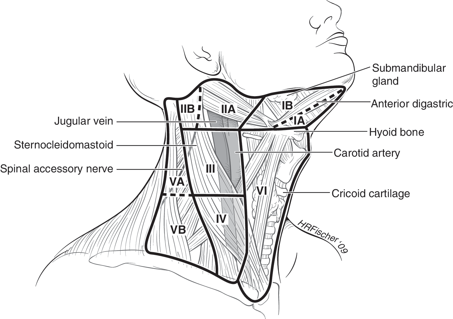
Lymph node compartments separated into levels and sublevels. Level VI contains the thyroid gland, and the adjacent nodes bordered superiorly by the hyoid bone, inferiorly by the innominate (brachiocephalic) artery, and laterally on each side by the carotid sheaths. The level II, III, and IV nodes are arrayed along the jugular veins on each side, bordered anteromedially by level VI and laterally by the posterior border of the sternocleidomastoid muscle. The level III nodes are bounded superiorly by the level of the hyoid bone, and inferiorly by the cricoid cartilage; levels II and IV are above and below level III, respectively. The level I node compartment includes the submental and submandibular nodes, above the hyoid bone, and anterior to the posterior edge of the submandibular gland. Finally, the level V nodes are in the posterior triangle, lateral to the lateral edge of the sternocleidomastoid muscle. Levels I, II, and V can be further subdivided as noted in the figure. The inferior extent of level VI is defined as the suprasternal notch. Many authors also include the pretracheal and paratracheal superior mediastinal lymph nodes above the level of the innominate artery (sometimes referred to as level VII) in central neck dissection (166).
Confirmation of malignancy in lymph nodes with a suspicious sonographic appearance is achieved by US-guided FNA aspiration for cytology and/or measurement of Tg in the needle washout. This FNA measurement of Tg is valid even in patients with circulating Tg autoantibodies (143,144).
Accurate staging is important in determining the prognosis and tailoring treatment for patients with DTC. However, unlike many tumor types, the presence of metastatic disease does not obviate the need for surgical excision of the primary tumor in DTC (145). Because metastatic disease may respond to RAI therapy, removal of the thyroid as well as the primary tumor and accessible locoregional disease remains an important component of initial treatment even in metastatic disease.
As US evaluation is uniquely operator dependent, alternative imaging procedures may be preferable in some clinical settings, though the sensitivities of CT, MRI, and PET for the detection of cervical lymph node metastases are all relatively low (30–40%) (146). These alternative imaging modalities, as well as laryngoscopy and endoscopy, may also be useful in the assessment of large, rapidly growing, or retrosternal or invasive tumors to assess the involvement of extrathyroidal tissues (147,148).
▪ RECOMMENDATION 21
Preoperative neck US for the contralateral lobe and cervical (central and especially lateral neck compartments) lymph nodes is recommended for all patients undergoing thyroidectomy for malignant cytologic findings on biopsy. US-guided FNA of sonographically suspicious lymph nodes should be performed to confirm malignancy if this would change management. Recommendation rating: B
▪ RECOMMENDATION 22
Routine preoperative use of other imaging studies (CT, MRI, PET) is not recommended. Recommendation rating: E
[B5] Measurement of serum Tg
There is limited evidence that high preoperative concentrations of serum Tg may predict a higher sensitivity for postoperative surveillance with serum Tg (149). Evidence that this impacts patient management or outcomes is not yet available.
▪ RECOMMENDATION 23
Routine preoperative measurement of serum Tg is not recommended. Recommendation rating: E
[B6] What is the appropriate operation for indeterminate thyroid nodules and DTC?
The goals of thyroid surgery can include provision of a diagnosis after a nondiagnostic or indeterminate biopsy, removal of the thyroid cancer, staging, and preparation for radioactive ablation and serum Tg monitoring. Surgical options to address the primary tumor should be limited to hemithyroidectomy with or without isthmusectomy, near-total thyroidectomy (removal of all grossly visible thyroid tissue, leaving only a small amount [<1 g] of tissue adjacent to the recurrent laryngeal nerve near the ligament of Berry), and total thyroidectomy (removal of all grossly visible thyroid tissue). Subtotal thyroidectomy, leaving >1 g of tissue with the posterior capsule on the uninvolved side, is an inappropriate operation for thyroid cancer (150).
[B7] Surgery for a nondiagnostic biopsy, a biopsy suspicious for papillary cancer or suggestive of “follicular neoplasm” (including special consideration for patients with other risk factors)
Amongst solitary thyroid nodules with an indeterminate (“follicular neoplasm” or Hürthle cell neoplasm) biopsy, the risk of malignancy is approximately 20% (151 –153). The risk is higher with large tumors (>4 cm), when atypical features (e.g., cellular pleomorphism) are seen on biopsy, when the biopsy reading is “suspicious for papillary carcinoma,” in patients with a family history of thyroid carcinoma, and in patients with a history of radiation exposure (66,154,155). For solitary nodules that are repeatedly nondiagnostic on biopsy, the risk of malignancy is unknown but is probably closer to 5–10% (63).
▪ RECOMMENDATION 24
For patients with an isolated indeterminate solitary nodule who prefer a more limited surgical procedure, thyroid lobectomy is the recommended initial surgical approach. Recommendation rating: C
▪ RECOMMENDATION 25
Because of an increased risk for malignancy, total thyroidectomy is indicated in patients with indeterminate nodules who have large tumors (>4 cm), when marked atypia is seen on biopsy, when the biopsy reading is “suspicious for papillary carcinoma,” in patients with a family history of thyroid carcinoma, and in patients with a history of radiation exposure. Recommendation rating: A
Patients with indeterminate nodules who have bilateral nodular disease, or those who prefer to undergo bilateral thyroidectomy to avoid the possibility of requiring a future surgery on the contralateral lobe, should also undergo total or near-total thyroidectomy. Recommendation rating: C
[B8] Surgery for a biopsy diagnostic for malignancy
Near-total or total thyroidectomy is recommended if the primary thyroid carcinoma is >1 cm (156), there are contralateral thyroid nodules present or regional or distant metastases are present, the patient has a personal history of radiation therapy to the head and neck, or the patient has first-degree family history of DTC. Older age (>45 years) may also be a criterion for recommending near-total or total thyroidectomy even with tumors <1–1.5 cm, because of higher recurrence rates in this age group (112,116,122,123,157). Increased extent of primary surgery may improve survival for high-risk patients (158 –160) and low-risk patients (156). A study of over 50,000 patients with PTC found on multivariate analysis that total thyroidectomy significantly improved recurrence and survival rates for tumors >1.0 cm (156). When examined separately, even patients with 1.0–2.0 cm tumors who underwent lobectomy, had a 24% higher risk of recurrence and a 49% higher risk of thyroid cancer mortality (p = 0.04 and p < 0.04, respectively). Other studies have also shown that rates of recurrence are reduced by total or near total thyroidectomy among low-risk patients (122,161,162).
▪ RECOMMENDATION 26
For patients with thyroid cancer >1 cm, the initial surgical procedure should be a near-total or total thyroidectomy unless there are contraindications to this surgery. Thyroid lobectomy alone may be sufficient treatment for small (<1 cm), low-risk, unifocal, intrathyroidal papillary carcinomas in the absence of prior head and neck irradiation or radiologically or clinically involved cervical nodal metastases. Recommendation rating: A
[B9] Lymph node dissection
Regional lymph node metastases are present at the time of diagnosis in 20–90% of patients with papillary carcinoma and a lesser proportion of patients with other histotypes (129,139). Although PTC lymph node metastases are reported by some to have no clinically important effect on outcome in low risk patients, a study of the Surveillance, Epidemiology, and End Results (SEER) database found, among 9904 patients with PTC, that lymph node metastases, age >45 years, distant metastasis, and large tumor size significantly predicted poor outcome on multivariate analysis (163). All-cause survival at 14 years was 82% for PTC without lymph node and 79% with lymph node metastases (p < 0.05). Another recent SEER registry study concluded that cervical lymph node metastases conferred an independent risk of decreased survival, but only in patients with follicular cancer and patients with papillary cancer over age 45 years (164). Also, the risk of regional recurrence is higher in patients with lymph node metastases, especially in those patients with multiple metastases and/or extracapsular nodal extension (165).
In many patients, lymph node metastases in the central compartment (166) do not appear abnormal preoperatively with imaging (138) or by inspection at the time of surgery. Central compartment dissection (therapeutic or prophylactic) can be achieved with low morbidity in experienced hands (167 –171), and may convert some patients from clinical N0 to pathologic N1a, upstaging patients over age 45 from American Joint Committee on Cancer (AJCC) stage I to III (172). A recent consensus conference statement discusses the relevant anatomy of the central neck compartment, delineates the nodal subgroups within the central compartment commonly involved with thyroid cancer, and defines the terminology relevant to central compartment neck dissection (173).
Comprehensive bilateral central compartment node dissection may improve survival compared to historic controls and reduce risk for nodal recurrence (174). In addition, selective unilateral paratracheal central compartment node dissection increases the proportion of patients who appear disease free with unmeasureable Tg levels 6 months after surgery (175). Other studies of central compartment dissection have demonstrated higher morbidity, primarily recurrent laryngeal nerve injury and transient hypoparathyroidism, with no reduction in recurrence (176,177). In another study, comprehensive (bilateral) central compartment dissection demonstrated higher rates of transient hypoparathyroidism compared to selective (unilateral) dissection with no reduction in rates of undetectable or low Tg levels (178). Although some lymph node metastases may be treated with radioactive iodine, several treatments may be necessary, depending upon the histology, size, and number of metastases (179).
RECOMMENDATION 27*
Therapeutic central-compartment (level VI) neck dissection for patients with clinically involved central or lateral neck lymph nodes should accompany total thyroidectomy to provide clearance of disease from the central neck. Recommendation rating: B
Prophylactic central-compartment neck dissection (ipsilateral or bilateral) may be performed in patients with papillary thyroid carcinoma with clinically uninvolved central neck lymph nodes, especially for advanced primary tumors (T3 or T4). Recommendation rating: C
Near-total or total thyroidectomy without prophylactic central neck dissection may be appropriate for small (T1 or T2), noninvasive, clinically node-negative PTCs and most follicular cancer. Recommendation rating: C
These recommendations (R27a–c) should be interpreted in light of available surgical expertise. For patients with small, noninvasive, apparently node-negative tumors, the balance of risk and benefit may favor simple near-total thyroidectomy with close intraoperative inspection of the central compartment with compartmental dissection only in the presence of obviously involved lymph nodes. This approach may increase the chance of future locoregional recurrence, but overall this approach may be safer in less experienced surgical hands.
Lymph nodes in the lateral neck (compartments II–V), level VII (anterior mediastinum), and rarely in Level I may also be involved by thyroid cancer (129,180). For those patients in whom nodal disease is evident clinically, on preoperative US and nodal FNA or Tg measurement, or at the time of surgery, surgical resection may reduce the risk of recurrence and possibly mortality (56,139,181). Functional compartmental en-bloc neck dissection is favored over isolated lymphadenectomy (“berry picking”) with limited data suggesting improved mortality (118,182 –184).
▪ RECOMMENDATION 28*
Therapeutic lateral neck compartmental lymph node dissection should be performed for patients with biopsy-proven metastatic lateral cervical lymphadenopathy. Recommendation rating: B
[B10] Completion thyroidectomy
Completion thyroidectomy may be necessary when the diagnosis of malignancy is made following lobectomy for an indeterminate or nondiagnostic biopsy. Some patients with malignancy may require completion thyroidectomy to provide complete resection of multicentric disease (185), and to allow RAI therapy. Most (186,187) but not all (185) studies of papillary cancer have observed a higher rate of cancer in the opposite lobe when multifocal (two or more foci), as opposed to unifocal, disease is present in the ipsilateral lobe. The surgical risks of two-stage thyroidectomy (lobectomy followed by completion thyroidectomy) are similar to those of a near-total or total thyroidectomy (188).
▪ RECOMMENDATION 29
Completion thyroidectomy should be offered to those patients for whom a near-total or total thyroidectomy would have been recommended had the diagnosis been available before the initial surgery. This includes all patients with thyroid cancer except those with small (<1 cm), unifocal, intrathyroidal, node-negative, low-risk tumors. Therapeutic central neck lymph node dissection should be included if the lymph nodes are clinically involved. Recommendation rating: B
▪ RECOMMENDATION 30
Ablation of the remaining lobe with radioactive iodine has been used as an alternative to completion thyroidectomy (189). It is unknown whether this approach results in similar long-term outcomes. Consequently, routine radioactive iodine ablation in lieu of completion thyroidectomy is not recommended. Recommendation rating: D
[B11] What is the role of postoperative staging systems and which should be used?
[B12] The role of postoperative staging
Postoperative staging for thyroid cancer, as for other cancer types, is used: 1) to permit prognostication for an individual patient with DTC; 2) to tailor decisions regarding postoperative adjunctive therapy, including RAI therapy and TSH suppression, to assess the patient's risk for disease recurrence and mortality; 3) to make decisions regarding the frequency and intensity of follow-up, directing more intensive follow-up towards patients at highest risk; and 4) to enable accurate communication regarding a patient among health care professionals. Staging systems also allow evaluation of differing therapeutic strategies applied to comparable groups of patients in clinical studies.
[B13] AJCC/UICC TNM staging
Application of the AJCC/International Union against Cancer (AJCC/UICC) classification system based on pTNM parameters and age is recommended for tumors of all types, including thyroid cancer (121,190), because it provides a useful shorthand method to describe the extent of the tumor (191) (Table 4). This classification is also used for hospital cancer registries and epidemiologic studies. In thyroid cancer, the AJCC/UICC stage does not take account of several additional independent prognostic variables and may risk misclassification of some patients. Numerous other schemes have been developed in an effort to achieve more accurate risk factor stratification, including CAEORTC, AGES, AMES, U of C, MACIS, OSU, MSKCC, and NTCTCS systems. (107,116,122,159,192
–195). These schemes take into account a number of factors identified as prognostic for outcome in multivariate analysis of retrospective studies, with the most predictive factors generally being regarded as the presence of distant metastases, the age of the patient, and the extent of the tumor. These and other risk factors are weighted differently among these systems according to their importance in predicting outcome, but no scheme has demonstrated clear superiority (195). Each of the schemes allows accurate identification of the majority (70–85%) of patients at low-risk of mortality (T1–3, M0 patients), allowing the follow-up and management of these patients to be less intensive than the higher-risk minority (T4 and M1 patients), who may benefit from a more aggressive management strategy (195). Nonetheless, none of the examined staging classifications is able to account for more than a small proportion of the uncertainty in either short-term, disease-specific mortality or the likelihood of remaining disease free (121,195,196). AJCC/IUCC staging was developed to predict risk for death, not recurrence. For assessment of risk of recurrence, a three-level stratification can be used: Low-risk patients have the following characteristics: 1) no local or distant metastases; 2) all macroscopic tumor has been resected; 3) there is no tumor invasion of locoregional tissues or structures; 4) the tumor does not have aggressive histology (e.g., tall cell, insular, columnar cell carcinoma) or vascular invasion; 5) and, if 131I is given, there is no 131I uptake outside the thyroid bed on the first posttreatment whole-body RAI scan (RxWBS) (197
–199). Intermediate-risk patients have any of the following: 1) microscopic invasion of tumor into the perithyroidal soft tissues at initial surgery; 2) cervical lymph node metastases or 131I uptake outside the thyroid bed on the RxWBS done after thyroid remnant ablation (200,201); or 3) tumor with aggressive histology or vascular invasion (202
–204). High-risk patients have 1) macroscopic tumor invasion, 2) incomplete tumor resection, 3) distant metastases, and possibly 4) thyroglobulinemia out of proportion to what is seen on the posttreatment scan (205).
TNM
Used with the permission of the American Joint Committee on Cancer (AJCC), Chicago, Illinois.
The original source for this material is the AJCC Cancer Staging Manual, Sixth Edition (435).
Since initial staging is based on clinico-pathologic factors that are available shortly after diagnosis and initial therapy, the AJCC stage of the patient does not change over time. However, depending on the clinical course of the disease and response to therapy, the risk of recurrence and the risk of death may change over time. Appropriate management requires an ongoing reassessment of the risk of recurrence and the risk of disease-specific mortality as new data are obtained during follow-up (206).
▪ RECOMMENDATION 31
Because of its utility in predicting disease mortality, and its requirement for cancer registries, AJCC/UICC staging is recommended for all patients with DTC. The use of postoperative clinico-pathologic staging systems is also recommended to improve prognostication and to plan follow-up for patients with DTC. Recommendation rating: B
[B14] What is the role of postoperative RAI remnant ablation?
Postoperative RAI remnant ablation is increasingly being used to eliminate the postsurgical thyroid remnant (122). Ablation of the small amount of residual normal thyroid remaining after total thyroidectomy may facilitate the early detection of recurrence based on serum Tg measurement and/or RAI WBS. Additionally, the posttherapy scan obtained at the time of remnant ablation may facilitate initial staging by identifying previously undiagnosed disease, especially in the lateral neck. Furthermore, from a theoretical point of view, this first dose of RAI may also be considered adjuvant therapy because of the potential tumoricidal effect on persistent thyroid cancer cells remaining after appropriate surgery in patients at risk for recurrence or disease specific mortality. Depending on the risk stratification of the individual patient, the primary goal of the first dose of RAI after total thyroidectomy may be 1) remnant ablation (to facilitate detection of recurrent disease and initial staging), 2) adjuvant therapy (to decrease risk of recurrence and disease specific mortality by destroying suspected, but unproven metastatic disease), or 3) RAI therapy (to treat known persistent disease). While these three goals are closely interrelated, a clearer understanding of the specific indications for treatment will improve our ability to select patients most likely to benefit from RAI after total thyroidectomy, and will also influence our recommendations regarding choice of administered activity for individual patients. Supporting the use of RAI as adjuvant therapy, a number of large, retrospective studies show a significant reduction in the rates of disease recurrence (107,159,160,207) and cause-specific mortality (159,160,207 –209). However, other similar studies show no such benefit, at least among the majority of patients with PTC, who are at the lowest risk for mortality (110,122,162,209 –212). In those studies that show benefit, the advantage appears to be restricted to patients with tumors >1.5 cm, or with residual disease following surgery, while lower-risk patients do not show evidence for benefit (122,159,213). The National Thyroid Cancer Treatment Cooperative Study Group (NTCTCSG) report (214) of 2936 patients found after a median follow-up of 3 years, that near-total thyroidectomy followed by RAI therapy and aggressive thyroid hormone suppression therapy predicted improved overall survival of patients with NTCTCSG stage III and IV disease, and was also beneficial for patients with NTCTCSG stage II disease. No impact of therapy was observed in patients with stage I disease. It should be noted that the NTCTCSG staging criteria are similar but not identical to the AJCC criteria. Thus, older patients with microscopic extrathyroidal extension are stage II in the NTCTCSG system, but are stage III in the AJCC system. There are recent data suggesting a benefit of RAI in patients with more aggressive histologies (215). There are no prospective randomized trials that have addressed this question (209). Unfortunately, many clinical circumstances have not been examined with regard to the efficacy of RAI ablative therapy. Table 5 presents a framework for deciding whether RAI is worthwhile, solely based on the AJCC classification, and provides the rationale for therapy and the strength of existing evidence for or against treatment.
Because of either conflicting or inadequate data, we cannot recommend either for or against RAI ablation for this entire subgroup. However, selected patients within this subgroup with higher risk features may benefit from RAI ablation (see modifying factors in the text).
In addition to the major factors listed in Table 5, several other histological features may place the patient at higher risk of local recurrence or metastases than would have been predicted by the AJCC staging system. These include worrisome histologic subtypes (such as tall cell, columnar, insular, and solid variants, as well as poorly differentiated thyroid cancer), the presence of intrathyroidal vascular invasion, or the finding of gross or microscopic multifocal disease. While many of these features have been associated with increased risk, there are inadequate data to determine whether RAI ablation has a benefit based on specific histologic findings, independent of tumor size, lymph node status, and the age of the patient. Therefore, while RAI ablation is not recommended for all patients with these higher risk histologic features, the presence of these features in combination with size of the tumor, lymph node status, and patient age may increase the risk of recurrence or metastatic spread to a degree that is high enough to warrant RAI ablation in selected patients. However, in the absence of data for most of these factors, clinical judgment must prevail in the decision-making process. For microscopic multifocal papillary cancer, when all foci are <1 cm, recent data suggest that RAI is of no benefit in preventing recurrence (216,217).
Nonpapillary histologies (such as follicular thyroid cancer and Hürthle cell cancer) are generally regarded as higher risk tumors. Expert opinion supports the use of RAI in almost all of these cases. However, because of the excellent prognosis associated with surgical resection alone in small follicular thyroid cancers manifesting only capsular invasion (without vascular invasion (so-called “minimally invasive follicular cancer”), RAI ablation may not be required for all patients with this histological diagnosis (112).
▪ RECOMMENDATION 32
RAI ablation is recommended for all patients with known distant metastases, gross extrathyroidal extension of the tumor regardless of tumor size, or primary tumor size >4 cm even in the absence of other higher risk features (see Table 5 for strength of evidence).
RAI ablation is recommended for selected patients with 1–4 cm thyroid cancers confined to the thyroid, who have documented lymph node metastases, or other higher risk features (see preceding paragraphs) when the combination of age, tumor size, lymph node status, and individual histology predicts an intermediate to high risk of recurrence or death from thyroid cancer (see Table 5 for strength of evidence for individual features). Recommendation rating: C (for selective use in higher risk patients)
RAI ablation is not recommended for patients with unifocal cancer <1 cm without other higher risk features (see preceding paragraphs). Recommendation rating: E
RAI ablation is not recommended for patients with multifocal cancer when all foci are <1 cm in the absence other higher risk features (see preceding paragraphs). Recommendation rating: E
[B15] How should patients be prepared for RAI ablation? (see Fig. 3)
Remnant ablation requires TSH stimulation. No controlled studies have been performed to assess adequate levels of endogenous TSH for optimal ablation therapy or follow-up testing. Noncontrolled studies suggest that a TSH of >30 mU/L is associated with increased RAI uptake in tumors (218), while studies using single dose exogenous TSH suggest maximal thyrocyte stimulation at TSH levels between 51 and 82 mU/L (219, 220). However, the total area under the TSH curve, and not simply the peak serum TSH concentration, is also potentially important for optimal RAI uptake by thyroid follicular cells. Endogenous TSH elevation can be achieved by two basic approaches to thyroid hormone withdrawal, stopping LT4 and switching to LT3 for 2–4 weeks followed by withdrawal of LT3 for 2 weeks, or discontinuation of LT4 for 3 weeks without use of LT3. Both methods of preparation can achieve serum TSH levels >30 mU/L in >90% of patients (220 –229). These two approaches have not been directly compared for efficiency of patient preparation (efficacy of ablation, iodine uptake, Tg levels, disease detection), although a recent prospective study showed no difference in hypothyroid symptoms between these two approaches (230). Other preparative methods have been proposed, but have not been validated by other investigators (231,232). Children with thyroid cancer achieve adequate TSH elevation within 14 days of LT4 withdrawal (233). A low serum Tg level at the time of ablation has excellent negative predictive value for absence of residual disease, and the risk of persistent disease increases with higher stimulated Tg levels (198,205,234).

Algorithm for initial follow-up of patients with differentiated thyroid carcinoma.
▪ RECOMMENDATION 33
Patients undergoing RAI therapy or diagnostic testing can be prepared by LT4 withdrawal for at least 2–3 weeks or LT3 treatment for 2–4 weeks and LT3 withdrawal for 2 weeks with measurement of serum TSH to determine timing of testing or therapy (TSH >30 mU/L). Thyroxine therapy (with or without LT3 for 7–10 days) may be resumed on the second or third day after RAI administration. Recommendation rating: B
[B16] Can rhTSH (Thyrogen™) be used in lieu of thyroxine withdrawal for remnant ablation?
For most patients, including those unable to tolerate hypothyroidism or unable to generate an elevated TSH, remnant ablation can be achieved with rhTSH (235,236). A prospective randomized study found that thyroid hormone withdrawal and rhTSH stimulation were equally effective in preparing patients for 131I remnant ablation with 100 mCi with significantly improved quality of life (237). Another randomized study using rhTSH showed that ablation rates were comparable with 50 mCi compared to 100 mCi with a significant decrease (33%) in whole-body irradiation (238). Finally, a recent study has shown that ablation rates were similar with either withdrawal or preparation with rhTSH using 50 mCi of 131I (239). In addition, short-term recurrence rates have been found to be similar in patients prepared with thyroid hormone withdrawal or rhTSH (240). Recombinant human TSH is approved for remnant ablation in the United States, Europe, and many other countries around the world.
▪ RECOMMENDATION 34
Remnant ablation can be performed following thyroxine withdrawal or rhTSH stimulation. Recommendation rating: A
[B17] Should RAI scanning be performed before RAI ablation?
RAI WBS provides information on the presence of iodine-avid thyroid tissue, which may represent the normal thyroid remnant or the presence of residual disease in the postoperative setting. In the presence of a large thyroid remnant, the scan is dominated by uptake within the remnant, potentially masking the presence of extrathyroidal disease within locoregional lymph nodes, the upper mediastinum, or even at distant sites, reducing the sensitivity of disease detection (241). Furthermore, there is an increasing trend to avoid pretherapy RAI scans altogether because of its low impact on the decision to ablate, and because of concerns over 131I-induced stunning of normal thyroid remnants (242) and distant metastases from thyroid cancer (243). Stunning is defined as a reduction in uptake of the 131I therapy dose induced by a pretreatment diagnostic activity. Stunning occurs most prominently with higher activities (5–10 mCi) of 131I (244), with increasing time between the diagnostic dose and therapy (245), and does not occur if the treatment dose is given within 72 hours of the scanning dose (246). However, the accuracy of low-activity 131I scans has been questioned, and some research has reported quantitatively the presence of stunning below the threshold of visual detection (247). Although comparison studies show excellent concordance between 123I and 131I for tumor detection, optimal 123I activity and time to scan after 123I administration are not known (248). Furthermore, 123I is expensive, is not universally available, its short half life (t½ = 13 hours) makes handling this isotope logistically more difficult (249), and stunning may also occur though to a lesser degree than with 131I (245). Furthermore, a recent study showed no difference in ablation rates between patients that had pretherapy scans with 123I (81%) compared to those who had received diagnostic scans using 2 mCi of 131I (74%, p > 0.05) (250). Alternatively, determination of the thyroid bed uptake, without scanning, can be achieved using 10–100 μCi 131I.
▪ RECOMMENDATION 35
Pretherapy scans and/or measurement of thyroid bed uptake may be useful when the extent of the thyroid remnant cannot be accurately ascertained from the surgical report or neck ultrasonography, or when the results would alter either the decision to treat or the activity of RAI that is administered. If performed, pretherapy scans should utilize 123I (1.5–3 mCi) or low-activity 131I (1–3 mCi), with the therapeutic activity optimally administered within 72 hours of the diagnostic activity. Recommendation rating: C
[B18] What activity of 131I should be used for remnant ablation?
Successful remnant ablation is usually defined as an absence of visible RAI uptake on a subsequent diagnostic RAI scan or an undetectable stimulated serum Tg. Activities between 30 and 100 mCi of 131I generally show similar rates of successful remnant ablation (251 –254) and recurrence rates (213). Although there is a trend toward higher ablation rates with higher activities (255,256), a recent prospective randomized study found no significant difference in the remnant ablation rate using 30 or 100 mCi of 131I (257). Furthermore, there are data showing that 30 mCi is effective in ablating the remnant with rhTSH preparation (258). In pediatric patients, it is preferable to adjust the ablation activity according to the patient's body weight (259) or surface area (260).
▪ RECOMMENDATION 36
The minimum activity (30–100 mCi) necessary to achieve successful remnant ablation should be utilized, particularly for low-risk patients. Recommendation rating: B
▪ RECOMMENDATION 37
If residual microscopic disease is suspected or documented, or if there is a more aggressive tumor histology (e.g., tall cell, insular, columnar cell carcinoma), then higher activities (100–200 mCi) may be appropriate. Recommendation rating: C
[B19] Is a low-iodine diet necessary before remnant ablation?
The efficacy of radioactive iodine depends on the radiation dose delivered to the thyroid tissue (261). Low-iodine diets (<50 μg/d of dietary iodine) and simple recommendations to avoid iodine contamination have been recommended prior to RAI therapy (261
–263) to increase the effective radiation dose. A history of possible iodine exposure (e.g., intravenous contrast, amiodarone use) should be sought. Measurement of iodine excretion with a spot urinary iodine determination may be a useful way to identify patients whose iodine intake could interfere with RAI remnant ablation (263). Information about low-iodine diets can be obtained at the Thyroid Cancer Survivors Association website (
▪ RECOMMENDATION 38
A low-iodine diet for 1–2 weeks is recommended for patients undergoing RAI remnant ablation, particularly for those patients with high iodine intake. Recommendation rating: B
[B20] Should a posttherapy scan be performed following remnant ablation?
Posttherapy whole-body iodine scanning is typically conducted approximately 1 week after RAI therapy to visualize metastases. Additional metastatic foci have been reported in 10–26% of patients scanned following high-dose RAI treatment compared with the diagnostic scan (264,265). The new abnormal uptake was found most often in the neck, lungs, and mediastinum, and the newly discovered disease altered the disease stage in approximately 10% of the patients, affecting clinical management in 9–15% (264 –266). Iodine 131 single photon emission computed tomography (SPECT)/CT fusion imaging may provide superior lesion localization after remnant ablation, but it is still a relatively new imaging modality (267).
▪ RECOMMENDATION 39
A posttherapy scan is recommended following RAI remnant ablation. This is typically done 2–10 days after the therapeutic dose is administered, although published data supporting this time interval are lacking. Recommendation rating: B
[B21] Postsurgery and RAI therapy early management of DTC
[B22] What is the role of TSH suppression therapy?
DTC expresses the TSH receptor on the cell membrane and responds to TSH stimulation by increasing the expression of several thyroid specific proteins (Tg, sodium-iodide symporter) and by increasing the rates of cell growth (268). Suppression of TSH, using supra-physiologic doses of LT4, is used commonly to treat patients with thyroid cancer in an effort to decrease the risk of recurrence (127,214,269). A meta-analysis supported the efficacy of TSH suppression therapy in preventing major adverse clinical events (RR = 0.73; CI = 0.60–0.88; p < 0.05) (269).
[B23] What is the appropriate degree of initial TSH suppression?
Retrospective and prospective studies have demonstrated that TSH suppression to below 0.1 mU/L may improve outcomes in high-risk thyroid cancer patients (127,270), though no such evidence of benefit has been documented in low-risk patients. A prospective cohort study (214) of 2936 patients found that overall survival improved significantly when the TSH was suppressed to undetectable levels in patients with NTCTCSG stage III or IV disease and suppressed to the subnormal to undetectable range in patients with NTCTCSG stage II disease; however, in the latter group there was no incremental benefit from suppressing TSH to undetectable levels. Suppression of TSH was not beneficial in patients with stage I disease. In another study, there was a positive association between serum TSH levels and the risk for recurrent disease and cancer-related mortality (271). Adverse effects of TSH suppression may include the known consequences of subclinical thyrotoxicosis, including exacerbation of angina in patients with ischemic heart disease, increased risk for atrial fibrillation in older patients (272), and increased risk of osteoporosis in postmenopausal women (273).
▪ RECOMMENDATION 40
Initial TSH suppression to below 0.1 mU/L is recommended for high-risk and intermediate-risk thyroid cancer patients, while maintenance of the TSH at or slightly below the lower limit of normal (0.1–0.5 mU/L) is appropriate for low-risk patients. Similar recommendations apply to low-risk patients who have not undergone remnant ablation, i.e., serum TSH 0.1–0.5 mU/L. Recommendation rating: B
[B24] Is there a role for adjunctive external beam irradiation or chemotherapy?
[B25] External beam irradiation
External beam irradiation is used infrequently in the management of thyroid cancer except as a palliative treatment for locally advanced, otherwise unresectable disease (274). There are reports of responses among patients with locally advanced disease (275,276) and improved relapse-free and cause-specific survival in patients over age 60 with extrathyroidal extension but no gross residual disease (277). It remains unknown whether external beam radiation might reduce the risk for recurrence in the neck following adequate primary surgery and/or RAI treatment in patients with aggressive histologic subtypes (278).
▪ RECOMMENDATION 41
The use of external beam irradiation to treat the primary tumor should be considered in patients over age 45 with grossly visible extrathyroidal extension at the time of surgery and a high likelihood of microscopic residual disease, and for those patients with gross residual tumor in whom further surgery or RAI would likely be ineffective. The sequence of external beam irradiation and RAI therapy depends on the volume of gross residual disease and the likelihood of the tumor being RAI responsive. Recommendation rating: B
[B26] Chemotherapy
There are no data to support the use of adjunctive chemotherapy in the management of DTC. Doxorubicin may act as a radiation sensitizer in some tumors of thyroid origin (279), and could be considered for patients with locally advanced disease undergoing external beam radiation.
▪ RECOMMENDATION 42
There is no role for the routine adjunctive use of chemotherapy in patients with DTC. Recommendation rating: F
[C1] DTC: Long-Term Management Guidelines
[C2] What are the appropriate features of long-term management?
Accurate surveillance for possible recurrence in patients thought to be free of disease is a major goal of long-term follow-up. Tests with high negative predictive value allow identification of patients unlikely to experience disease recurrence, so that less aggressive management strategies can be used that may be more cost effective and safe. Similarly, patients with a higher risk of recurrence are monitored more aggressively because it is believed that early detection of recurrent disease offers the best opportunity for effective treatment. A large study (280), found that the residual life span in disease-free patients treated with total or near-total thyroidectomy and 131I for remnant ablation and, in some cases, high dose 131I for residual disease, was similar to that in the general Dutch population. In contrast, the life expectancy for patients with persistent disease was reduced to 60% of that in the general population but varied widely depending upon tumor features. Age was not a factor in disease-specific mortality when patients were compared with aged matched individuals in the Dutch population. Treatment thus appears safe and does not shorten life expectancy. Although an increased incidence of second tumors in thyroid cancer patients has been recognized (157,281) this elevated risk was not found to be associated with the use of 131I in another study (282), and RAI therapy in low-risk patients did not affect median overall survival in another (210). Patients with persistent or recurrent disease are offered treatment to cure or to delay future morbidity or mortality. In the absence of such options, therapies to palliate by substantially reducing tumor burden or preventing tumor growth are utilized, with special attention paid to tumors threatening critical structures.
A second goal of long-term follow-up is to monitor thyroxine suppression or replacement therapy, to avoid underreplacement or overly aggressive therapy (283).
[C3] What is the appropriate method for following patients after surgery with or without remnant ablation?
See Fig. 4 for an algorithm for the first 6–12 months of management.
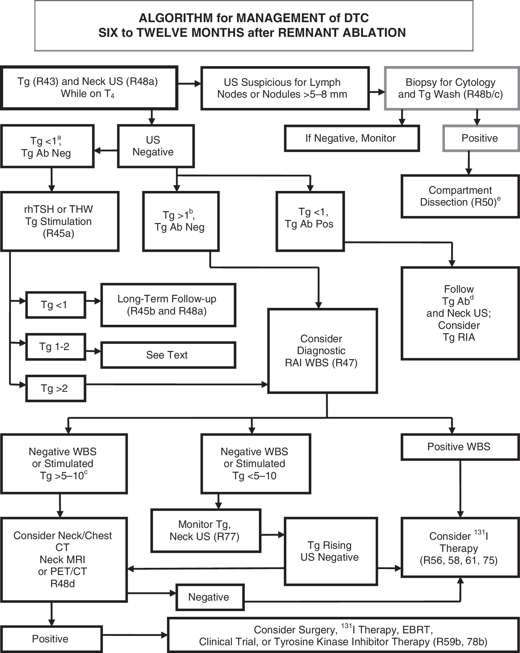
Longer term follow-up of patients with differentiated thyroid carcinoma.
[C4] What are the criteria for absence of persistent tumor?
In patients who have undergone total or near-total thyroidectomy and thyroid remnant ablation, disease-free status comprises all of the following: no clinical evidence of tumor, no imaging evidence of tumor (no uptake outside the thyroid bed on the initial posttreatment WBS, or, if uptake outside the thyroid bed had been present, no imaging evidence of tumor on a recent diagnostic scan and neck US), and undetectable serum Tg levels during TSH suppression and stimulation in the absence of interfering antibodies.
[C5] What is the role of serum Tg assays in the follow up of DTC?
Measurement of serum Tg levels is an important modality to monitor patients for residual or recurrent disease. Most laboratories currently use immunometric assays to measure serum Tg, and it is important that these assays are calibrated against the CRM-457 international standard. Despite improvements in standardization of thyroglobuin assays, there is still a twofold difference between some assays (149), leading to the recommendation that measurements in individual patients over time be performed in the same assay. Immunometric assays are prone to interference from Tg autoantibodies, which commonly cause falsely low serum Tg measurements. Radioimmunoassays may be less prone to antibody interference, but are not as widely available, and their role in the clinical care of patients is uncertain. In the absence of antibody interference, serum Tg has a high degree of sensitivity and specificity to detect thyroid cancer, especially after total thyroidectomy and remnant ablation, with the highest degrees of sensitivity noted following thyroid hormone withdrawal or stimulation using rhTSH (284). Serum Tg measurements obtained during thyroid hormone suppression of TSH, and, less commonly during TSH stimulation, may fail to identify patients with relatively small amounts of residual tumor (197,285,286). Conversely, even TSH-stimulated Tg measurement may fail to identify patients with clinically significant tumor, due to anti-Tg antibodies or less commonly to defective or absent production and secretion of immunoreactive Tg by tumor cells (286). Tg levels should be interpreted in light of the pretest probability of clinically significant residual tumor. An aggressive or poorly differentiated tumor may be present despite low basal or stimulated Tg; in contrast, a minimally elevated stimulated Tg may occur in patients at low risk for clinically significant morbidity (287). Nevertheless, a single rhTSH-stimulated serum Tg <0.5 ng/mL in the absence of anti-Tg antibody has an approximately 98–99.5% likelihood of identifying patients completely free of tumor on follow-up (288,289).
Follow-up of low-risk patients who have undergone total or near-total thyroidectomy alone without 131I remnant ablation or hemithyroidectomy alone may represent a challenge. A cohort of 80 consecutive patients with very low-risk papillary thyroid microcarcinoma who had undergone near-total thyroidectomy without postoperative RAI treatment was studied over 5 years (290). The rhTSH-stimulated serum Tg levels were ≤1 ng/mL in 45 patients (56%) and >1 ng/mL in 35 (44%) patients in whom rhTSH-stimulated Tg levels were as high as 25 ng/mL. The diagnostic WBS (DxWBS) revealed uptake in the thyroid bed but showed no pathological uptake in any patient, and thyroid bed uptake correlated with the rhTSH-stimulated serum Tg levels (p < 0.0001). Neck ultrasonography identified lymph node metastases in both Tg-positive and Tg-negative patients. The authors concluded that for follow-up of this group of patients: 1) WBS was ineffective in detecting metastases; 2) neck ultrasonography as the main surveillance tool was highly sensitive in detecting node metastases; and 3) detectable rhTSH-stimulated serum Tg levels mainly depended upon the size of thyroid remnants.
Initial follow-up for low-risk patients (about 85% of postoperative patients) who have undergone total or near-total thyroidectomy and 131I remnant ablation should be based mainly on TSH-suppressed Tg and cervical US, followed by TSH-stimulated serum Tg measurements if the TSH-suppressed Tg testing is undetectable (197,285). However, a Tg assay with a functional sensitivity of 0.1 ng/mL may reduce the need to perform TSH-stimulated Tg measurements during the initial follow-up of some patients. In one study of this assay, a T4-suppressed serum Tg <0.1 ng/mL was only rarely (2.5%) associated with an rhTSH-stimulated Tg >2 ng/mL; however, 61.5% of the patients had baseline Tg elevation >0.1 ng/mL, but only one patient was found to have residual tumor (291). In another study of the same assay (292), a TSH-suppressed serum Tg level was >0.1 ng/mL in 14% of patients, but the false-positive rate was 35% using an rhTSH-stimulated Tg cutoff of >2 ng/mL, raising the possibility of unnecessary testing and treatment. The only prospective study also documented increased sensitivity of detection of disease at the expense of reduced specificity (293).
Approximately 20% of patients who are clinically free of disease with serum Tg levels <1 ng/mL during thyroid hormone suppression of TSH (285) will have a serum Tg level >2 ng/mL after rhTSH or thyroid hormone withdrawal at 12 months after initial therapy with surgery and RAI. In this patient population, one third will have identification of persistent or recurrent disease and of increasing Tg levels, and the other two thirds will remain free of clinical disease and will have stable or decreasing stimulated serum Tg levels over time (294). There is good evidence that a Tg cutoff level above 2 ng/mL following rhTSH stimulation is highly sensitive in identifying patients with persistent tumor (285,295 –300). However, the results of serum Tg measurements made on the same serum specimen differ among assay methods (149). Therefore, the Tg cutoff may differ significantly among medical centers and laboratories. Further, the clinical significance of minimally detectable Tg levels is unclear, especially if only detected following TSH stimulation. In these patients, the trend in serum Tg over time will typically identify patients with clinically significant residual disease. A rising unstimulated or stimulated serum Tg indicates disease that is likely to become clinically apparent (294,301).
The presence of anti-Tg antibodies, which occur in approximately 25% of thyroid cancer patients (302) and 10% of the general population (303), will falsely lower serum Tg determinations in immunometric assays (304). The use of recovery assays in this setting to detect significant interference is controversial (201,304). Serial serum anti-Tg antibody quantification using the same methodology may serve as an imprecise surrogate marker of residual normal thyroid tissue or tumor (305, 306).
▪ RECOMMENDATION 43
Serum Tg should be measured every 6–12 months by an immunometric assay that is calibrated against the CRM-457 standard. Ideally, serum Tg should be assessed in the same laboratory and using the same assay, during follow-up of patients with DTC who have undergone total or near total thyroidectomy with or without thyroid remnant ablation. Thyroglobulin antibodies should be quantitatively assessed with every measurement of serum Tg. Recommendation rating: A
▪ RECOMMENDATION 44
Periodic serum Tg measurements and neck ultrasonography should be considered during follow-up of patients with DTC who have undergone less than total thyroidectomy, and in patients who have had a total thyroidectomy but not RAI ablation. While specific cutoff levels during TSH suppression or stimulation that optimally distinguish normal residual thyroid tissue from persistent thyroid cancer are unknown, rising Tg values over time are suspicious for growing thyroid tissue or cancer. Recommendation rating: B
▪ RECOMMENDATION 45
In low-risk patients who have had remnant ablation and negative cervical US and undetectable TSH-suppressed Tg within the first year after treatment, serum Tg should be measured after thyroxine withdrawal or rhTSH stimulation approximately 12 months after the ablation to verify absence of disease. Recommendation rating: A
The timing or necessity of subsequent stimulated testing is uncertain for those found to be free of disease, because there is infrequent benefit in this patient cohort from repeated TSH-stimulated Tg testing (289).
Low-risk patients who have had remnant ablation, negative cervical US, and undetectable TSH-stimulated Tg can be followed primarily with yearly clinical examination and Tg measurements on thyroid hormone replacement. Recommendation rating: B
[C6] What are the roles of diagnostic whole-body RAI scans, US, and other imaging techniques during follow-up of DTC?
[C7] Diagnostic whole-body RAI scans
There are two main issues that affect the use of DxWBS during follow-up: stunning (described above) and accuracy. A DxWBS is most useful during follow-up when there is little or no remaining normal thyroid tissue. Disease not visualized on the DxWBS, regardless of the activity of 131I employed, may occasionally be visualized on the RxWBS images done after larger, therapeutic amounts of 131I (285,307 –310). Following RAI ablation, when the posttherapy scan does not reveal uptake outside the thyroid bed, subsequent DxWBS have low sensitivity and are usually not necessary in low-risk patients who are clinically free of residual tumor and have an undetectable serum Tg level on thyroid hormone and negative cervical US (197,285,309,311).
▪ RECOMMENDATION 46
After the first RxWBS performed following RAI remnant ablation, low-risk patients with an undetectable Tg on thyroid hormone with negative antithyrogolublin antibodies and a negative US do not require routine DxWBS during follow-up. Recommendation rating: F
▪ RECOMMENDATION 47
DxWBS, either following thyroid hormone withdrawal or rhTSH, 6–12 months after remnant ablation may be of value in the follow-up of patients with high or intermediate risk of persistent disease (see risk stratification system under AJCC/UICC TNM staging), but should be done with 123I or low activity 131I. Recommendation rating: C
[C8] Cervical ultrasonography
Cervical ultrasonography is highly sensitive in the detection of cervical metastases in patients with DTC (139,290,312). Recent data suggest that measurement of Tg in the needle washout fluid enhances the sensitivity of FNA of cervical nodes that are suspicious on US (313,314). Cervical metastases occasionally may be detected by neck ultrasonography even when TSH-stimulated serum Tg levels remain undetectable (201,296).
▪ RECOMMENDATION 48
Following surgery, cervical US to evaluate the thyroid bed and central and lateral cervical nodal compartments should be performed at 6–12 months and then periodically, depending on the patient's risk for recurrent disease and Tg status. Recommendation rating: B
If a positive result would change management, ultrasonographically suspicious lymph nodes greater than 5–8 mm in the smallest diameter should be biopsied for cytology with Tg measurement in the needle washout fluid. Recommendation rating: A
Suspicious lymph nodes less than 5–8 mm in largest diameter may be followed without biopsy with consideration for intervention if there is growth or if the node threatens vital structures. Recommendation rating: C
[C9] 18FDG-PET scanning
For many years, the primary clinical application of 18FDG-PET scanning in thyroid cancer was to localize disease in Tg-positive (>10 ng/mL), RAI scan–negative patients (315). When used for this indication, insurance providers have usually required documentation that the patient had a follicular derived thyroid cancer with suppressed or stimulated Tg >10 ng/mL in the setting of a negative DxWBS. Still, the impact of 18FDG-PET imaging on biochemical cure, survival, or progression-free survival in this setting are not well defined.
More recently, publications provide data that support the use of 18FDG-PET scanning for indications beyond simple disease localization in Tg-positive, RAI scan–negative patients (315,316).
Current additional clinical uses of 18FDG-PET scanning may include: Initial staging and follow-up of high-risk patients with poorly differentiated thyroid cancers unlikely to concentrate RAI in order to identify sites of disease that may be missed with RAI scanning and conventional imaging. Initial staging and follow-up of invasive or metastatic Hürthle cell carcinoma. As a powerful prognostic tool for identifying which patients with known distant metastases are at highest risk for disease-specific mortality. As a selection tool to identify those patients unlikely to respond to additional RAI therapy. As a measurement of posttreatment response following external beam irradiation, surgical resection, embolization, or systemic therapy.
As can be seen from the list of indications above, low-risk patients are very unlikely to require 18FDG-PET scanning as part of initial staging or follow-up. Additionally, inflammatory lymph nodes, suture granulomas, and increased muscle activity are common causes of false-positive 18FDG-PET findings. Therefore, cytologic or histologic confirmation is required before one can be certain that an 18FDG-positive lesion represents metastatic disease.
The sensitivity of 18FDG-PET scanning may be marginally improved with TSH stimulation (especially in patients with low Tg values), but the clinical benefit of identifying these additional small foci is yet to be proven (316).
In addition to its proven role in the localization of disease in Tg-positive, RAI scan–negative patients, 18FDG-PET scanning may be employed 1) as part of initial staging in poorly differentiated thyroid cancers and invasive Hürthle cell carcinomas, especially those with other evidence of disease on imaging or because of elevated serum Tg levels, and 2) as a prognostic tool in patients with metastatic disease to identify those patients at highest risk for rapid disease progression and disease-specific mortality, 3) and as an evaluation of posttreatment response following systemic or local therapy of metastatic or locally invasive disease. Recommendation rating: C
[C10] What is the role of thyroxine TSH suppression during thyroid hormone therapy in the long-term follow-up of DTC?
A meta-analysis has suggested an association (269) between thyroid hormone suppression therapy and reduction of major adverse clinical events. The appropriate degree of TSH suppression by LT4 is still unknown, especially in high-risk patients rendered free of disease. One study found that a constantly suppressed TSH (≤0.05 mU/L) was associated with a longer relapse-free survival than when serum TSH levels were always 1 mU/L or greater, and that the degree of TSH suppression was an independent predictor of recurrence in multivariate analysis (270). Conversely, another large study found that disease stage, patient age, and 131I therapy independently predicted disease progression, but that the degree of TSH suppression did not (127). A third study showed that during LT4 therapy the mean Tg levels were significantly higher when TSH levels were normal than when TSH levels were suppressed (<0.5 mU/L) but only in patients with local or distant relapse (317). A fourth study of 2936 patients found that overall survival improved significantly when the TSH was suppressed to <0.1 mU/L in patients with NTCTCSG stage III or IV disease and to 0.1 to about 0.5 range in patients with NTCTCSG stage II disease; however, there was no incremental benefit from suppressing TSH to undetectable levels in stage II patients and suppression of TSH was of no benefit in patients with stage I disease (214). Another recent study found that a serum TSH threshold of 2 mU/L differentiated best between patients free of disease and those with relapse or cancer-related mortality (271). No prospective studies have been performed examining the risk of recurrence and death from thyroid cancer associated with varying serum TSH levels, based on the criteria for the absence of tumor at 6–12 months postsurgery and RAI ablation outlined above in [C3].
▪ RECOMMENDATION 49
In patients with persistent disease, the serum TSH should be maintained below 0.1 mU/L indefinitely in the absence of specific contraindications. Recommendation rating: B
In patients who are clinically and biochemically free of disease but who presented with high risk disease, consideration should be given to maintaining TSH-suppressive therapy to achieve serum TSH levels of 0.1–0.5 mU/L for 5–10 years. Recommendation rating: C
In patients free of disease, especially those at low risk for recurrence, the serum TSH may be kept within the low normal range (0.3–2 mU/L). Recommendation rating: B
In patients who have not undergone remnant ablation who are clinically free of disease and have undetectable suppressed serum Tg and normal neck US, the serum TSH may be allowed to rise to the low normal range (0.3–2 mU/L). Recommendation rating: C
[C11] What is the most appropriate management of DTC patients with metastatic disease?
Metastases discovered during follow-up are likely manifestations of persistent disease that has survived initial treatment. Some patients will have a reduction in tumor burden with additional treatments that may offer a survival or palliative benefit (318 –322). The preferred hierarchy of treatment for metastatic disease (in order) is surgical excision of locoregional disease in potentially curable patients, 131I therapy for RAI-avid disease, external beam radiation, watchful waiting with patients with stable or slowly progressive asymptomatic disease, and experimental trials, especially for patients with significantly progressive macroscopic refractory disease. Experimental trials may be tried before external beam radiation in special circumstances, in part because of the morbidity of external beam radiation and its relative lack of efficacy. A small fraction of patients may benefit from radiofrequency ablation (323), ethanol ablation (324), or chemo-embolization (325). Additionally, surgical therapy in selected incurable patients is important to prevent complications in targeted areas, such as the central nervous system (CNS) and central neck compartment. Conversely, watchful waiting may be appropriate for selected patients with stable asymptomatic local metastatic disease, and most patients with stable asymptomatic non-CNS distant metastatic disease.
[C12] What is the surgical management of locoregional metastases?
Surgery is favored for locoregional (i.e., cervical lymph nodes and/or soft tissue tumor in the neck) recurrences, when distant metastases are not present. Approximately one third to one half of patients may become free of disease in short-term follow-up (288). It is not clear that treatment of locoregional disease is beneficial in the setting of untreatable distant metastases, except for possible palliation of symptoms or prevention of airway or aerodigestive obstruction. Impalpable metastatic lymph nodes, visualized on US or other anatomic imaging modality, that have survived initial 131I therapy should be considered for resection. Conversely, the benefit to removing asymptomatic small (<5–8 mm) metastatic lymph nodes towards improving gross clinical disease recurrences or disease-specific survival is unproven. When surgery is elected, most surgeons endorse comprehensive or selective ipsilateral compartmental dissection of previously unexplored compartments with clinically significant persistent or recurrent disease (i.e., lymph nodes >0.8 cm in diameter,) while sparing vital structures (e.g., ipsilateral central neck dissection [level VI], selective neck dissection levels II–IV, or modified neck dissection [levels II–V sparing the spinal accessory nerve, the internal jugular vein, and sternocleidomastoid muscle] (326) as opposed to “berry picking,” limited lymph node resection procedures, or ethanol ablation (324), because microscopic lymph node metastases are commonly more extensive than would appear from imaging studies alone (183,327,328). Conversely, compartmental surgical dissections may not be feasible in the setting of compartments that have been previously explored due to extensive scarring, and only a more limited or targeted lymph node resection may be possible.
▪ RECOMMENDATION 50
Therapeutic comprehensive compartmental lateral and/or central neck dissection, sparing uninvolved vital structures, should be performed for patients with persistent or recurrent disease confined to the neck. Recommendation rating: B
Limited compartmental lateral and/or central compartmental neck dissection may be a reasonable alternative to more extensive comprehensive dissection for patients with recurrent disease within compartments having undergone prior comprehensive dissection and/or external beam radiotherapy. Recommendation rating: C
[C13] What is the surgical management of aerodigestive invasion?
For tumors that invade the upper aerodigestive tract, surgery combined with additional therapy such as 131I and/or external beam radiation is generally advised (329,330). Patient outcome is related to complete resection of all gross disease with the preservation of function, with techniques ranging from shaving tumor off the trachea or esophagus for superficial invasion, to more aggressive techniques when the trachea is more deeply invaded (e.g., direct intraluminal invasion) including tracheal resection and anastomosis (331 –333) or laryngopharyngoesophagectomy. Patients who are not curable may undergo less aggressive local treatment in cases of asphyxia or significant hemoptysis, and as a preliminary step prior to subsequent radical or palliative treatments (330).
▪ RECOMMENDATION 51
When technically feasible, surgery for aerodigestive invasive disease is recommended in combination with RAI and/or external beam radiotherapy. Recommendation rating: B
[C14] What is the nature of RAI therapy for locoregional or distant metastatic disease?
For regional nodal metastases discovered on DxWBS, RAI may be employed, although surgery is typically used in the presence of bulky disease or disease amenable to surgery found on anatomic imaging such as US, CT scanning, or MRI. Radioiodine is also used adjunctively following surgery for regional nodal disease or aerodigestive invasion if residual RAI avid disease is present or suspected.
[C15] Dose and methods of administering 131I for locoregional or metastatic disease
Despite the apparent effectiveness of 131I therapy in many patients, the optimal therapeutic activity remains uncertain and controversial (334). There are three approaches to 131I therapy: empiric fixed amounts, therapy determined by the upper bound limit of blood and body dosimetry, and quantitative tumor dosimetry (335). Dosimetric methods are often reserved for patients with distant metastases or unusual situations such as renal insufficiency (336,337) or when therapy with rhTSH stimulation is deemed necessary. Comparison of outcome among these methods from published series is difficult (334). No prospective randomized trial to address the optimal therapeutic approach has been published. Arguments in favor of higher activities cite a positive relationship between the total 131I uptake per tumor mass and outcome (225), while others have not confirmed this relationship (338). In the future, the use of 123I or 131I with modern SPECT/CT or 124I PET-based dosimetry may facilitate whole-body and lesional dosimetry (339,340).
The maximum tolerated radiation absorbed dose (MTRD), commonly defined as 200 rads (cGy) to the blood, is potentially exceeded in a significant number of patients undergoing empiric treatment with various amounts of 131I. In one study (341) 1–22% of patients treated with 131I according to dosimetry calculations would have theoretically exceeded the MTRD had they been empirically treated with 100–300 mCi of 131I. Another study (342) found that an empirically administered 131I activity of 200 mCi would exceed the MTRD in 8–15% of patients younger than age 70 and 22–38% of patients aged 70 years and older. Administering 250 mCi empirically would have exceeded the MTRD in 22% of patients younger than 70 and 50% of patients 70 and older.
▪ RECOMMENDATION 52
In the treatment of locoregional or metastatic disease, no recommendation can be made about the superiority of one method of RAI administration over another (empiric high dose vs. blood and/or body dosimetry vs. lesional dosimetry.) Recommendation rating: I
Empirically administered amounts of 131I exceeding 200 mCi that often potentially exceed the maximum tolerable tissue dose should be avoided in patients over age 70 years. Recommendation rating: A
No randomized trial comparing thyroid hormone withdrawal therapy to rhTSH-mediated therapy for treatment of metastatic disease has been reported but there is, despite a growing body of nonrandomized studies regarding this use (343 –352), one small comparative study that showed the radiation dose to metastatic foci is lower with rhTSH than that following withdrawal (353). Many of these case reports and series report disease stabilization or improvement in some patients following rhTSH-mediated 131I therapy. The use of rhTSH does not eliminate and may even increase the possibility of rapid swelling of metastatic lesions (348,354 –356).
▪ RECOMMENDATION 53
There are currently insufficient outcome data to recommend rhTSH-mediated therapy for all patients with metastatic disease being treated with 131I. Recommendation rating: D
▪ RECOMMENDATION 54
Recombinant human TSH–mediated therapy may be indicated in selected patients with underlying comorbidities making iatrogenic hypothyroidism potentially risky, in patients with pituitary disease who are unable to raise their serum TSH, or in patients in whom a delay in therapy might be deleterious. Such patients should be given the same or higher activity that would have been given had they been prepared with hypothyroidism or a dosimetrically determined activity. Recommendation rating: C
[C16] Use of lithium in 131I therapy
Lithium inhibits iodine release from the thyroid without impairing iodine uptake, thus enhancing 131I retention in normal thyroid and tumor cells (357). One study (358) found that lithium increased the estimated 131I radiation dose in metastatic tumors an average of more than twofold, but primarily in those tumors that rapidly cleared iodine. On the other hand, another more recent study was unable to document any clinical advantage of lithium therapy on outcome in patients with metastatic disease, despite an increase in RAI uptake in tumor deposits (359).
▪ RECOMMENDATION 55
Since there are no outcome data that demonstrate a better outcome of patients treated with lithium as an adjunct to 131I therapy, the data are insufficient to recommend lithium therapy. Recommendation rating: I
[C17] How should distant metastatic disease to various organs be treated?
The overall approach to treatment of distant metastatic thyroid cancer is based upon the following observations and oncologic principles: Morbidity and mortality are increased in patients with distant metastases, but individual prognosis depends upon factors including histology of the primary tumor, distribution and number of sites of metastasis (e.g., brain, bone, lung), tumor burden, age at diagnosis of metastases, and 18FDG and RAI avidity (320,351,360
–366). Improved survival is associated with responsiveness to surgery and/or RAI (320,351,360
–366). In the absence of demonstrated survival benefit, certain interventions can provide significant palliation or reduce morbidity (325,367
–369). In the absence of improved survival, palliative benefit, or reduced potential morbidity, the value of empiric therapeutic intervention is significantly limited by the potential for toxicity. Treatment of a specific metastatic area must be considered in light of the patient's performance status and other sites of disease; e.g., 5–20% of patients with distant metastases die from progressive cervical disease (366,370). Longitudinal re-evaluation of patient status and continuing re-assessment of potential benefit and risk of intervention is required. The overall poor outcome of patients with radiographically evident or symptomatic metastases that do not respond to RAI, the complexity of multidisciplinary treatment considerations and the availability of prospective clinical trials should encourage the clinician to refer such patients to tertiary centers with particular expertise.
[C18] Treatment of pulmonary metastases
In the management of the patient with pulmonary metastases, key criteria for therapeutic decisions include 1) size of metastatic lesions (macronodular typically detected by chest radiography; micronodular typically detected by CT; lesions beneath the resolution of CT); 2) avidity for RAI and, if applicable, response to prior RAI therapy; and 3) stability (or lack thereof ) of metastatic lesions. Pulmonary pneumonitis and fibrosis are rare complications of high-dose radioactive iodine treatment. Dosimetry studies with a limit of 80 mCi whole-body retention at 48 hours and 200 cGy to the red bone marrow should be considered in patients with diffuse 131I pulmonary uptake (371). If pulmonary fibrosis is suspected, then appropriate periodic pulmonary function testing and consultation should be obtained. The presence of pulmonary fibrosis may limit the ability to further treat metastatic disease with RAI.
▪ RECOMMENDATION 56
Pulmonary micrometastases should be treated with RAI therapy, and repeated every 6–12 months as long as disease continues to concentrate RAI and respond clinically, because the highest rates of complete remission are reported in these subgroups (360,365,372,373). Recommendation rating: A
▪ RECOMMENDATION 57
The selection of RAI activity to administer for pulmonary micrometastases can be empiric (100–200 mCi) or estimated by dosimetry to limit whole-body retention to 80 mCi at 48 hours and 200 cGy to the red bone marrow. Recommendation rating: B
Macronodular pulmonary metastases may also be treated with RAI if demonstrated to be iodine avid. How many doses of RAI to give and how often to give it is a decision that must be individualized based on the disease response to treatment, the rate of disease progression in between treatments, age of the patient, the presence or absence of other metastatic lesions, and the availability of other treatment options including clinical trials (360,365).
▪ RECOMMENDATION 58
Radioiodine-avid macronodular metastases should be treated with RAI and treatment should be repeated when objective benefit is demonstrated (decrease in the size of the lesions, decreasing Tg), but complete remission is not common and survival remains poor. The selection of RAI activity to administer can be made empirically (100–200 mCi) or estimated by lesional dosimetry or dosimetry to limit whole-body retention to 80 mCi at 48 hours and 200 cGy to the red bone marrow. Recommendation rating: B
[C19] Non–RAI-avid pulmonary disease
Radioiodine is of no benefit in patients with non–RAI-avid disease. In the setting of a negative diagnostic RAI scan, micronodular pulmonary metastases may demonstrate a positive posttreatment scan and measurable benefit to RAI therapy, whereas this is unlikely in the setting of macronodular metastases. In one study, administration of 200–300 mCi of RAI to 10 patients with pulmonary macrometastases who had negative 3 mCi diagnostic scans was associated with a fivefold increase in the median TSH-suppressed Tg, and death was reported in several patients within 4 years of treatment (374). Although not specifically limited to pulmonary lesions, patients with increasing volumes of 18FDG-avid disease seen on PET scans were less likely to respond to RAI and more likely to die during a 3-year follow-up compared with 18FDG-negative patients (375). Another study found that RAI therapy of metastatic lesions that were positive on 18FDG-PET scanning was of no benefit (376). In other studies of 18FDG-PET imaging, however, insufficient details exist in patients known to have pulmonary metastases to assess the utility of this modality to predict treatment response or prognosis (377). A study (378) that retrospectively examined the clinical course of 400 thyroid cancer patients with distant metastases who had undergone 18FDG-PET scanning found that although age, initial tumor stage, histology, Tg level, RAI uptake, and PET outcomes all correlated with survival by univariate analysis, only age and PET results were strong predictors of survival. There were significant inverse relationships between survival and both the glycolytic rate of the most active lesion and the number of 18FDG-avid lesions. The study found tumors that did not concentrate 18FDG had a significantly better prognosis after a median follow-up of about 8 years than did tumors that avidly concentrated 18FDG.
Most studies evaluating systemic therapy for metastatic disease have focused on patients with pulmonary metastases. Traditional cytotoxic chemotherapeutic agents, such as doxorubicin and cisplatin, are generally associated with no more than 25% partial response rates, complete remission has been rare, and toxicities from these treatments are considerable (379). Doxorubicin monotherapy, which remains the only treatment for metastatic thyroid carcinoma approved by the U.S. Food and Drug Administration, is occasionally effective when dosed appropriately (60–75 mg/m2 every 3 weeks) (380 –383), but durable responses are uncommon. Most studies of combination chemotherapy show no increased response over single agent doxorubicin and increased toxicity (384). Some specialists recommend consideration of single agent doxorubicin or paclitaxel, or a combination of these agents, based on limited data in anaplastic thyroid carcinoma (385). One recent study evaluated the effect of combination chemotherapy (carboplatinum and epirubicin) under TSH stimulation (endogenous or rhTSH) (386), demonstrating an overall rate of complete and partial response of 37%. These data need to be confirmed prior to consideration for general use. Recently published phase II trials suggest that anti-angiogenic therapies may produce partial response rates of up to 31% and stabilize another 40–50% of patients with progressive metastatic disease (387 –391). Clinical benefit lasting at least 24 weeks was observed in about half of patients. The orally available anti-angiogenic tyrosine kinase inhibitors (axitinib, motesanib, and sorafenib) have numerous common side effects, including hypertension, diarrhea, fatigue, skin rashes and erythema, and weight loss, and various drug-specific toxicities have been reported as well. These side effects, although often mild and responsive to supportive care measures, justify suggesting that treatment with these agents should be limited to specialists experienced in their use. Similar results are also being reported with use of sunitinib, but phase II studies are still ongoing. Serum TSH levels may increase with the use of these agents. Serum TSH should be monitored, and the thyroxine dose increased as needed. Multiple other agents are in clinical trials, targeting pathways involved in angiogenesis, cell cycle regulation, and tumor differentiation.
If the patient qualifies for a clinical trial, they should consider bypassing traditional chemotherapy and moving directly to clinical trials. However, often patients cannot participate in clinical trials because of the time and expense required, or failure to meet strict eligibility criteria. Most available trials can be found listed at
▪ RECOMMENDATION 59
Evidence of benefit of routine treatment of non–RAI-avid pulmonary metastases is insufficient to recommend any specific systemic therapy. For many patients, metastatic disease is slowly progressive and patients can often be followed conservatively on TSH-suppressive therapy with minimal evidence of radiographic or symptomatic progression. For selected patients, however, other treatment options need to be considered, such as metastasectomy, endobronchial laser ablation, or external beam radiation for palliation of symptomatic intrathoracic lesions (e.g., obstructing or bleeding endobronchial masses), and pleural or pericardial drainage for symptomatic effusions. Referral for participation in clinical trials should be considered. Recommendation rating: C
Referral for participation in clinical trials should be considered for patients with progressive or symptomatic metastatic disease. For those patients who do not participate in clinical trials, treatment with tyrosine kinase inhibitors should be considered. Recommendation rating: B
[C20] Treatment of bone metastases
In the management of the patient with bone metastases, key criteria for therapeutic decisions include 1) the presence of or the risk for pathologic fracture, particularly in a weight-bearing structure; 2) risk for neurologic compromise from vertebral lesions; 3) presence of pain; 4) avidity of RAI uptake; and 5) potential significant marrow exposure from radiation arising from RAI-avid pelvic metastases.
▪ RECOMMENDATION 60
Complete surgical resection of isolated symptomatic metastases has been associated with improved survival and should be considered, especially in patients <45 years old with slowly progressive disease (320,363). Recommendation rating: B
▪ RECOMMENDATION 61
RAI therapy of iodine-avid bone metastases has been associated with improved survival and should be employed (320,365), although RAI is rarely curative. The RAI activity administered can be given empirically (100–200 mCi) or determined by dosimetry (225). Recommendation rating: B
▪ RECOMMENDATION 62
When skeletal metastatic lesions arise in locations where acute swelling may produce severe pain, fracture, or neurologic complications, external radiation and the concomitant use of glucocorticoids to minimize potential TSH-induced and/or radiation-related tumor expansion should be strongly considered (392). Recommendation rating: C
▪ RECOMMENDATION 63
Painful lesions that cannot be resected can also be treated by several options individually or in combination, including RAI, external beam radiotherapy, intra-arterial embolization (325,393), radiofrequency ablation (394), periodic pamidronate or zoledronate infusions (with monitoring for development of possible mandibular osteonecrosis) (369), or verteboplasty or kyphoplasty (395). While many of these modalities have been shown to relieve bone pain in cancer, they have not necessarily been reported to have been used in thyroid cancer patients. Recommendation rating: C
▪ RECOMMENDATION 64
Evidence is insufficient to recommend treatment of asymptomatic, non–RAI-responsive, stable lesions that do not threaten nearby critical structures. Recommendation rating: I
[C21] Treatment of brain metastases
Brain metastases typically occur in older patients with more advanced disease and are associated with a poor prognosis (351). Surgical resection and external beam radiotherapy traditionally have been the mainstays of therapy (351,396). There are few data showing efficacy of RAI.
▪ RECOMMENDATION 65
Complete surgical resection of CNS metastases should be considered regardless of RAI avidity, because it is associated with significantly longer survival. Recommendation rating: B
▪ RECOMMENDATION 66
CNS lesions that are not amenable to surgery should be considered for external beam irradiation. Optimally, very targeted approaches (such as radiosurgery) are employed to limit the radiation exposure of the surrounding brain tissue. Whole brain and spine irradiation could be considered if multiple metastases are present. Recommendation rating: C
▪ RECOMMENDATION 67
If CNS metastases do concentrate RAI, then RAI could be considered. If RAI is being considered, prior external beam radiotherapy and concomitant glucocorticoid therapy are strongly recommended to minimize the effects of a potential TSH-induced increase in tumor size and the subsequent inflammatory effects of the RAI (392). Recommendation rating: C
[C22] What is the management of complications of RAI therapy?
While RAI appears to be a reasonably safe therapy, it is associated with a cumulative dose-related low risk of early- and late-onset complications such as salivary gland damage, dental caries (397), nasolacrimal duct obstruction (398), and secondary malignancies (157,281,399,400). Therefore, it is important to ensure that the benefits of RAI therapy, especially repeated courses, outweigh the potential risks. There is probably no dose of RAI that is completely safe nor is there any maximum cumulative dose that could not be used in selected situations. However, with higher individual and cumulative doses there are increased risks of side effects as discussed previously.
For acute transient loss of taste or change in taste and sialadentitis, recommended measures to prevent damage to the salivary glands have included amifostine, hydration, sour candies, and cholinergic agents (401), but evidence is insufficient to recommend for or against these modalities. One recent study suggested sour candy may actually increase salivary gland damage when given within 1 hour of RAI therapy, as compared to its use until 24 hours posttherapy (402). For chronic salivary gland complications, such as dry mouth and dental caries, cholinergic agents may increase salivary flow (401).
▪ RECOMMENDATION 68
The evidence is insufficient to recommend for or against the routine use of preventive measures to prevent salivary gland damage after RAI therapy. Recommendation rating: I
▪ RECOMMENDATION 69
Patients with xerostomia are at increased risk of dental caries and should discuss preventive strategies with their dentists. Recommendation rating: C
▪ RECOMMENDATION 70
Surgical correction should be considered for nasolacrimal outflow obstruction, which often presents as excessive tearing (epiphora) but also predisposes to infection. Recommendation rating: B
[C23] What is the risk of second malignancies and leukemia from RAI therapy?
Most long-term follow-up studies variably report a very low risk of secondary malignancies (bone and soft tissue malignancies, including breast, colorectal, kidney, and salivary cancers, and myeloma and leukemia) in long-term survivors (157,281). A meta-analysis of two large multicenter studies showed that the risk of second malignancies was significantly increased at 1.19 (95% CI: 1.04–1.36; p < 0.010), relative to thyroid cancer survivors not treated with RAI (403). The risk of leukemia was also significantly increased in thyroid cancer survivors treated with RAI, with a relative risk of 2.5 (95% CI: 1.13–5.53; p < 0.024) (403). The risk of secondary malignancies is dose related (157), with an excess absolute risk of 14.4 solid cancers and of 0.8 leukemias per gigabecquerel of 131I at 10,000 person-years of follow-up. Cumulative 131I activities above 500–600 mCi are associated with a significant increase in risk. There appears to be an increased risk of breast cancer in women with thyroid cancer (281,399,404). It is unclear whether this is due to screening bias, RAI therapy, or other factors. An elevated risk of breast cancer with 131I was not observed in another study (282). The use of laxatives may decrease radiation exposure of the bowel, and vigorous oral hydration will reduce exposure of the bladder and gonads (15).
▪ RECOMMENDATION 71
Because there is no evidence demonstrating a benefit of more intensive screening, all thyroid cancer patients should be encouraged to seek age-appropriate screenings for cancer according to routine health maintenance recommendations. Patients who receive a cumulative 131I activity in excess of 500–600 mCi should be advised that they may have a small excess risk of developing leukemia and solid tumors in the future. Recommendation rating: C
[C24] What are other risks to the bone marrow from RAI therapy?
Published data indicate that when administered activities are selected to remain below 200 cGy to the bone marrow, minimal transient effects are noted in white blood cell and platelet counts (371). However, persistent mild decrements in white blood cell count and/or platelets are not uncommon in patients who have received multiple RAI therapies. Further, radiation to the bone marrow is impacted by several factors, including renal function.
▪ RECOMMENDATION 72
Patients receiving therapeutic doses of RAI should have baseline CBC and assessment of renal function. Recommendation rating: C
[C25] What are the effects of RAI on gonadal function and in nursing women?
Women about to receive radioactive iodine therapy should first undergo pregnancy testing. Gonadal tissue is exposed to radiation from RAI in the blood, urine, and feces. Temporary amenorrhea/oligomenorrhea lasting 4–10 months occurs in 20–27% of menstruating women after 131I therapy for thyroid cancer. Although the numbers of patients studied are small, long-term rates of infertility, miscarriage, and fetal malformation do not appear to be elevated in women after RAI therapy (405 –407). One large retrospective study suggested that pregnancy should be postponed for 1 year after therapy because of an increase in miscarriage rate (408), although this was not confirmed in another similarly designed study (409). Ovarian damage from RAI therapy may result in menopause occurring approximately 1 year earlier than the general population, but this result was not associated with cumulative dose administered or the age at which the therapy was given (410). In men, RAI therapy may be associated with a temporary reduction in sperm counts and elevated serum follicle-stimulating hormone (FSH) levels (411,412). Higher cumulative activities (500–800 mCi) in men are associated with an increased risk of persistent elevation of serum FSH levels, but fertility and risks of miscarriage or congenital abnormalities in subsequent pregnancies are not changed with moderate RAI activities (∼200 mCi) (413,414). Permanent male infertility is unlikely with a single ablative activity of RAI, but theoretically there could be cumulative damage with multiple treatments. It has been suggested that sperm banking be considered in men who may receive cumulative RAI activities ≥400 mCi (412). Gonadal radiation exposure is reduced with good hydration, frequent micturition to empty the bladder, and avoidance of constipation (415).
▪ RECOMMENDATION 73
Women receiving RAI therapy should avoid pregnancy for 6–12 months. Recommendation rating: C
▪ RECOMMENDATION 74
Radioactive iodine should not be given to nursing women. Depending on the clinical situation, RAI therapy could be deferred until a time when lactating women have stopped breast-feeding for at least 6–8 weeks. Recommendation rating: B
Dopaminergic agents might be useful in decreasing breast exposure in recently lactating women, although caution should be exercised given the risk of serious side effects associated with their routine use to suppress postpartum lactation. Recommendation rating: C
[C26] What is the management of Tg-positive, RAI scan–negative patients?
If the unstimulated Tg is or becomes detectable, or increases over time, or if stimulated Tg levels rise to greater than 2 ng/mL, imaging of the neck and chest should be performed to search for metastatic disease, typically with neck US and with thin cut (5–7 mm) helical chest CT. Iodinated contrast should be avoided if RAI therapy is planned within the subsequent few months, although intravenous contrast may aid in identification of cervical and mediastinal disease. In addition, for patients with a prior history of metastatic cervical lymph nodes in the anterior compartments, cross-sectional imaging with either neck CT or MRI should be considered to evaluate the retropharyngeal lymph nodes that cannot be imaged by sonography. If imaging is negative for disease that is potentially curable by surgery, or the serum Tg appears out of proportion to the identified surgically resectable disease, then whole-body 18FDG-PET imaging may be obtained if the stimulated serum Tg is >10 ng/mL. If the 18FDG PET scan is negative, then empiric therapy with RAI (100–200 mCi) should be considered to aid localization or for therapy of surgically incurable disease (Fig. 5). This approach may identify the location of persistent disease in approximately 50% of patients (307,416) with a wide range of reported success. Some investigators have reported a fall in serum Tg after empiric RAI therapy in patients with negative DxWBS (417,418), but there is no evidence for improved survival with empiric therapy in this setting (374,418). On the other hand, Tg levels may decline without specific therapy during the first years of follow-up (418).

Considerations for empiric treatment with radioiodine.
When the RxWBS after empiric 131I therapy is negative, 18FDG-PET scanning is indicated if not already obtained. Integrated 18FDG-PET/CT is able to improve diagnostic accuracy of 18FDG-PET in patients with iodine-negative tumors. In a study of 40 such patients, in whom PET and CT images were scored blindly, the diagnostic accuracy was 93% for integrated 18FDG-PET/CT and 78% for PET alone (p < 0.5) (419). In 74% of the patients with suspicious 18FDG foci, integrated 18FDG-PET/CT added relevant information to the side-by-side interpretation of PET and CT images by precisely localizing the lesions. 18FDG-PET/CT fusion studies led to a change of therapy in 48% of the patients. In another study, 18FDG-PET/CT changed the clinical management of 44% of 61 patients, including surgery, radiation therapy, or chemotherapy (420). The rate of PET scan positivity is low (11–13%) in patients with stimulated Tg levels <10 ng/mL (421,422). Some have argued that 18FDG-PET scanning should be performed prior to empiric RAI therapy (423), since tumors that are 18FDG-PET positive do not generally concentrate RAI (376), and RAI therapy is unlikely to alter the poorer outcome in such patients (378).
A cutoff value of Tg above which a patient should be treated with an empiric dose of RAI is difficult to determine, due in part to the wide variation in available Tg assays (including those used in reports suggesting benefit of such therapy) and the differences in Tg levels based on method and degree of TSH stimulation or suppression. Recent studies have reported primarily on patients with Tg levels after T4 withdrawal of 10 ng/mL or higher, and it has been suggested that a corresponding level after rhTSH stimulation would be 5 ng/mL (308,374,416,418,424). A Tg level that is rising may warrant greater concern for the need for empiric therapy, although data regarding the appropriate rate of change are minimal (301). However a detectable but low Tg level at 9–12 months following remnant ablation may not warrant further therapy.
▪ RECOMMENDATION 75
Empiric radioactive iodine therapy (100–200 mCi) might be considered in patients with elevated (Tg levels after T4 withdrawal of 10 ng/mL or higher, or a level of 5 ng/mL or higher after rhTSH stimulation) or rising serum Tg levels in whom imaging has failed to reveal a potential tumor source. If the posttherapy scan is negative, no further RAI therapy should be administered. Recommendation rating: C
▪ RECOMMENDATION 76
If persistent nonresectable disease is localized after an empiric dose of RAI, and there is objective evidence of significant tumor reduction, then RAI therapy should be repeated until the tumor has been eradicated or the tumor no longer responds to treatment. The risk of repeated therapeutic doses of RAI must be balanced against uncertain long-term benefits. Recommendation rating: C
▪ RECOMMENDATION 77
In the absence of structurally evident disease, stimulated serum Tg <10 ng/mL with thyroid hormone withdrawal or <5 ng/mL with rhTSH can be followed with continued LT4 therapy alone, reserving additional therapies for those patients with rising serum Tg levels over time or other evidence of structural disease progression. Recommendation rating: C
[C27] What is the management of patients with a negative RxWBS?
▪ RECOMMENDATION 78
If an empiric dose (100–200 mCi) of RAI fails to localize the persistent disease, 18FDG-PET/CT scanning should be considered, especially in patients with unstimulated serum Tg levels >10–20 ng/mL or in those with aggressive histologies, in order to localize metastatic lesions that may require treatment or continued close observation (425,426). Recommendation rating: B
Stimulation with endogenous TSH following thyroxine withdrawal or rhTSH (316) and CT fusion (427) may minimally enhance the sensitivity and specificity of 18FDG-PET scanning.
Tg-positive, RxWBS-negative patients with disease that is incurable with surgery and is structurally evident or visualized on 18FDG-PET/CT scan can be managed with thyroid hormone suppression therapy, external beam radiotherapy, chemotherapy, radiofrequency ablation, chemo-embolization, or monitoring without additional therapy if stable. Clinical trials should also be considered. Recommendation rating: C
▪ RECOMMENDATION 79
Tg-positive, RxWBS-negative patients with no structural evidence of disease can be followed with serial structural imaging studies and serial Tg measurements, with both performed more frequently if the Tg level is rising. When and how often to repeat 18FDG-PET/CT imaging in this setting is less certain. Recommendation rating: C
[C28] What is the role of external beam radiotherapy in treatment of metastatic disease?
▪ RECOMMENDATION 80
External beam radiation should be used in the management of unresectable gross residual or recurrent cervical disease, painful bone metastases, or metastatic lesions in critical locations likely to result in fracture, neurological, or compressive symptoms that are not amenable to surgery (e.g., vertebral metastases, CNS metastases, selected mediastinal or subcarinal lymph nodes, pelvic metastases) (277). Recommendation rating: B
[D1] What are Directions for Future Research?
[D2] Novel therapies and clinical trials
While surgery and the judicious use of RAI, as described in these guidelines, is sufficient treatment for the majority of patients with DTC, a minority of these patients experience progressive, life-threatening growth and metastatic spread of the disease. The recent explosion of knowledge regarding the molecular and cellular pathogenesis of cancer has led to the development of a range of targeted therapies, now undergoing clinical evaluation. Efficacy has already been demonstrated for several agents in phase II studies, including axitinib, motesanib, sorafenib, pazopanib, and thalidomide, whereas many others are in ongoing trials. Randomized phase III trials to demonstrate improved survival, improved progression free survival, or superiority of one therapy over another have not been performed, however, and none of these drugs have been specifically approved for treatment of metastatic thyroid carcinoma. These therapies can be grouped into a number of categories.
[D3] Inhibitors of oncogenic signaling pathways
Tyrosine kinase inhibitors of interest in thyroid carcinoma usually target transmembrane tyrosine kinase receptors that initiate signaling through the MAP kinase pathway. This signaling pathway is activated in the majority of PTCs. Inhibitors of RET, RAS, RAF, and MEK kinases target various members of the same signaling pathway. Several of these agents are in development with several clinical trials completed or underway. Specific oncogene targeting for follicular thyroid cancer and Hürthle cell cancer awaits better understanding of the pathways involved in initiation of these tumor types, although responses in patients with these subtypes have been reported in clinical trials.
[D4] Modulators of growth or apoptosis
Key components of growth and apoptotic pathways are targeted by PPARγ activators, including COX2 inhibitors; rexinoids, which activate RXR; bortezomib, which inactivates the cancer proteasome; and derivatives of geldanomycin, which target the hsp-90 protein. Clinical trials in thyroid cancer of each of these agents are available.
[D5] Angiogenesis inhibitors
Targeting of vascular endothelial growth factor (VEGF) receptors and other members of the signaling cascades responsible for neoangiogenesis may limit the growth of cancers by restricting their blood supply. Many of the kinase inhibitors that have been studied to date are very potent inhibitors of the tyrosine kinase of the VEGF receptors. Trials of several of these agents are currently underway in all subtypes of thyroid cancer.
[D6] Immunomodulators
Stimulation of the immune response to cancer may be achieved by augmenting the activity of antigen-presenting dendritic cells. This approach has shown possible benefits in phase I clinical trials, but has not yet been studied in thyroid cancer. The apparent immunogenicity of thyroid cells makes this an attractive approach for future clinical trials.
[D7] Gene therapy
Preclinical studies have demonstrated some efficacy in thyroid cancer cell lines. Approaches include introducing toxic genes under the control of thyroid-specific promoters, or restoration of the p53 tumor suppressor gene in anaplastic thyroid cancer cell lines. Problems with gene delivery limit the clinical utility of these approaches, which have not yet reached clinical trials in thyroid cancer.
Each of these targeted approaches holds promise for our future ability to treat patients with life-threatening disease unresponsive to traditional therapy. In the meantime, for appropriate patients, entry into one of the available clinical trials may be an attractive option.
[D8] Better understanding of the long-term risks of RAI
With the more widespread use of RAI in the management of thyroid cancer, and the normal life expectancy of most patients with the disease, it is imperative that we have a better understanding of the long-term risks associated with its use. Research that focuses on how to minimize the impact of RAI on the salivary glands in order to prevent sialadenitis and xerostomia would provide a significant benefit to patients. A better understanding of the long-term effects of RAI on reproductive issues in men and women is also an important topic. Finally, while the risk of second malignancies appears small following the usual activities of RAI used for remnant ablation, we need better understanding of the long-term risks for salivary gland tumors, bladder tumors, and colon cancers when repeated doses of RAI are needed in young patients who are potentially long-term survivors of thyroid cancer.
[D9] Clinical significance of persistent low levels of serum Tg
After initial surgery and RAI therapy some patients will have persistently detectable stimulated serum Tg when evaluated 9–12 months later. Most of these patients have stimulated Tg levels in the range of 1–10 ng/mL, levels typically associated with a small volume of tissue. Some of these patients demonstrate a subsequent spontaneous fall in Tg over time, others remain stable, while still others demonstrate rising Tg levels. The optimal management of these patients is unknown. How often should they undergo neck US or stimulated serum Tg testing? Will sensitive Tg assays combined with neck US replace stimulation testing? Which (if any) of these patients should undergo chest CT, PET, or empiric RAI therapy? Can we improve our abilities to predict and monitor which patients are likely to be harmed by their disease as opposed to those who will live unaffected by theirs? Does metastatic disease in small local lymph nodes have the potential to metastasize to distant sites during observation while on TSH suppression therapy? The current impetus to test and treat all of these patients is based on the argument that early diagnosis may lead to early treatment of residual disease when treatment is more likely to be effective, as opposed to less effective treatment when the tumor is more bulky, more extensive, or has spread to inoperable locations. However, there is no current proof that aggressive treatment of minimal residual disease improves patient outcome. This is brought into focus by the fact that only about 5% of all PTC patients die of their disease, yet 15–20% of low-risk PTC patients are likely to have persistent disease based on persistent measurable Tg with stimulation testing.
[D10] The problem of Tg antibodies
Anti-Tg antibodies are a common clinical problem in patients with DTC (305). The presence of these antibodies usually interferes with serum Tg measurement and recovery assays do not appear to accurately predict this interference (305,428). Decreasing antibody levels are correlated with “disease-free” status while increasing levels suggest persistent disease (306,429). However, there are clear exceptions to this “rule.” These patients are therefore a challenge to manage or study because one often can not be certain of their disease status. This problem limits definitive investigation which, in turn, hampers development of evidence-based guidelines such as these to assist clinicians. Measurement of Tg mRNA in the blood may be a sensitive marker for persistent thyroid cells even in the presence of anti-Tg antibodies (430 –432), but RNA extraction is not well standardized and some studies question the specificity of this marker (433,434). Future studies optimizing the measurement of Tg mRNA and perhaps other thyroid-related substances in blood from DTC patients with anti-Tg antibodies are needed to better monitor this challenging subgroup of DTC patients. This goal would also be enhanced by development of Tg assays that have limited interference by anti-Tg antibodies and by methods to clear anti-Tg antibodies prior to Tg measurement.
[D11] Small cervical lymph node metastases
The rates of cervical lymph node metastases generally range from about 20% to 50% in most large series of DTC, with higher rates in children or when micrometastases are considered. The location and number of lymph node metastases is often difficult to identify before, during, or after surgery, especially micrometastases. Although postoperative 131I given to ablate the thyroid remnant undoubtedly destroys some micrometastases, the most common site of recurrence is in cervical lymph nodes, which comprise the majority of all recurrences. Future research must consider the dilemma of minimizing iatrogenic patient harm versus preventing cancer morbidity and (perhaps) mortality. Perhaps techniques will be developed to safely remove or destroy small cervical nodal metastases, which in some cases would otherwise progress to overt, clinically significant metastases. Conversely, the clinical significance of very small (<0.5 cm) nodal metastases needs to be clarified by long-term follow-up studies. Development of a cost-effective method to determine which metastases can be safely followed without intervention would be of great benefit.
[D12] Improved risk stratification
Current risk stratification schemes rely almost exclusively on clinical, pathological, and radiological data obtained during the initial evaluation and therapy of the patient. However, none of the commonly used risk stratification schemes adequately incorporate the prognostic implications of the very detailed pathological descriptions that are provided (e.g., various histological subtypes of thyroid cancer, frequent mitoses, areas of tumor necrosis, minor degrees of extrathyroidal extension, or capsular invasion) or the molecular characteristics of the primary tumor. Furthermore, current staging systems are static representations of the patient at the time of presentation and are not easily modifiable over time as new data become available during follow-up. Therefore, a risk stratification system that incorporates all the important information available at presentation and also evolves over time as new data become available would be useful in providing ongoing risk assessments that would optimize management throughout the life of the patient.
Footnotes
Acknowledgments
The taskforce wishes to thank Ms. Bobbi Smith, Executive Director, American Thyroid Association, and Ms. Sheri Slaughter, Assistant to the Taskforce, for their constant help and support. We also wish to thank Sally Carty, M.D., Quan-Yang Duh, M.D., Gregory Randolph, M.D., David Steward, M.D., David Terris, M.D., Ralph Tufano, M.D., and Robert Udelsman, M.D., for their help in developing recommendations related to central neck dissection.
Disclaimer
It is our goal in formulating these guidelines, and the ATA's goal in providing support for the development of these guidelines, that they assist in the clinical care of patients, and share what we believe is current, rational, and optimal medical practice. In some circumstances, it may be apparent that the level of care recommended may be best provided in limited centers with specific expertise. Finally, it is not the intent of these guidelines to replace individual decision making, the wishes of the patient or family, or clinical judgment.
Disclosure Statement
These guidelines were funded by the American Thyroid Association without support from any commercial sources.
GMD is a consultant for MedTronic ENT. BRH has received honoraria from Genzyme and grant/research support from Veracyte. RTK has received grant/research support from Genzyme, Bayer-Onyx, Eisai, and Veracyte; is a consultant for Genzyme, Bayer-Onyx, Abbott, and Veracyte; and is on the Speakers Bureau for Genzyme and Abbott. He has received no honoraria for commercial speaking since November 2006 and all commercial consulting since that time has been approved by the ATA Board of Directors, the ATA Ethics Committee, and has been without financial compensation. SLL has received grant/research support from Bayer and is a consultant for Abbott, Onyx, and Bayer. SJM has received grant/research support from Veracyte and has been a CME speaker for Genzyme. ELM is on the Speakers Bureau for Genzyme. FP has received grant/research support from Amgen, Exelixis, and AstraZeneca and is a consultant and on the Speakers Bureau for Genzyme. MS has received grant/research support from Genzyme, Amgen, AstraZeneca, Bayer, Exelixis, and Eisai; is a consultant for Genzyme, AstraZeneca, Bayer, and Exelixis; and is on the Speakers Bureau for Genzyme, AstraZeneca, and Exelixis. SIS has received grant/research support from Genzyme, Amgen, AstraZeneca, and Eisai; is a consultant for AstraZeneca, Eisai, Exelixis, Plexxikon, Oxigene, Semalore, Celgene, and Eli Lily; is on the Speakers Bureau for Genzyme; and has received honoraria from Abbott. DLS has received grant/research support from Veracyte, Wyeth, Astra-Zeneca, and Gyrus. RMT is a consultant for Genzyme, Abbott, and Eli Lily, and has received honoraria from Genzyme and Abbott. DSC and BM report that no competing financial interests exist.
*
R27a, 27b, 27c, and 28 were developed in collaboration with an ad hoc committee of endocrinologists (David S. Cooper, M.D., Richard T. Kloos, M.D., Susan J. Mandel, M.D., M.P.H., and R. Michael Tuttle, M.D.), otolaryngology-head and neck surgeons (Gregory Randolph, M.D., David Steward, M.D., David Terris, M.D. and Ralph Tufano, M.D.), and endocrine surgeons (Sally Carty, M.D., Gerard M. Doherty, M.D., Quan-Yang Duh, M.D., and Robert Udelsman, M.D., M.B.A.)