




Abstract

The incidental discovery of a thyroid nodule by sonography occurs in 19–67% of the general population. Occasionally, a Zenker's diverticulum may be mistaken for a left-sided thyroid nodule on ultrasonography (1 –8). We report a patient with a Zenker's diverticulum presenting as a thyroid nodule, and review the literature of this uncommon presentation.
The patient was a 54-year-old woman who presented with a left neck mass. Ultrasonography demonstrated a heterogeneous 2.0 × 1.2 × 1.2 cm hypoechoic nodule in the posterior aspect of the left thyroid lobe (Fig. 1). A fine-needle aspiration (FNA) biopsy of this suspected thyroid nodule was interpreted by two different pathologists. One reported follicular cells, scant colloid, acute and chronic inflammation, filamentous bacteria, and cellular debris consistent with an infected cyst. The second analysis reported follicular cells, numerous filamentous bacteria with foreign material, a background of inflammation with neutrophils, and scattered lymphocytes, histiocytes, and squamous cells. These findings were considered nondiagnostic. A follow-up computed tomography scan showed air and calcium within the nodule (Fig. 2). A second FNA yielded filamentous bacteria, vegetable matter, fungal organisms, squamous epithelium, and material consistent with food. The patient then underwent a neck exploration, left thyroid lobectomy, resection of a 3.5 cm Zenker's diverticulum, and an esophagomyotomy. Pathology revealed thyroid tissue with lymphocytic infiltrate and vegetative material as well as a Zenker's diverticulum with chronic inflammatory infiltrate and vegetative material.
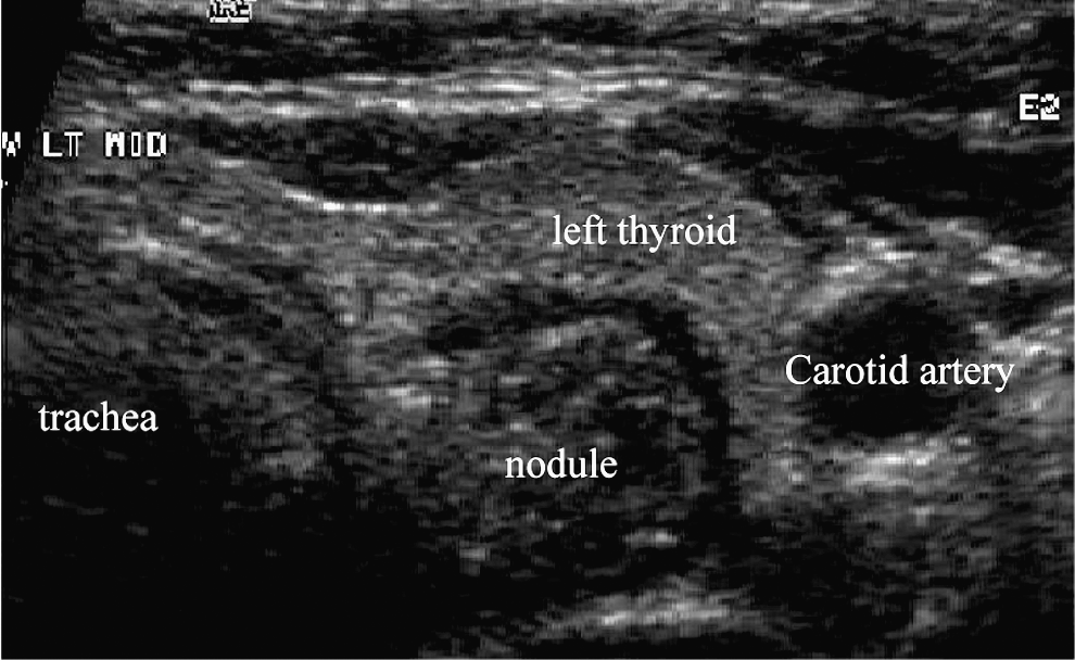
Thyroid ultrasonography showing a Zenker's diverticulum resembling a 2 cm left thyroid nodule.
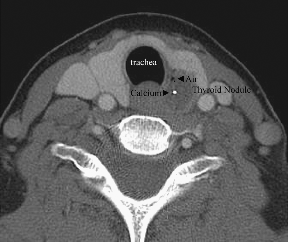
Computed tomography scan of the neck showing a Zenker's diverticulum resembling a calcified left thyroid nodule.
The first patient with a Zenker's diverticulum was described by Albert Ludlow in 1764, but the eponym recognizes Fredreich Albert Zenker for his thorough delineation in 1867 of all previous reports of hypopharyngeal diverticula. A Zenker's diverticulum is a herniation of mucosa and submucosa at Killian's triangle, a natural area of weakness at the junction of the thyropharyngeus and cricopharyngeus muscles in the posterior hypopharynx. The etiology is still unclear, but it is believed that these diverticula are pulsion diverticula occurring as a result of spasm of the cricopharyngeus muscle, incoordination of the pharyngeal muscles, or congenital muscle weakness. Zenker's diverticula protrude posteriorly, and typically project to the left. The diverticulum can appear to be close to the thyroid and may present as a thyroid abnormality on ultrasonography (2,3,5,7). A few articles have examined the sonographic similarities and differences between a Zenker's diverticulum versus a true thyroid abnormality.
On ultrasonography, many Zenker's diverticula show horizontal lines or strong echogenic foci that may appear similar to microcalcifications (1,6,8). However, these findings in fact represent air within the diverticulum. Transient changes of internal echo and size of the lesion are consistent with changes in the contents of the diverticulum. Performing real-time ultrasonography when the patient swallows water, or during compressing the thyroid is extremely helpful in differentiating a Zenker's diverticulum from a true thyroid nodule. The majority of reported Zenker's diverticula found on ultrasonography presented in the posterior aspect of the left lobe of the thyroid, consistent with the observation that large Zenker's tend to protrude to the left. A multilayered pattern may also be seen at the border of the lesion, which is suggestive of the layers of the alimentary tract (1,4,6,8).
FNA biopsy of a left-sided thyroid nodule may be revealing. The absence of follicular cells and presence of squamous cells as well as bacterial or fungal organisms or vegetable matter from fine-needle biopsies should raise the suspicion of a Zenker's diverticulum, thyroglossal duct fistula, or branchial cleft fistula (3,7).
In summary, we present a woman with a sonographically detectable left thyroid nodule that turned out to be a Zenker's diverticulum. Combined with several other reports, she illustrates that sonographic left-sided thyroid nodules that exhibit squamous cells, bacteria, or foreign material on FNA biopsy should raise the suspicion of an occult Zenker's diverticulum.