
Abstract

18Fluorodeoxyglucose (FDG) accumulation in normal thyroid tissue is usually low to absent, while focal 18FDG uptake is reported to be associated with thyroid malignancy (1). Diffuse 18FDG thyroid uptake is typically observed in autoimmune thyroiditis (AIT) due to glucose consumption by lymphocytes and fibroblasts infiltrating the thyroid (2,3). Selenium supplementation has been shown to reduce intrathyroid inflammation and to decrease anti-thyroid peroxidase antibody (TPOAb) titers without affecting thyroid function (4,5). Here we describe a patient with evidence of autoimmune thyroid disease who had a marked decrease in thyroid 18FDG uptake associated with selenium supplementation.
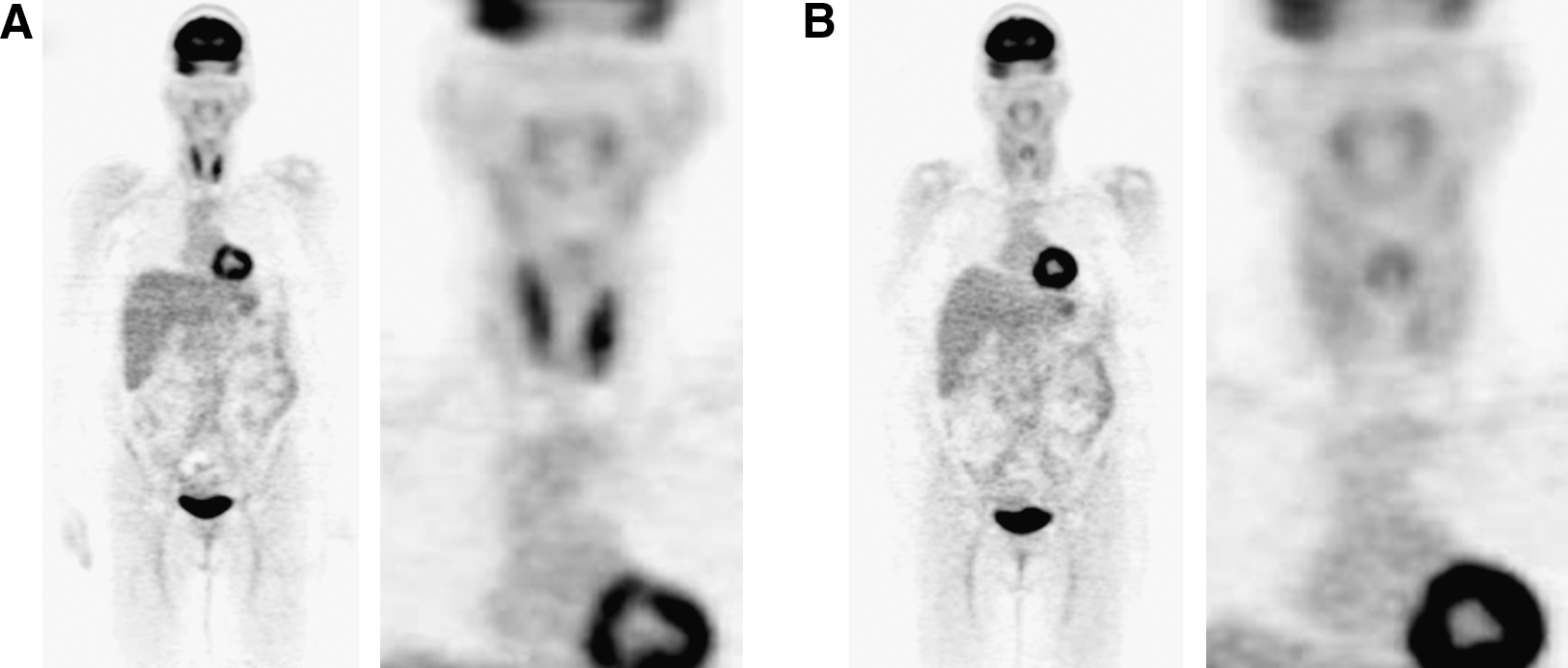
A 47-year-old woman underwent an 18FDG–positron emission tomography/computed tomography (PET/CT) examination in our center to stage a previously resected malignant melanoma of the right arm. The 18FDG-PET/CT was negative for evidence of metastasis, but there was diffuse 18FDG uptake in the thyroid, particularly the left lobe (Fig. 1A). The serum thyroid-stimulating hormone was 1.9 mUI/L (range, 0.4–4.0 mUI/L), and TPOAb was 2860 IU/mL (normal range, 0–9 IU/mL). A thyroid ultrasonography showed a slightly enlarged and diffusely hypoechoic thyroid. Euthyroid AIT was diagnosed and selenium supplementation byL-seleniomethionine (200 μg/d) was started 2 weeks after 18FDG-PET/CT examination. Twelve months later, the 18FDG-PET/CT was repeated for evaluation of metastasis from a possible recurrent melanoma. Again it was negative for evidence of malignancy. There was a marked reduction in thyroid 18FDG uptake, however (Fig. 1B). The serum TPOAb titer was 795 IU/mL, a value above one-third that previously, and there was a slight decrease in thyroid volume and amelioration in echogenic pattern on thyroid ultrasonography (images not shown). The thyroid-stimulating hormone was 1.7 mUI/L. Plasma selenium concentrations, determined by atomic absorption spectrometry, were 0.9 and 1.3 μmol/L, respectively, before and 12 months after L-seleniomethionine supplementation.
18FDG thyroid uptake (
Selenium is an important component in the immune response and selenium deficiency influences the peroxidation of thyroid cell components, leading to an increased rate of thyroid cell necrosis and invasion of macrophages in patients affected by AIT (6). The immune modulatory effects of selenium-dependent enzymes such as glutathione peroxidase and thioredoxin reductase are involved in the organ-specific immune response (i.e., TPOAb synthesis) (6). On the other hand, 18FDG uptake in both fibroblasts and macrophages increases when these cells are exposed to inflammatory cytokines (7). The nuclear factor kappa-B signaling pathway has been associated with enhanced inflammatory response, and its activation has been significantly correlated with interleukin-6 and tumor necrosis factor-alpha production. Selenium may inhibit the activation of nuclear factor kappa-B, thereby attenuating the inflammatory process (8).
In the patient with AIT reported here we noted, for the first time to the best of our knowledge, a dramatic fall in thyroid 18FDG uptake in association with selenium supplementation. Although this report concerns only one patient, the temporal relationship between selenium supplementation and the decrease in thyroid 18FDG uptake as well as theoretical considerations relating to AIT suggest that the selenium supplementation may have caused this patient's decline in 18FDG uptake. Further studies, perhaps performed in conjunction with 18FDG-PET/CT in thyroid cancer patients with and without AIT, would be needed to confirm this hypothesis. The hypothesis, if correct, might lead to the development of methods to evaluate the activity of thyroid inflammation in AIT. However, it is notable that the decline in 18FDG uptake in our patient appeared to be greater than the decline in TPOAb. For this and other reasons it seems unlikely that a decline in 18FDG uptake with selenium supplementation, even if consistently observed, will reflect all aspects of thyroid autoimmunity.
Footnotes
Disclosure Statement
No funds were received for this work.