
Abstract
Background:
Exophthalmos associated with goiter and/or symptoms of hyperthyroidism have been known since antiquity. It was not until around 1800 that a number of studies described this disorder in more detail.
Summary:
For many years the nature of the disease remained unclear and it was appreciated as either a cardiac or neurological disease. There was no agreement on treatment. Surgery on the thyroid, orbit, autonomous nerve system, and pituitary has been employed. Medical treatment was introduced around World War II. Later, as a consequence of the discovery of long-acting thyroid stimulator, it became apparent that Graves–Basedow's disease was an autoimmune disease and so was the exophthalmos, though many details of the pathophysiology remain in doubt.
Conclusions:
This article presents a brief review of the history of the exophthalmos associated with thyroid disease.
The Early Period
Ibn Sina or Abu Ali al-Husayn ibn Abd Allah ibn Sina (980–1037) was a Persian physician, in the West better known as Avicenna. In his most renowned work, Al-Qanoon, from the early 11th century, he gives detailed description of exophthalmos and goiter with symptoms today recognized as evidence of hyperfunction of the thyroid (3). Although Avicenna is not widely recognized in the West, he is undoubtedly a major figure in the history of medicine and of considerable importance in the European academic world until after the Renaissance.
It would be centuries before the combination of symptoms again attracted attention. Caleb Hillier Parry (1755–1822) (Fig. 1) spent most his life as a general practitioner in Bath, England. One of his major contributions was that he recognized the nature of angina pectoris and published a study on this issue: “Inquiry into the Symptoms and Causes of Syncope Anginosa, commonly called Angina Pectoris” (4). The detailed notes of his clinical observations and many of his experiments were not published until 3 years after his death (5). In these notes he described dilation of the colon in newborns (now known as Hirschsprung's disease).
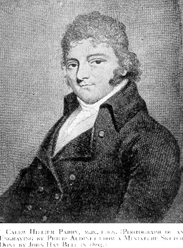
Caleb Hillier Parry (1755–1822).
In the chapter on diseases of the heart, he gave an account of five patients with goiter and obvious hyperthyroidism: “Enlargement of the Thyroid Gland in Connection with Enlargement or Palpitation of the Heart.” One was Mrs. Grace B, 37 years old when first seen by Parry in August 1786. She suffered from palpitations and had an irregular pulse of 156. Her thyroid was enormous “projecting; forwards before the margin of the lower jaw … the eyes were protruded from their sockets and the countenance exhibited an appearance of agitation and distress, especially on any muscular exertion, which I have rarely seen equalled.” The patient was treated with bleedings and a preparation of squill and quicksilver but developed anasarca with nightly orthopnea. Parry was unaware of the later course but assumed that the patient soon “paid her debt to nature.” This brief description is usually taken to be the first on thyroid-associated exophthalmos.
Chronologically, the next description of exophthalmos concurrent with thyroid disease was published anonymously in 1816 in the Medico-Chirurgical Journal and Review. As quoted by Legg (6), “A young lady, 22 years old, had palpitation of the heart, and a swelling on each side of the neck as large as a goose's egg. The eyes had a prominence as though they were about to start from their sockets. She was very nervous, tall, and extremely plethoric. Port wine and bark had done her no good. She was put on a strict antiphlogistic system; she was bled, given nothing but water for drink, and no animal food, calomel, digitalis, &c. Seven months after she was met by chance in the street, quite well.” Legg was able to refer to a few more cases of thyroid tumor associated with exophthalmos published 1820–1828.
Robert Graves (1796–1853) (Fig. 2) (7) in 1834–1835 at the University Hospital in Dublin delivered a series of lectures. One* described three patients with goiter and palpitation (8). The fourth patient was “a lady aged twenty, became affected with some symptoms which were supposed to be hysterical.” She developed tachycardia, swelling of the thyroid and nervousness; “the eyeballs were visibly enlarged, to such a degree the eyelids were unable to shut during sleep and when trying to close the eye. When the eyes were open the white of the eyes could be seen in the breadth of several lines around all of cornea.”

Robert James Graves (1796–1853).
From 1822, Carl Adolph von Basedow (1799–1854) (Fig. 3) practiced medicine in a small town, Merseburg, between Leipzig and Halle in Germany (9). He saw three patients with goiter within a few years (10,11). The first two (Madame G and Madame F) presented with symptoms of florid hyperthyroidism and exophthalmos. In the course of the disease, both were thought to have become insane, and actually Madame F was admitted to a lunatic asylum. One of the women had what may well have been pretibial myxedema or dermopathy.

Carl Adolph von Basedow (1799–1854).
The third patient, Herr M, was a 50-year-old man who in 1832 began feeling malaise and having diarrhea. He suffered from “a heat of the blood,” intense sweating, and oppression of the chest. He had a pale puffy countenance and protruding eyes; the thyroid was enlarged; the patient was emaciated in spite of good appetite and continued to suffer from loose bowels. The course was cruel. After keratitis the patient developed purulent infection in both eyes. Basedow proposed a large incision in the eyeballs to evacuate pus, but the patient rejected this option. Eventually, vision was lost on both sides and, as Basedow noted, “the poor patient suffered unspeakable pain.” As if this was not enough, he developed severe cardiac problems with tachycardia and respiratory difficulties due to the large goiter. The patient died suddenly in 1843 (12).
Legg was not very impressed by Basedow's contribution, apparently being irked by a statement in a German textbook that “Basedow was plainly the true founder of all our knowledge of exophthalmic goitre.” Instead, Legg eulogized Stokes for having made references to Parry's observation. In addition, he called attention to Sir Henry Marsh, who addressed the Pathological Society of Dublin, on January 2, 1841, saying that “it would be, perhaps, in the recollection of many of the members that he had, last year [i.e. 1840] described a singular variety of disease of the heart”; the symptoms included “rapid, violent, and irregular action of the heart, and these, in every instance, co-existing with enlargement and swelling of the thyroid gland.” He had mentioned also that, in the majority of these cases, there was a remarkable prominence and protrusion of the eyeballs, so as to give to the group of symptoms by which this disease was characterized a very striking feature. It seems that Legg was neither fair nor rational when he concluded that “if we are to search for the true founders of our knowledge of exophthalmic goitre, it seems that we must look to Dublin—to Graves, to Marsh, and to Stokes; not to Merseburg.”
In 1877, Yeo (13) first reported on two patients with monolateral exophthalmos (Fig. 4). The reason for this phenomenon remains as enigmatic today as it was more than 130 years ago.

Unilateral exophthalmos and large goiter [from Yeo 1877 (13)].
In this context, two additional names are occasionally brought up. The Italians Flajani and Testa are said to have published similar observations in Italian in 1802 and 1810, respectively (9). Their studies are readily available, however.
Pathological Anatomy
Basedow performed the first autopsy on a patient with thyroid-associated exophthalmos—in fact, on his own patient, Herr M. His findings were certainly not characteristic, as both eyeballs were fibrotic and reduced to half their normal size. There were abundant quantities of yellow fat in the orbits. Jean-Martin Charcot (1825–1893) (14) summed up his results of postmortem studies on the orbits in patients with exophthalmos, confirming that the globe appeared normal but that an extraordinary mass of fat had accumulated within the orbit. Armand Trousseau (1801–1867) (15) cited a Danish article, which reviewed reports that had been published on orbital tissues in thyroid-associated exophthalmos. When looked for, considerable deposits of fat were found in the orbits. Trousseau was able to confirm this finding in one of his own patients.
Moore in 1920 performed a postmortem dissection of the orbits in a patient dying with exophthalmos and Graves–Basedow's disease (16). He summarized the main hypotheses at that time as to the cause of exophthalmos: “It has been variously attributed to three entirely different causes: (1) irritation of the sympathetic nerve producing contraction of unstriped muscle tissue in the orbit; (2) engorgement of the orbit with blood; and (3) increase of the orbital fat.” He argued that as exophthalmos does not disappear postmortem, it could not be caused by sympathetic activity or by vascular disorders. “In this case, however, the orbit was certainly full to overflowing with it, and nothing else abnormal was present; the proptosis was clearly due to the excess of fat. … It seems to me undeniable that increase of orbital fat is the usual cause of the exophthalmos of Graves's disease, and that at present there is no really satisfactory evidence that there is any other cause of it.”
In 1933, Howard Christian Naffziger (1884–1961) (17) published a lengthy account of many studies on orbital structures in exophthalmos—some trying to explain it as being caused by widened or varicose veins or dilated arteries in the orbits or myositis of the extraocular muscles. Naffziger pointed out that in his own material, enlargement of the extraocular muscles had been striking.
Rundle and Pochin (18) conducted a thorough study on the properties of the different tissues within the orbit in exophthalmos. They concluded that the increase in fat in the orbital tissue is responsible for most of the increase in bulk.
Cause
The century following Graves and Basedow saw a large number of publications on the concurrent presence of goiter, heart symptoms, and proptosis. The authors tried to explain the cause of this strange association of symptoms.
Cooper (19) in one of the earliest reviews of protrusion of the eyes and goiter pointed out that two of his five patients erroneously had been diagnosed with hydrophthalmia, obviously meaning glaucoma, and “much mischief was the result of a mercurial course, to which the patients had been subjected.” Cooper did not venture into any attempt to establish the course of the disease but was eager to present his views on treatment: “If there are indications of anaemia, as the bruit in the veins of the neck, if there is palpitation, and, in addition, enlargement of the thyroid gland, there will be strong evidence in favour of the condition of the eyes being secondary; and this view will be strengthened if the patient makes no complaint of pain and uneasiness in the eyes, or of imperfection of vision. The most efficacious remedies are, iron, aloes, and myrrh, with sedatives, ablution of the body with cold salt-and-water, followed by friction, and if there are indications of hysteria, friction along the spine with a stimulating liniment.”
Although it is easy to scoff at theories and ideas advanced more than a century ago under conditions completely different from those today, some of the notions must indeed even at that time have appeared remarkable, almost preposterous. Thus, Jones in 1860 (20) wrote: “The fundamental malady was debility, especially of the nervous system, which, by affecting various vasomotor nerves, gave rise to the several symptoms. Thus effusion behind the globe would cause proptosis; hyperæmia and increased action of the cell elements of the thyroid would produce goitre; and paresis of the vagi would give rise to palpitation and vomiting. The benefit derived from tonic remedies strongly corroborated this view.”
Many authors pointed to cardiac or neurological disturbances as the likely cause. The notion that “exophthalmic goiter” was a neurological disorder was brought forward in 1861 (21). The famed French physicians as Trousseau (15) and Charcot (14) supported this view. So did Pierre Marie (1853–1940)—in an article on the characteristic tremor that Marie thought had been overlooked by most previous investigators (22).
In 1890, Mackenzie (23) published two clinical lectures on Graves–Basedow's disease (completely failing to mention Basedow). Mackenzie referred to Charles Darwin's description of “man in intense fear.” “[With] the condition resulting from severe terror [in mind], we have only to imagine the condition to become prolonged by a failure of the nervous system to recover its balance and to right itself, and we have a more or less complete clinical picture of Graves' disease. We have all the well-known symptoms—trembling, palpitation, flushing, sweating, exophthalmos, and relaxation of the bowels.” Mackenzie concluded: “Such, gentlemen, is what I would suggest to you as the origin and development of this very curious disease. It is likely that the alteration of the function of the thyroid body whose importance in connexion with nutrition and the transmission of nerve force has been amply demonstrated has a good deal to do with many of the secondary symptoms to which I have called attention, but the real disease is a widely distributed derangement of the emotional nervous system.”
Paul Julius Möbius (1853–1907) in a brief but cogent article derided previous notions that Basedow's disease was a blood disease, a heart disease, or a neurological disease (24). He confessed that at a time he himself had speculated that Basedow's disease was a disorder closely related to hysteria. He noted that the symptoms of myxedema in many aspects were exactly opposite to those of hyperthyroidism and suggested that Basedow's disease was an illness of the thyroid gland. He refuted arguments—popular at that time—that the thyroid was a redundant organ or that its function was to regulate cerebral blood flow. He offered no explanation of exophthalmos.
In 1893, George Redmayne Murray (1865–1939)—who in April 1891 first administered thyroid extract to a patient with hypothyroidism (25)—published a judicious article (26). He surmised that “some of the symptoms of exophthalmic goitre are due to over-activity of the thyroid gland.” He argued that “a lessening of its secretory activity would improve the condition of the patient, and this has been found to be the case.” He quoted reports that surgical resection of the thyroid gland had brought about complete cure.
Eventually, it became obvious that the cause of exophthalmos could not be ascribed exclusively to hypersecretion from the thyroid. In some patients cured from hyperthyroidism after thyroid surgery, the exophthalmos remained unchanged or even worsened. In addition, administration of thyroid extract failed to produce exophthalmos.
Hirsch published a review on exophthalmos in which he expressed views that today—just a generation later—have been completely abandoned (27). Thus, he distinguished between “malignant exophthalmos” and “exophthalmos associated with diffuse, toxic goiter”: “Malignant exophthalmos is a clinical entity distinct from exophthalmos as generally seen accompanying thyrotoxicosis. These conditions may be closely related etiologically; yet the diverse clinical pictures they present and the difference in their management make it necessary that the clinician be acquainted with the diagnostic criteria of the disease and the surgeon be familiar with the underlying pathologic process on which the treatment is based.”
It has become clear that in many patients with Graves–Basedow's disease with no clinical orbitopathy, magnetic resonance imaging (MRI) will reveal subtle changes within the extraocular muscles. Although most patients with proptosis have hyperthyroidism, a few will be euthyroid or even hypothyroid (28). The reasons for the variation in clinical manifestations are unclear. Mechanical and/or exogenous factors could play a role—thus, the effect of smoking (29,30) and treatment with radioiodine are well known.
In 1946, Iason (31) was able to list more than 25 proposed causes of exophthalmic goiter. Some were slightly strange (e.g., rupture of the jugular vessels from drinking snow or ice water or adrenal insufficiency).
Solomon and Kleeman have given an excellent account of the various concepts of pathogenesis in Graves' disease (32).
Pituitary Involvement
In 1887—when virtually nothing was known about the function of the pituitary—Minkowski published an article on the relationship between the gland and acromegaly (33). This study would have a decisive impact. Very likely it was crucial for prompting interest in the pituitary leading to a wealth of studies in the first years of the 20th century. Less than a generation later the function of the pituitary as an organ controlling vital endocrine functions had been ascertained; both hyper- and hypofunction were well recognized and pituitary surgery was routinely performed (34).
Studies from around World War I suggested that in larvae and amphibians the pituitary secreted a thyrotrophic substance and this was confirmed to be the case also in mammalians (35 –37). It was speculated that this hormone might play a role in the development of exophthalmic goiter (38). Eventually, Werner in 1953 in a careful review (39) came to the conclusion that thyrotropin (TSH) was not involved in the pathogenesis of Graves' disease. Nonetheless, hypophysectomy was still performed for exophthalmos a decade later (40,41).
The structure of TSH was established in 1971 (42) and proved to be a glycoprotein with α- and β-chains as in gonadotropins. TSH exerts its effect by binding to a protein (TSH receptor) on the surface of the thyroid cell.
Long-Acting Thyroid Stimulator
In 1953, a brief article by Adams and Purves reported that in patients with hyperthyroidism a substance with function similar to that of TSH was present. This study precipitated a flurry of important articles reviewed by Adams in 1965 (43). He was able to conclude that this substance called long-acting thyroid stimulator (LATS) had a longer duration of action than TSH from which it was chemically different, was not produced in the pituitary, and probably was a gamma globulin. It appeared likely that a causal relationship existed between LATS and Graves–Basedow's disease.
In 1964, Meek et al. (44) noted the possibility that the “long-acting thyroid stimulator is indeed an antibody, as the present work indicates, (and) it becomes necessary to consider the presence of LATS as representing a disorder of the body's immune mechanism. In contrast to the usual protective function of antibodies, LATS stimulates the thyroid gland.” Kriss et al. came to the same conclusion and made the important observation that administration of glucocorticosteroids caused a steep decrease in the amount of circulating LATS (45).
At present, a main hypothesis to explain the association between the thyroid component of Graves–Basedow's disease and the orbital manifestations is cross-reactivity of antibodies against antigens shared between the thyroid gland and the orbit. Details are still incompletely understood.
Garrity and Bahn (46) have recently reviewed current concepts of the pathogenesis of the orbitopathy. The orbital fibroblast appears to be the major target cell of the autoimmune process. Some of these cells are capable of producing hyaluronic acid and differentiating into mature adipocytes. Enlargement of the extraocular muscles and expansion of the orbital fatty connective tissues result from abnormal hyaluronic acid accumulation and edema within these tissues and expansion of the orbital adipose tissues.
Lehmann et al. (47) in a later survey made similar conclusions: “The key elements involve binding and activation of orbital fibroblasts by autoantibodies, produced by autoreactive B cell–derived plasma cells. Trafficking of T cells to the orbit ultimately leads to complex interactions of fibroblasts with these T cells and the deposition of extracellular matrix molecules. Proliferation and differentiation of fibroblast subsets into either fat-laden adipocytes or scar-forming myofibroblasts culminates in the increased orbital connective tissue volume and remodeling that underlie the clinical manifestations of thyroid eye disease.”
Therapy
Thyroid surgery
Thyroid surgery has been performed since antiquity. A procedure was recorded as early as 952 AD, when a Moorish physician, Khalaf Egn Abbas (or Albulcasis), removed a large goiter, and for this purpose used hot branding irons! (48). In Europe, thyroid surgery was performed well before 1200 (49). It is agreed that the French surgeon Pierre Joseph Desault in 1791 removed a large thyroid adenoma in a young woman who recovered (whereas his second patient died from disastrous bleeding) (50). Until about 1880 surgeons worried about the vascularity of the thyroid, and at almost any cost avoided surgery on the gland—large suffocating goiters seem to be the only potential exception. The experience of the famous surgeon Theodor Billroth (1829–1894) is telling. From 1860 to 1876 he performed 36 operations for goiter. Thirteen patients did not survive, a mortality rate of 36%. From 1877 to 1881, when antisepsis was firmly in place and surgical techniques had improved, Billroth's operative mortality had been reduced to 8%. If those patients requiring tracheostomy were excluded, the operative mortality was 2.3% (51).
Ludwig Rehn (1849–1930) in 1884 (52) reported on partial thyroidectomy in four patients with goiter, exophthalmos, and cardiac symptoms. This article is very impressive. All four patients were in miserable condition—one seems to have been in overt thyrotoxic storm at the time of operation with “cardiac delirium” (atrial fibrillation with extreme tachycardia), but all were cured. Regularly, Rehn is being cited as the first surgeon to perform surgery for hyperthyroidism. Actually, there are a number of descriptions predating Rehn's publication. Dreesmann (53) reported strange procedures performed on two patients with hyperthyroidism, goiter, and exophthalmos. One was carried out by Macnaughton Jones, who in August 1872 cauterized the thyroid with a chlorine preparation. Ollier in June 1877 also applied cauterization but in addition inserted a canula into the thyroid gland through which over a period of time tincture of iodine was instilled. In both patients the course was claimed to be utterly satisfactory.
Although Joseph Lister (1827–1912) is one of the most renowned surgeons, it is not widely known that he also was one of the very first physicians to operate on a patient with exophthalmic goiter. On July 18, 1877, Lister operated on a young woman with a goiter sufficiently large to cause swallowing problems and periodical severe dyspnea and feeling of being choked. It may well have been these symptoms that persuaded Lister to operate on the patient who also had obvious hyperthyroidism and exophthalmos. “The pulse rate before the operation was about 130. Five days afterwards it had fallen to 72, and the irregularity of the heart's action had disappeared. In a few weeks the exophthalmos had reduced itself very distinctly. The operation, therefore, had relieved all the symptoms of exophthalmic goitre.” Some months postoperatively the patients began having bouts of convulsions (hypoparathyroidism?). It is somewhat odd that Lister did not report this operation, which was not made public until 10 years later at a meeting of a medical society in Edinburgh (54). At that time the patient was alive though she had contracted phthisis.
The French surgeon Tillaux in 1880 successfully performed thyroid surgery in a patient with what he called “exophthalmic goiter,” though his description favors a diagnosis of a simple toxic goiter with suffocation and cardiac symptoms (55).
A major figure in the annals of surgery (and endocrinology) is Theodor Kocher (1841–1917) (Fig. 5) of Berne, Switzerland. Soon after Rehn's publication, he began performing the same procedure and in the following 20 years carried out about 250 such operations (56). In 1907 he had operated on 3333 patients with nontoxic goiter. The operative mortality was 0.3% versus 4.5% in patients with toxic goiter (50). Eventually, in 1909 he was awarded the Nobel Prize.

Theodor Kocher (1841–1917).
Charles Mayo (1865–1939) at the Mayo Clinic (at that time St. Mary's Hospital) in the years after 1889 performed 5000 such operations, of which 2295 were for exophthalmic goiters (57). The cure rate was 75%. Mayo took the occasion to discuss the widespread theory that intoxication was a crucial factor in the pathogenesis (hence the term thyrotoxicosis)—probably via the intestinal canal. He quoted an experiment in which a goat was given boiled water contaminated with residue of feces from a patient who recently developed goiter. The goat promptly developed goiter!
Thus, at the end of the 19th century surgery was an accepted therapy for the goiter and hyperthyroidism in exophthalmic goiter. Briner (58) has given a detailed account of the early surgical procedures. He also mentioned the reported benefits on exophthalmos from application of electrical current.
A major improvement was the introduction of iodine administration preoperatively, often as Lugol's solution. It was introduced in 1923 and caused an immediate decrease in operative mortality (59).
Following the initial report on the benefit of beta-blockers in hyperthyroidism (60) the effect of preoperative administration of the β-blocking agent propranolol was demonstrated (61). Although β-blockers remain important in the treatment of hyperthyroidism, local application into the eye, which initially was advocated as beneficial (62), is no longer in common use.
After the introduction of therapy with radioiodine during World War II, thyroid surgery for Graves–Basedow's disease is being used less frequently, especially in the United States, though surgery still plays an important and sometimes crucial role in special circumstances (63).
Surgery on the sympathetic system
Abadie in 1899 (64) performed section of the cervical sympathetic in patients with severe bilateral exophthalmos. He claimed the operation a huge success with complete cure. He had no doubt that the course proved the procedure to be valuable in the treatment of exophthalmos. Within 1–2 years several other, particularly French, authors (Jaboulay, Jonnesco, Poncet, Reclus, and Faure) had taken up this procedure (65), which in fact was performed well into the 1920s as some still maintained that exophthalmic goiter was a neurological disorder. Cannon et al. insisted that they were able to induce hyperthyroidism in cats by stimulation of the sympathetic nerves (66). As late as 1939 experiments were performed to prove this assertion—though unsuccessfully. The publication by Brain in 1939 (67) seems to have put an end to speculations that exophthalmos is a nervous disorder.
Orbital surgery
Exophthalmos can be an agonizing disease, not only extremely disfiguring but also painful and threatening vision as first described by Basedow. Thus, it is not surprising that the medical society at an early stage pondered how to remedy its course. As early as 1867 v. Graefe proposed local surgery to contain the eyeballs and bring about symptomatic relief (68). On October 20, 1910, Dollinger in Budapest performed the first major operation with the explicit purpose of relieving exophthalmos in Graves–Basedow's disease (69). The patient had no clinical evidence of thyroid hyperfunction but had severe proptosis of his right eye with keratitis (Fig. 6A). Despite large doses of morphine he was in extreme agony. The lateral orbital wall was removed with swift, marked, and lasting improvement (Fig. 6B). This approach was based on a technique developed by Rudolf Ulrich Kroenlein (1847–1910)—one of the imposing surgeons in the latter half of the 20th century (70).

(
It might have been foreseen that within the 100 years after Dollinger's operation, agreement on orbital surgery would have been reached. Oskar Hirsch (1877–1965) (71) in 1930—20 years after his pioneer work on pituitary surgery (72,73)—described removal of the inferior floor. Naffziger (74), who also was a pioneer within pituitary surgery, introduced removal of the orbital roof. Dollinger's article was apparently forgotten and resection of the lateral wall was performed in a few studies 1936–1940 in the United States in the belief that it was a novel procedure (75).
Many other surgical procedures on the bony borders of the orbit have been devised, some including resection of three or four walls of the orbit—indeed hardly a bone of the orbit has not been included in a procedure.
Soft tissue decompression would appear a logical alternative in view of the early recognition that accumulation of fat is an important feature of thyroid-associated exophthalmos. In fact, an early publication (16) reported highly satisfactory outcome following “picking away piecemeal with forceps as much orbital fat as possible.” Hirsch applied the same principle in combination with removal of the inferior floor of the orbit. This principle is still being employed as reviewed by Adenis et al. (76). In addition, various techniques have been developed to adjust affected external ocular muscles and eyelids.
As late as 2009, Leong et al. remarked (77) that “there are myriad techniques in practice for decompressing the orbit, but no one technique has yet to achieve consistently good outcome and low complication rates.” They were able to retrieve from the literature 56 articles in English on this subject and published in the period January 1990–April 2006. Fifteen different surgical techniques were broadly identified.
Radiotherapy
Various modalities of radiation therapy have been used in patients with Graves–Basedow's disease. American surgeons were the first in 1902 to advocate external radiation therapy of the neck. From 1904 several reports were published from Europe (78). Apparently, in some countries radiation became the most frequently employed mode of therapy for goiter and hyperthyroidism.
At the beginning of World War II, trials were begun in the United States with radioactive iodine for treatment of Graves–Basedow's disease and other hyperthyroid states by Hamilton and Lawrence (79) and Hertz and Roberts (80). The first studies, the outcome of which was published in 1946 (81), used a mixture of I130 (about 90%) and I131 (about 10%). The effect in larger series was published a few years later (82,83). In the majority of patients I131 therapy will cure the hyperthyroidism within months (84). Most patients develop permanent hypothyroidism and a minority of patients are quite resistant to radioiodine (84). Moreover, radioiodine therapy causes an increase in the amount of TSH-receptor antibodies in serum (85) that may be associated with worsening of the orbital disease (86). Radioiodine treatment in patients with Graves–Basedow's disease is still a somewhat controversial issue—reflected in the fact that I131 is used much more frequently for this disorder in the United States than in Europe.
A further mode of radiation treatment was first reported in 1929 by Mandeville (87): radiotherapy delivered to the orbit. The outcome only in a small number of patients was reported and the results were conflicting. The first long-term follow-up of the outcome of orbital radiotherapy was not published until almost 30 years later (88), soon to be followed by a blinded comparison between therapy with radiation and with glucocorticosteroid (89).
Medical therapy
It was a major breakthrough when in 1943 Astwood reported that he had tested 106 chemical compounds and found that thiourea derivatives were effective in inhibiting thyroid function (90). Thiouracil was soon widely used in treatment of hyperthyroidism (91).
It appeared that there is a certain relationship between the course of exophthalmos and the treatment of the hyperthyroidism. Often, but not invariably, the eye symptoms improve in parallel to the remission of autoimmunity accompanying normalization of thyroid function during therapy with antithyroid drugs (92). It became apparent that specific therapeutic steps might be necessary in some cases of exophthalmos. With the finding that exophthalmic goiter apparently is an autoimmune disorder, it followed that glucocorticoid treatment might be of value. However, two of the very first trials of steroid treatment were undertaken on the assumption that TSH (or an exophthalmos producing factor different from but similar to TSH) was involved in the pathogenesis and that steroid would inhibit the secretion of this factor from the pituitary (93,94). The Medical Research Council Committee in 1955 conducted a trial with cortisone or corticotropin (ACTH) (95). The results were disappointing—in all probability because the doses of cortisone given were far too small. Systemic glucocorticoid administration proved to be effective, but the amount of steroid necessary was very high (96), a fact that has remained a major problem since. Recently, Marcocci et al. in a randomized study reported that intravenous (instead of oral) administration of steroid yields a more favorable balance between effect on orbitopathy and side effects (97).
Garber reported on the effect of methylprednisolone in a modest dose administered into the subconjuntival space. He concluded that “the method is simple, harmless, and effective” (98). In a later investigation this form of treatment was found to be not very effective (99), but it is still in use (100,101).
Wiersinga and his collaborators first described in a controlled study the effect of steroid versus cyclosporine in treatment of Graves–Basedow's orbitopathy (102).
Thyroid eponymy
It is appropriate for a disease that—though known for centuries but still unsettled as far as exact etiology and optimal treatment are concerned—not even its name can be agreed upon. It seems beyond discussion that Parry was the first to describe—although very briefly—the essential symptoms in exophthalmic goiter. For a good reason then, Sir William Osler maintained that “if the name of any physician is to be associated with the disease, undoubtedly it should be that of the distinguished old Bath physician” (103). Basedow was one of the early authors who gave far the most complete description of the clinical symptoms of exophthalmos in exophthalmic goiter. Sawin (104) has vividly explained why it came to pass that Graves—and not Basedow—was the man who lent his name to the disease in English-speaking countries. Early, it appeared settled that the disorder should be named after Basedow, and Charcot had agreed. From 1862 the issue was discussed at many meetings in the French Academy of Medicine. A decision was made that the most used term “exophthalmic goiter” was misleading, inasmuch as neither exophthalmos nor goiter is invariably present in the patients. Trousseau, the doyen of French medicine, was in favor of the eponym Graves' disease (“il nous faudrait substituer au nom de Basedow celui de Graves … serait dit maladie de Graves”) and managed to get his way.
Worldwide, the term “Graves' disease” is the most used name of the disease, though in parts of the world—for example, in the German-speaking part of the European continent—the formal designation still is Basedow's disease. Obviously, at present it would be completely futile even to contemplate introducing the use of Parry's name, which according to the facts would be the just solution. Seen in the mirror of history, Graves–Basedow's disease would be a fair alternative.
Through the ages, various designations have been used for the exophthalmic component and some are still in use. The most recent term “orbitopathy” should then be termed Graves–Basedow's orbitopathy, appreciating that this is part of Graves–Basedow's disease, but not “thyroid associated” as some patients present with orbital affection exclusively and no, or only marginal, thyroid abnormalities. Graves–Basedow's orbitopathy (not ophthalmopathy) would be an adequate designation as the anatomical substrate of the disease certainly is the orbits, not the globes.
Footnotes
Acknowledgment
We are grateful for the efficient assistance of Mr. Jakob Borg Andersen, Medical Library, Aarhus University Hospital, Aalborg.
Disclosure Statement
The authors declare that no competing financial interests exist.
*
This lecture, “Newly Observed Affection of the Thyroid Gland in Females,” apparently appeared in London Med Surg J 1835;71:516–520. At that time the publisher and the editor had a falling-out, resulting in two versions of the journal being published, causing much confusion and erroneous references. The lecture is re-printed in Medical Classics 1940;5:33–36.