
Abstract
Background:
The risk of second primary malignancies in patients with well-differentiated thyroid cancer is of special interest because of the common use of radioactive iodine (RAI) ablation and/or treatment of these patients and the theoretical risk of subsequent nonthyroid malignancies associated with the radiation exposure. This brief report focuses specifically on the occurrence of second primary malignancies of the salivary glands. RAI residency within salivary tissues is known to have both acute and chronic consequences on salivary function, but secondary neoplasia is quite unusual.
Summary:
We present a very rare case of a patient with papillary thyroid cancer treated with 600 mCi of RAI, who subsequently developed salivary gland cancer.
Conclusions:
We recommend salivary gland protection to diminish potential side effects after the exposure to radioiodine. On the basis of our experience we suggest administration of sialogogues (such as lemon juice) continuously, every 30–60 minutes for 24 hours, after RAI administration.
Introduction
In addition to thyroid follicular cells and the salivary glands, other tissues that transport iodine and therefore might be at risk for RAI-related SPM include gastric mucosa, choroid plexus, and breast. A possible association of breast cancer with thyroid disease, in general (9 –11), and RAI therapy, in particular, has been considered (12). Other organs will be exposed to RAI without having an active iodide transport process during the therapeutic administration of RAI for WDTC. The excretory system would be at risk for collateral damage by ionizing radiation because the isotope makes its way through the kidneys, ureters, and bladder. The risk of SPM in patients with thyroid cancer has been reviewed recently (13), and the rarity of an SPM of the salivary glands after high doses of RAI administration is remarkable (3,4). We present a very rare case of a patient with papillary thyroid cancer (PTC) treated with 600 mCi of RAI, who subsequently developed salivary gland cancer.
Patient Presentation
The patient is a 47-year-old Caucasian woman referred to our medical center in 2009. Her history dates to May 1990, when she presented with a small nodule in the right lobe of the thyroid and underwent a partial right lobectomy. She was advised that nodule was benign and treated with L-thyroxine because of documented hypothyroidism. The patient did well until April 2005, when a lump was noted in her neck and a thyroid ultrasonography documented a 1-cm nodule in the left lobe, and her dose of L-thyroxine increased. In January 2006, a repeat ultrasonography showed no significant change, but fine-needle aspiration of the nodule was interpreted as follicular neoplasm, and a completion thyroidectomy was performed in April 2006. The surgical pathology report described a multinodular goiter with background Hashimoto's thyroiditis, Hurthle cell adenoma, and a 1-cm PTC in the left lobe. The patient was classified as stage I (T1, N0, M0), started on L-thyroxine 0.15 mg, and prepared for radioiodine ablation using recombinant human thyrotropin (rhTSH; Thyrogen®). On May 17, 2006, an rhTSH-stimulated thyroglobulin (Tg) was 3.9 ng/mL with positive anti-Tg antibodies (anti-Tg Ab) of 39 IU/mL (normal <40) and positive anti-thyroid peroxidase antibodies (anti-TPO Ab) of 332 IU/mL (normal <35). A diagnostic scan post-rhTSH showed some intense uptake in the left thyroid bed and some mild uptake in the right thyroid bed. The patient received rhTSH for the radioiodine therapy and was given 143 mCi on June 14, 2006. Five months later on December 13, 2006, her rhTSH-stimulated Tg was undetectable at <0.2 ng/mL with strongly positive anti-Tg Ab at 86 IU/mL (normal <60) and anti-TPO Ab >1300 IU/mL (normal <60). A scan showed a positive intense focus in the lower thyroid bed. Because of this finding, she was treated again with 149 mCi on April 18, 2007. The next evaluation on October 3, 2007, included undetectable Tg and positive anti-Tg Ab, and a post-rhTSH scan showed positive uptake in the lower neck. She was administered a third dose of radioiodine of 152 mCi on January 16, 2008, and 2 months later on August 6, 2008, a post-rhTSH scan again showed positive uptake in the lower neck with a Tg of <0.2 ng/mL with interfering anti-Tg Ab 82 IU/mL and anti-TPO Ab 1274 IU/mL. The persistent positive neck uptake led to her fourth dose of radioiodine of 150 mCi on September 4, 2008.
Laboratory studies on February 9, 2009, after rhTSH stimulation included a TSH of 85, Tg <0.2, anti-Tg Ab 84 IU/mL, and anti-TPO Ab 553 IU/mL. The whole-body scan (WBS) showed a focus of uptake in the right paracentral neck, with the suggestion that this might be a lymph node. On March 18, 2009, an [18F]-fluorodeoxyglucose (FDG) positron emission tomography (PET) scan showed no foci of FDG uptake corresponding to the focus of radiodine uptake described in WBS, but showed a positive uptake in the left parotid gland with standard uptake value 5.3 (Fig. 1). This finding was consistent with physical examination, which had revealed a 1.5-cm mass in the area of the parotid gland that was nontender to palpation.

Positron emission tomography/computed tomography of the head with uptake in the left parotid gland, with standard uptake value 5.3.
The patient underwent fine-needle aspiration of the parotid gland, which was read as “possible monomorphic adenoma.” Subsequently, in October 2009, a left parotidectomy was performed with a histopathological diagnosis consistent with carcinoma ex-pleomorphic adenoma (CaExPA). The greatest dimension of the tumor was 1.5 cm, Grade G2– moderately differentiated, N0 (2 lymph nodes examined). Distance of the tumor from closest margin was 0.1 cm, with no venous/lymphatic or perineural invasion observed (Fig. 2). Nevertheless, because of aggressive behavior of CaExPA, external beam radiotherapy was initiated.
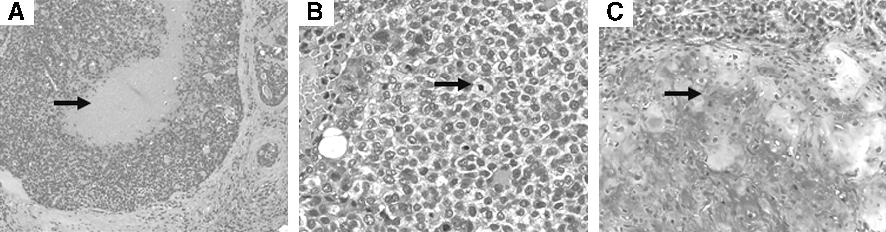
Histopathology of patient's carcinoma ex-pleomorphic adenoma of the left parotid gland. (
Discussion
The risks of damage to the salivary glands after RAI therapy for thyroid cancer have been well described (14 –16). The patient described in this report raises the more unique question of the possible relationship of RAI treatment to development of a secondary malignancy of the salivary glands.
Whether it was appropriate to treat this patient with stage I PTC with four doses of ∼150 mCi to reach a cumulative activity 600 mCi of RAI is beyond the scope of this report, but does raise questions about a link between this therapeutic approach and the development of her salivary gland cancer. Current guidelines for thyroid cancer treatment underline the necessity of individualized treatment with RAI. For most patients with a unifocal 1-cm tumor, without lymph node metastases and with histopathology of classic PTC, RAI treatment would not be recommended (17). In this patient, the repetitive RAI therapies were based on the findings on WBS, because Tg measurements were not reliable due to interfering anti-Tg Ab. Nevertheless, the differential diagnosis for persistent uptake in the midline lower neck in a low risk patient should have included false-positive findings such as an esophageal diverticulum, and imaging procedures to rule this out were warranted.
The occurrence of a CaExPA in the parotid gland after 600 mCi RAI therapy is the issue of concern. High-dose RAI administration has been linked to an increased risk of tumors of the salivary gland (2 –4). An indisputable etiologic relationship between RAI therapy and SPM of the salivary glands is difficult to establish definitively because of the very low numbers of patients reported (1–2 patients in some cases). Other studies have not shown a significant relationship between RAI treatment and increased risk of salivary gland cancer (5,8,18). In a modified meta-analysis of all SPMs (not confined to salivary neoplasms), pooled analysis of three European cohorts noted a dose–response relationship between RAI and development of SPMs (Table 1). The authors proposed that repeated use of RAI should be restricted only to thyroid cancer patients in whom it may be beneficial (4). We would underscore that these analyses indicating a relationship of SPMs to therapy for thyroid cancer should be interpreted with caution because of limitations of their retrospective design and possible surveillance bias, misclassification bias, and the inability often to standardize histology review.
RAI, radioactive iodine; SPMs, second primary malignancies.
A recent meta-analysis including two multicenter studies concluded that there is a slightly increased risk of an SPM, including salivary gland cancer, in thyroid cancer survivors treated with RAI compared to patients not treated with RAI (13). While this may be true, these patient populations historically may reflect radiation exposures in excess of those experienced by patients receiving RAI therapy today due to increasing use of diuretics, laxatives, and rhTSH-mediated ablation with less radiation exposure than experienced after thyroxine-withdrawal-mediated therapy (19).
Nevertheless, any risk from radiation exposure should be avoided and it is important to consider any and all means to prevent the salivary gland damage from administered RAI in view of the probable connection between that exposure and the subsequent development of the salivary gland cancer. This problem was not sufficiently addressed in the recently published guidelines of the American Thyroid Association (ATA) for the management of thyroid cancer, possibly due to the lack of randomized trials showing the effect of salivary gland protection strategies (20). Indeed, there is some controversy over current approaches to salivary gland radiation protection. Taking into consideration the physiologic mechanism of radioiodine uptake in salivary gland tissues—extraction of iodide from blood in the periductal capillaries, concentration by the ductal epithelium, and secretion into the ductal lumen and then into the oral cavity—the reasonable approach would be to suppress salivary radioiodine uptake and to stimulate salivation (21,22).
Nakada et al. studied 116 consecutive patients who were instructed to suck one or two lemon candies every 2–3 hours in the daytime for 5 days starting within 1 hour (group 1; 116 patients) or 24 hours (group 2; 139 patients) after RAI therapy (23). The incidence of salivary gland side-effect was determined prospectively over >24 months. The authors reported an increased rate of acute sialoadenitis and taste dysfunction in patients who sucked lemon candies within 1 hour after therapy compared to those with the delayed start. The authors proposed that induction of salivation early, when the level of radioiodine in the blood was high, delivered more radioiodine to the salivary glands.
On the other hand, Van Nostrand et al. in one case study demonstrated a clear potential benefit from early and repeated salivary stimulation after 131-therapy. The single administration of lemon juice reduced the salivary gland radiation exposure in the first hour by 30%–38% and repeated administration in a second hour by 61%–67% (24). This study suggested that sialogogues should be administered continuously, every 30–60 minutes, after RAI administration.
Other approaches to the reduction of salivary radiation exposure have been taken. Bohuslavizki et al. and Mendoza et al. reported salivary gland protection using the sialogogue, amifostine, 500 mg/m2 intravenously or subcutaneously (25,26), but no benefit was seen in a randomized trial that employed 300 mg/m2 doses of amifostine (27). Convincing evidence of radiation protection by any of the above approaches remains to be determined.
In regard to the etiology of the parotid cancer in our patient, we cannot know with certainty that the tumor was a consequence of her RAI treatment. Such malignancies after RAI therapy are very rare—with only 14 documented cases (1). Moreover, the specific histopathology of CaExPA has not been directly linked to a history of radiation. In the largest reported series of 73 patients with CaExPA, only 3 cases were identified as having prior radiation therapy as a possible etiologic factor (28). The CaExPA tumor accounts for 4.5%–15% of all salivary gland cancers and arises from pleomorphic adenomas, which are composed of both epithelial cells and myoepithelial cells that produce a mesenchymal stromal component. CaExPA is typically a high-grade carcinoma with frequent metastases and poor clinical outcome (29 –33) and may be difficult to diagnose because the mixed tumor component is often small and overlooked. Based upon the degree of invasion present, CaExPA can be subdivided into three prognostic subtypes: noninvasive (in situ, intracapsular), minimally invasive (extracapsular invasion <1.5 mm), and invasive (extracapsular invasion >1.5 mm). Depending on the subtype or degree of invasiveness, the 5-year survival rate can range from >90% (in situ) to <20% (invasive >5 mm).
The clinical presentation of CaExPA is extremely variable ranging from an asymptomatic parotid mass in 86% to a painful mass with facial nerve paresis or paralysis and skin ulceration. The last three symptoms significantly affect survival. Management depends on prognostic factors with surgery alone recommended for a carcinoma in situ, whereas adjunctive external beam radiation therapy would be recommended in case of invasion and has been shown to effectively prevent local recurrences (34 –36).
Conclusions
The case presented underscores the necessity of proper risk–benefit estimation in patients treated with high cumulative dosages of radioiodine, considering that cumulative activities above 500–600 mCi are associated with significant increased risk of SPMs. Recommendation 71 of the recently revised ATA guidelines for management of thyroid cancer indicates that all patients should be encouraged to seek age-appropriate screenings for cancer. There is no evidence of a benefit of more intensive screening. The guidelines also suggest that patients who received the cumulative activity of radioiodine in excess of 500–600 mCi should be advised that they may have increased risk of leukemia and solid tumors in the future (17).
There is no ATA recommendation for or against the routine use of sialogogues or measures of salivary gland radiation exposure to prevent salivary gland damage. The latter is a consequence of the lack of sufficient evidence-based data. In our opinion, efforts should be encouraged to reduce radiation exposure from radioiodine to the thyroid cancer patients. On the basis of our experience, we suggest administration of sialogogues (such as lemon juice) continuously, every 30–60 minutes for 24 hours, after RAI administration (24).
Footnotes
Disclosure Statement
No competing financial interests exist.