
Abstract
Background:
Killian-Jamieson diverticulum (KJD) is a rare esophageal diverticulum that protrudes through the muscular gap in the anterolateral wall of the proximal cervical esophagus inferior to the cricopharyngeus. Although several patients with bilateral KJD have been reported, high-resolution ultrasound (US) image of bilateral KJD has never been reported.
Summary:
A 71-year-old man visited our hospital for annual follow-up of known thyroid nodules. In addition to the previously noted thyroid nodules, two arc-shaped hyperechoic lesions with reverberation artifacts, which suggested air-containing lesions, were unexpectedly seen posterior to bilateral thyroid lobes. Although the connection between the esophagus and the lesions was not definitely visible on US, our first differential diagnosis was esophageal diverticula. They were confirmed to be bilateral KJD on the following pharyngoesophagography.
Conclusions:
If US features are not enough to differentiate esophageal diverticulum from suspicious thyroid nodule, pharyngoesophagography can be performed instead of invasive fine-needle aspiration to obtain a definite diagnosis of incidentally detected esophageal diverticulum.
Introduction
Patient
A 71-year-old man visited our hospital for annual follow-up of known thyroid nodules. US was performed using a real-time linear array unit with 5–12 MHz transducer (iU22; Philips Medical Systems). In addition to the previously noted thyroid nodules, two arc-shaped hyperechoic lesions with reverberation artifacts (Fig. 1) were unexpectedly seen posterior to bilateral thyroid lobes. These hyperechoic lesions had a hypoehoic boundary and changed in position and shape when the patient was swallowing. They did not have connection to the esophageal wall when visualized, but the US feature of two arc-shaped hyperechoic lesions with reverberation artifacts suggested air-containing lesions such as esophageal diverticula. Pharyngoesophagography was performed after informed consent and it revealed two outpouching structures protruding from the lateral walls of the cervical esophagus on both sides (Fig. 2), consistent with bilateral KJD.
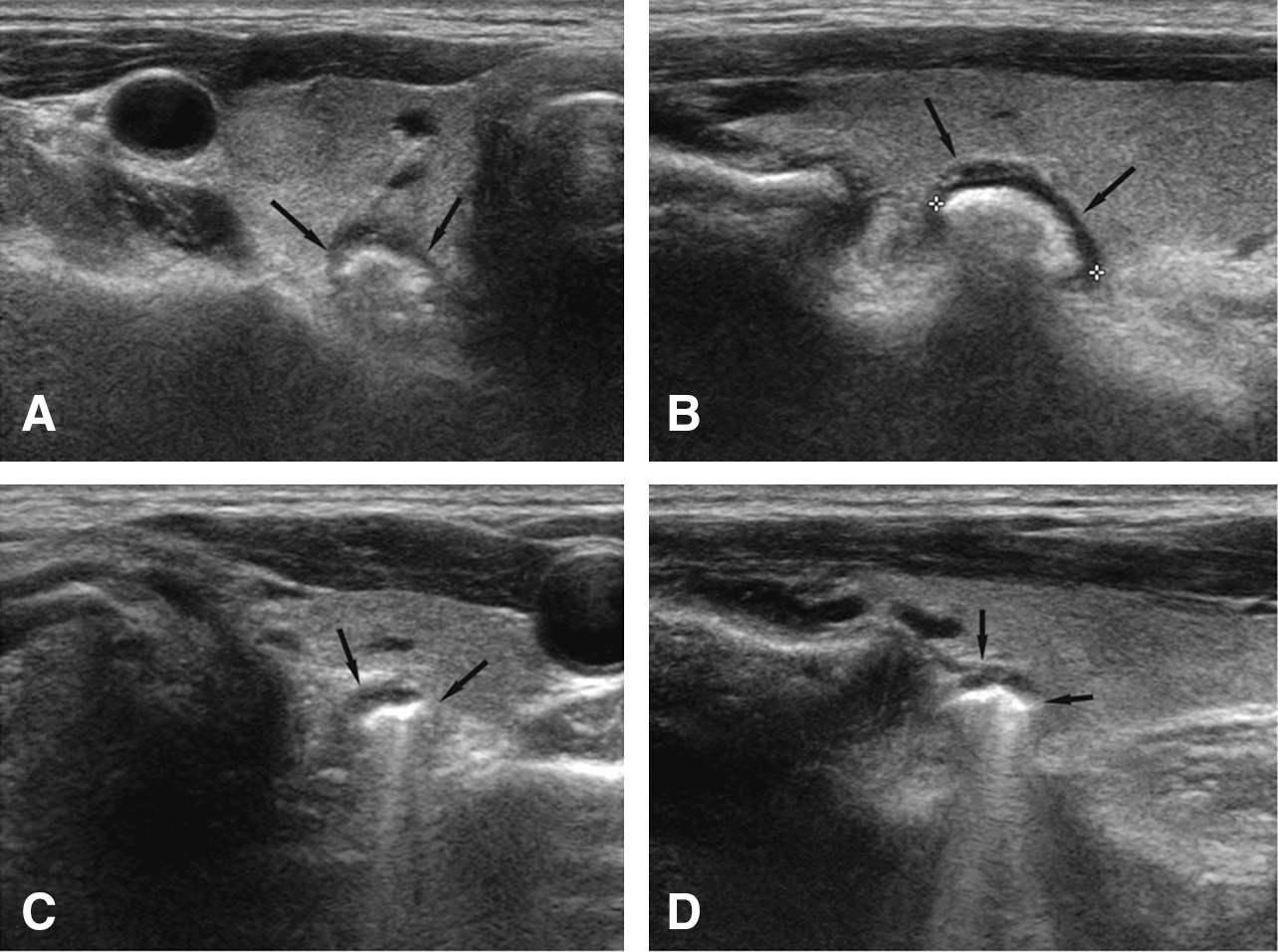
Ultrasonography (

Pharyngoesophagography shows two outpouching structures protruding from the lateral walls of the cervical esophagus on both sides, which suggests bilateral Killian-Jamieson diverticula.
Discussion
KJD is a rare esophageal diverticulum that protrudes through the muscular gap in the anterolateral wall of the proximal cervical esophagus inferior to the cricopharyngeus. This gap is known as Killian-Jamieson space (2). On pharyngoesophagography, it can be differentiated from Zenker's diverticulum, which has an outpouching from the muscular gap in the posterior portion of the cricopharyngeus (also known as Killian's dehiscence) (1,2). Zenker's diverticulum is four times more common than KJD (1).
With increased use of US for thyroid nodules, the frequency of incidentally detected extrathyroidal lesions has also increased (3). Extrathyroidal lesions sometimes mimic malignant thyroid nodules on US, leading to unnecessary fine-needle aspiration, as noted by Oertel et al. (4). Air bubbles in a diverticulum may mimic calcifications within a thyroid nodule and the hypoechoic mural portion of a diverticulum may mimic a hypoechoic solid portion of thyroid nodule. If the lesion is an esophageal diverticulum, it may be in contiguity with the cervical esophagus, move separately from the thyroid gland, or show changes in shape while the patient is swallowing (5). If US features are not enough to differentiate esophageal diverticulum from suspicious thyroid nodule, pharyngoesophagography can be performed instead of invasive fine-needle aspiration to obtain a definite diagnosis of incidentally detected esophageal diverticulum (5).
Footnotes
Disclosure Statement
The authors declare that no competing financial interests exist.