
Abstract
Background:
Substernal goiters are frequently associated with compressive symptoms. Compression of the trachea and esophagus are common, whereas thoracic duct compression is a rare occurrence.
Methods:
We report a rare case of a 72-year-old woman with thoracic duct compression by a large substernal goiter that presented with shortness of breath. After undergoing thoracentesis multiple times, the patient was treated with thyroidectomy.
Results:
Transcervical thyroidectomy was performed without sternotomy. This led to resolution of her symptoms. Confirmation of chylothorax resolution was obtained with postoperative computed tomography of the chest.
Conclusion:
Chylothorax is a rare sequela of substernal goiters. It can be managed with thyroidectomy. Sternotomy was avoided in this case.
Introduction
Case Report
A 72-year-old woman was referred to the otolaryngology clinic for evaluation of her substernal goiter. She had a 6-month history of worsening shortness of breath. Computed tomography (CT) of the chest showed a pleural effusion on the right (Fig. 1). Initial differential diagnosis included infections process versus malignant effusion. An initial thoracentesis was performed and evaluated with cytology and gram stain. Gram stain did not reveal bacteria and cytology showed evidence of lymphocytes without evidence of lymphoma or other malignant cells. The effusion returned rapidly over several days as well as dyspnea. Multiple thoracenteses were performed over the next few months and chylous leak was confirmed with elevated triglycerides at 212 mg/dL. Thoracenteses were required approximately every 3 weeks due to progressive shortness of breath despite the patient adhering to low-fat diet. After undergoing an additional preoperative thoracentesis, a transcervical approach to removal of her substernal thyroid goiter was performed (Fig. 2). A sternotomy was not required for removal and no thoracic duct repair or ligation was necessary. A closed suction drain was left in the wound overnight. There were no signs of chyle leak at the time of surgery or in the postoperative period. She had an uneventful recovery with resolution of her shortness of breath, and no further thoracentesis was required. Postoperative CT at 3 months showed no residual chylothorax (Fig. 3).
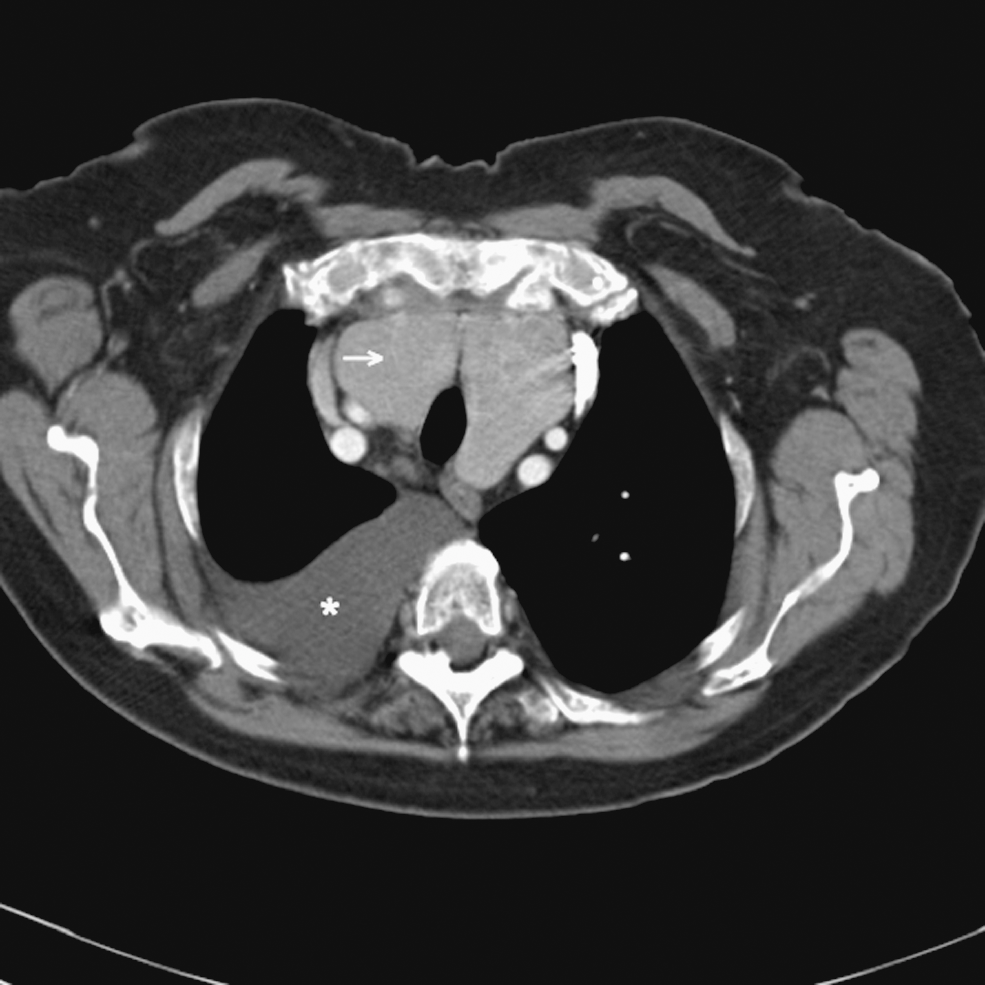
Preoperative computed tomography of chest. Shown are large chylous pleural effusion (asterisk) in the right hemithorax as well as large thyroid goiter (arrow) located behind the manubrium.
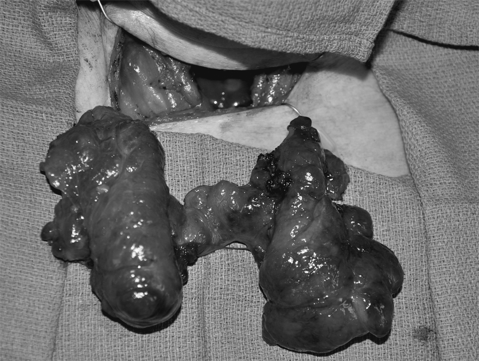
Operative photo of removed substernal thyroid goiter.
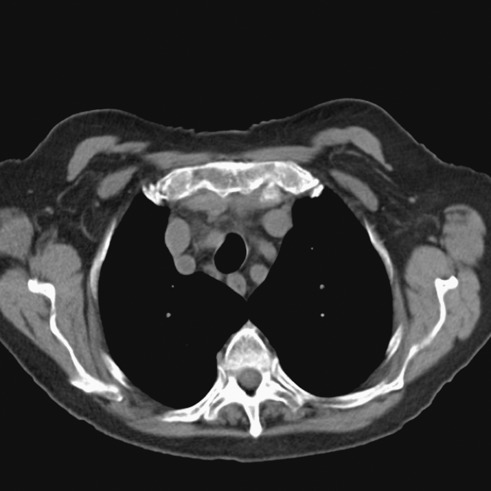
Postoperative computed tomography of chest. There is now resolution of chylous pleural effusion.
Discussion
Chylous pleural effusion can be caused by congentital, traumatic, and neoplastic processes (5). In association with thyroid disease, it has been frequently described after thyroid surgery (6 –8). It has also been described in association with Graves' disease in patients with thyrotoxic cardiomyopathy (9). However, it is rarely described in association with substernal goiters with only four cases reported in the English literature (10 –13). Of the four cases described, treatment required a sternotomy and even thoracic duct repair in one case (10,12–13). One patient was treated with I131 (11). Radioactive iodine treatment has been shown to decrease the volume of nontoxic thyroid goiters by more than one-half at 2 years (14). This reduction of gland volume is the likely reason for decrease external compression of the thoracic duct though this may take several months to achieve a significant size reduction. The current case highlights this rare complication of substernal goiter and the potential surgical treatment. The patient presented with several months of shortness of breath requiring frequent thoracenteses. The presentation was similar to other reported cases of goiter-associated chylothoraces with dyspnea.
The presumed mechanism is external compression of the thoracic duct. The thoracic duct travels along the right side of the esophagus before crossing to the left side in the mediastinum at the fifth or sixth thoracic vertebrae. It then travels superiorly along the esophagus before turning left and draining at the junction of the left internal jugular and subclavian veins. Compression can occur at any point along the course. A substernal goiter can cause compression if it has significant posterior extension, especially if it extends inferiorly into the thoracic inlet. The current case had significant extension in both the posterior and inferior directions. The external compression can be relieved with removal of the goiter and thoracic duct repair is not necessary, at least in this case. Likely, with benign disease and careful dissection, thoracic duct repair is not necessary in these patients. That must be determined at the time of surgery depending on whether a leak is identified. If the mechanism of the chylothorax is by external compression, then it is unlikely that a chyle leak will be identified in the neck. However, if a leak is identified at the time of surgery, it may be repaired. This repair can be performed through a transcervical or mediastinal approach depending on the location of the leak. Typically, this involves ligation of the thoracic duct. If the chylothorax persists after removal of the external compression, other measures may be necessary. These include thoracic duct ligation in the chest or pleurodesis and/or pleurectomy (15). More recently, there has been success with lymphangiography and embolization in patiets with postoperative chyle leaks (16). In the patient reported here there was no need for thoracic duct ligation and the goiter was completely removed through a transcervical approach without the need for a sternotomy as is the case with the vast majority of substernal goiters. There have been are two other cases of goiter-associated pleural effusions treated with thyroidectomy; both required sternotomy (10,12). The patient described here was the first to be treated with pure transcervical excision. Successful resolution was documented with postoperative CT.
Conclusion
Substernal goiters often present with compressive symptoms. Thoracic duct compression occurs rarely and may require thoracentesis to relieve the shortness of breath. Transcervical removal of the goiter without thoracic duct repair may be an appropriate treatment to relieve symptoms.
Footnotes
Disclosure Statement
The authors declare that no competing financial interests exist.