
Abstract
Background:
Percutaneous ethanol injection (PEI) is used to treat cystic or mixed benign thyroid nodules. This treatment can result in rare complications, and three cases of Graves' disease (GD) without Graves' ophthalmopathy (GO) have been reported after PEI treatment of toxic thyroid adenomas. Here we present a 55-year-old male patient who developed GD and severe GO after PEI treatment of a mixed cystic-solid, nontoxic thyroid nodule.
Patient Findings:
Six months after PEI, the nodule volume had decreased from 8.9 to 3.0 mL, but we observed severe hyperthyroidism with elevated serum free triiodothyronine, free thyroxine, and thyrotropin receptor antibody levels. We also observed ophthalmopathy with symmetrical orbit and soft tissue involvement (grade b/c) and a clinical activity score of 4/7. The diagnosis of GO was confirmed by bilateral corneal damage, increased intraocular pressure on upgaze, and inconstant diplopia. A computed tomography scan showed that the inferior, medial, and superior extraocular muscles were bilaterally enlarged, the perineural space at the orbital cone was slightly reduced and the ophthalmic vein was congested.
Summary:
A cause-effect relationship between PEI and GD/GO was likely in this patient because of the temporal sequence. Although the mechanism was unknown, we speculated that the thyroid tissue damage caused by PEI released a large amount of antigenic materials from follicular thyroid cells, including thyrotropin receptor protein, which triggered the autoimmune inflammatory response against the thyroid itself and the orbital soft tissues. The patient did not have any risk factors for either GD or GO.
Conclusions:
This observation raises the concern, therefore, that unpredictable and severe complications, such as GD and GO, may occur in a few patients treated with PEI.
Introduction
Levothyroxine therapy is the most widely used approach and is based upon suppression of thyrotropin (TSH) secretion as a potential growth factor for the nodule. Levothyroxine therapy has low efficacy, and safety may be compromised when TSH-suppressive therapy is administered (2 –4). All other techniques are based upon destruction or damage of the nodular tissue. Radioiodine is the treatment of choice for toxic nodules and may also be indicated to reduce compressive symptoms from nontoxic nodular goiter. After radioiodine therapy of toxic nodular goiter, Graves' disease (GD) may develop in up to 5% of patients (5).
The other nonsurgical options (PLA and PEI) are not commonly used because they require specifically trained medical personnel and are restricted to selected patients (6). Thermal ablation by photocoagulation of solid thyroid nodules was first introduced in 2000 (7). Although small clinical trials have supported its efficacy and safety, PLA has not been widely used (8 –10). Mild to intense pain is the most frequent side effect, which results from parenchymal edema and thyroid capsule thermal damage (about 20% of treated cases) (11). Interestingly, one study showed that 2/122 PLA-treated patients had a transient thyrotropin receptor antibody (TRAb) peak with hyperthyroidism one month after PLA. Both patients required treatment with either methimazole or radioiodine (11).
PEI was first introduced in 1990 (12) as a possible therapy for autonomously functioning thyroid nodules, but PEI is no longer recommended for solid nodules. Currently, PEI is only used for cystic or mixed cystic-solid benign thyroid nodules with a large fluid component (13 –18). PEI is a relatively safe technique, and the most common side effects are local pain, flushing, dizziness, and transient dysphonia. In three rare cases, GD was reported after PEI treatment of an autonomous thyroid adenoma (19 –21), but all cases were without Graves' ophthalmopathy (GO). Here we present a patient with normal thyroid function who developed hyperthyroidism with severe GO after PEI treatment of a mixed cystic-solid thyroid nodule.
Patient
A 55-year-old man was referred to our thyroid clinic because of hyperthyroidism and thyroid-related orbitopathy that rapidly developed after PEI treatment of a benign thyroid nodule (performed in another hospital). The patient had no family history of thyroid disease. The patient's thyroid history was significant with a nontoxic nodular goiter, which had been diagnosed 10 years earlier when a 30×27×34-mm (volume 14.3 mL), mixed cystic-solid nodule in the left thyroid lobe with a large intranodular fluid component was documented in an ultrasound scan. TSH, free triiodothyronine (FT3), and free thyroxine (FT4) serum levels were in the normal range. Antithyroglobulin (AbTg) and antithyroid peroxidase antibodies (AbTPO) were absent. Fine needle aspiration of the nodule indicated a follicular lesion. Surgery was suggested, but the patient refused surgery and was placed on levothyroxine therapy (112.5 μg/day) that was maintained for 8 years and resulted in a small reduction in the nodule volume. FT4 and FT3 serum levels were always in the normal range, and serum TSH values ranged between <0.01 and 0.3 mU/L, which indicated persistent subclinical hyperthyroidism due to levothyroxine “suppressive” treatment. Because of potential side effects of subclinical hyperthyroidism due to this treatment in an aging patient, PEI was proposed as an alternative therapy. Cyst evacuation (3 mL) plus ultrasound-guided PEI was performed by a skilled operator, and 2 mL ethanol was injected. Biochemical data, the nodule volume, and levothyroxine therapy before and after PEI are shown in Table 1.
FT3, free triiodothyronine; FT4, free thyroxine; AbTg, antithyroglobulin antibody; AbTPO, antithyroid peroxidase antibody; TSH, thyrotropin; TRAb, thyrotropin receptor antibody; LT4, levothyroxine.
Two weeks after PEI, the patient complained of excessive tearing and swelling of his eyelids. Levothyroxine therapy was reduced to 75 μg/day, but a blood measurement 3 months later showed that TSH was suppressed (<0.01 mU/L). FT3 and FT4 were in the normal range, and the patient was negative for AbTg and AbTPO. Because of the eye symptoms, we examined the TRAb level and found that it was slightly elevated (25.0 U/L, normal values 0–10). The nodule volume was reduced to 5.7 mL. Levothyroxine treatment was stopped after 4 weeks, but one month later the serum TSH level was still <0.01 mU/L and serum FT3 had abnormally increased to 4.8 pg/mL; however, FT4 was still normal (16.2 pg/mL). Moreover, TRAb levels had increased to 62.0 U/L.
The patient was referred to our center. Six months after PEI, he presented with fatigue, tachycardia, sweating, photophobia, excess tearing, grittiness, and diplopia. Laboratory data indicated that serum TSH was <0.01 mU/L, serum FT3 and FT4 levels were elevated (8.0 and 28.8 pg/mL, respectively) and TRAb levels had increased to 81.8 U/L. In addition, AbTg and AbTPO were still absent. An I-131 thyroid scan showed a diffuse tracer uptake in a normal size thyroid gland with no evidence of the nodule, and thyroid radioiodine uptake values were within the normal range. Echocolordoppler revealed increased gland vascularization with a nodule volume of 3 mL. FNA indicated a benign lesion.
Ophthalmopathy evaluation showed symmetrical eyelid edema, caruncle and plica swelling, and conjunctival redness with relevant chemosis (>30% of the conjunctival surface was involved). According to the EUGOGO Atlas, soft tissue involvement was classified as grade b/c bilaterally (22). The clinical activity score was 4/7, which indicated an active phase of autoimmune orbital soft tissue involvement. Hertel measurements were 19 and 18 mm for the right and left eyes, respectively, and eye movements were reduced by about 30% on upgaze. According to the Gorman score, the patient had inconstant diplopia on upgaze.
Confocal microscopy (Nidek–CS4 confocal microscope) demonstrated the presence of initial corneal layer damage with a significant reduction in cell count and morphological changes, including cell polymegathism >80% (normal values <30%) and pleomorphism <45% (normal values >59.6%) (23). Visual acuity was 10/10 according to Snellen lines. No significant abnormality of the visual field (Humphrey, with a sensitivity threshold of 30–2) was detected, but intraocular pressure was increased on upgaze (22 mm Hg bilaterally).
Using computer tomography with a Lightspeed Multidetector (GE Medical Systems, Milwaukee, WI) and a helicoidal capture technique of imaging and scanning orbits with contiguous 1.25-mm-thick slices (200 mA, 120 kV, pitch 0.5) and electronic reconstruction of 2-mm thickness on a coronal plane in respect to the neuro-optic axis, we found a bilateral enlargement of the inferior, medial, and superior extraocular muscles. The perineural space at the orbital cone was slightly reduced, and the ophthalmic vein was congested. No area of fat degeneration was detected inside the involved orbital muscles (Fig. 1).
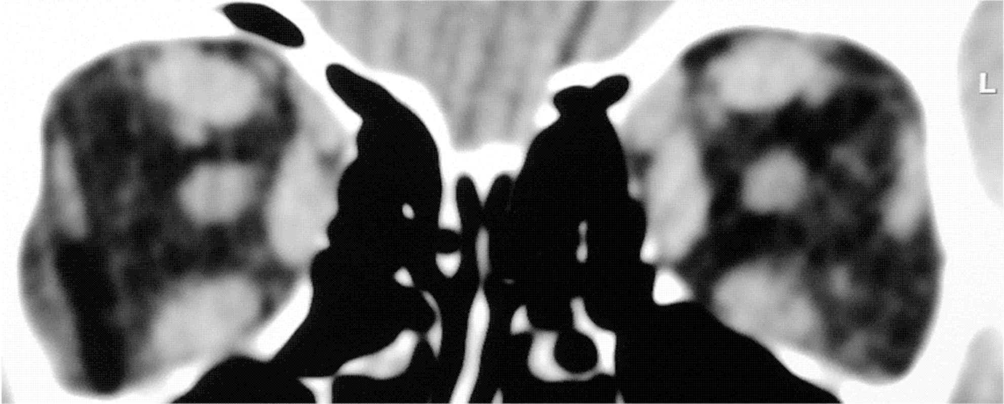
This computed tomography scan shows a bilateral enlargement of the inferior, medial, and superior extraocular muscles. The perineural space at the orbital cone is slightly reduced, and the ophthalmic vein is congested.
Based upon clinical, laboratory, and radiological findings, a diagnosis of GD with associated severe GO was made. Methimazole, 30 mg/day, and a beta-adrenergic inhibitor were administered for hyperthyroidism. Two methylprednisolone pulse therapy courses (9-g cumulative dose) were also given with one retrobulbar radiotherapy course (10 Gy for eye) for ophthalmopathy. At the last observation, which was 2 years after PEI, the patient was euthyroid without antithyroid therapy and had inactive moderate ophthalmopathy with moderate involvement of the soft tissues and deficit of the ocular motility on upgaze. Informed consent was obtained by the patient for the use of these data in a scientific publication.
Discussion
Graves' hyperthyroidism is caused by autoantibody (TRAb) activation of the thyrotropin receptor (TSHr) on thyroid follicular cells. Most patients with GO have increased levels of TRAb, which suggests that immunoreactivity against the TSHr is a link between GD and GO. The TSHr protein, which is the major target antigen in GD, is also expressed in orbital fibroblasts and adipocytes. When activated, T-lymphocytes react with TSHr, which is expressed in thyroid and orbital tissue, and a cascade of events is triggered that produces lymphocytic infiltration, differentiation of orbital fibroblasts into adipocytes, hydrophilic glycosaminoglycan accumulation, and expansion of retrobulbar soft tissues (i.e., the signs of orbital autoimmune inflammation) (24).
Several risk factors for GO development have been identified: gender, genetic background, environment, mechanical factors, and factors related to thyroid function (25). In addition, women have a greater risk of developing GO (2:1 ratio). Genetic factors might influence susceptibility to the disease, and there may be multiple susceptibility alleles that could interact with nongenetic factors to contribute to the different phenotypic expression of GO in different patients. Other studies have demonstrated a correlation between the amount of cigarette smoking and the development of GO. The risk for GO development and exacerbation is also documented after radioiodine treatment of GD (15%–39% of cases). Anatomical characteristics of the orbital walls, elevated serum FT3 values, and positive TRAb are additional risk factors for GO development and/or exacerbation (25). Interestingly, none of these risk factors were present in our patient. Our patient was a male subject with no family history of autoimmune disease. Moreover, he had no clinical signs of hyperthyroidism before PEI, and he had normal serum FT3 and FT4 values during levothyroxine therapy. The suppressed TSH serum value was likely due to the “suppressive” levothyroxine treatment. In addition, AbTg and AbTPO were absent. Unfortunately, the TRAb level was not measured before PEI because there was no reason to measure it in the absence of any signs or symptoms. Symptoms related to hyperthyroidism and GO started a few weeks after PEI, which suggested that the development of GD and GO in this patient was causally linked to PEI. Although the spontaneous appearance of GD cannot be ruled out, the temporal sequence of clinical and laboratory signs supported a direct correlation between the thyroid nodule PEI procedure and the onset of GD and severe GO. Interestingly, GD and GO are complications that can be expected after procedures, including PEI, that extensively damage follicular thyroid cells.
The mechanism for the causal relationship between PEI, GD, and GO development is not known. We speculated that damage to thyroid tissue (by large needle sections and ethanol) in subjects with a genetic susceptibility for autoimmune reactions, even when risk factors, such as antithyroid antibodies, smoking, and family history, are absent, may release a large amount of antigenic materials, including TSHr protein, from follicular thyroid cells. This antigenic material may trigger an autoimmune inflammatory response against the thyroid itself and the orbital soft tissues that could lead to both GD and GO. This pathogenetic sequence may be applicable to all causes of thyroid tissue trauma and damage (26,27), including radioiodine treatment of GD and nodular goiter (5), PLA treatment of thyroid nodules (11), and even cosmetic botulinum toxin injection (28).
However, in this patient ethanol injection did not trigger AbTg appearance, as it might have expected because of thyrocytes destruction (20). Therefore, a pre-existent form of subclinical GD with low circulating levels of TRAb cannot be ruled out.
Our patient is the first case of both GD and GO observed after PEI treatment of a nonfunctioning thyroid nodule because all three previous cases occurred after PEI treatment of hyperfunctioning thyroid nodules, and no GO was observed (19 –21). Because no risk factor for either GD or GO was present before PEI, these complications are unpredictable and may also occur in euthyroid patients without any risk factors. Although serious complications, such as GD and GO, are rare, they should be considered in patients who undergo the PEI procedure.
Footnotes
Disclosure Statement
The authors declare that no competing financial interests exist.