




Abstract
Background:
The occurrence of thyroid carcinoma in patients with congenital hypothyroidism (CH) caused by dyshormonogenesis is very rare, and has only been reported in one patient harboring mutations in the thyroid peroxidase (TPO) gene.
Patient Findings:
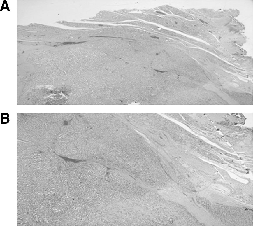
We report on a 29-year follow-up of two consanguineous siblings with CH due to total iodide organification defect who also had sensorineural hearing loss. Molecular analysis revealed a novel biallelic mutation of the TPO gene in which phenylalanine substitutes serine at codon 292 (c.875C>T, p.S292F) in exon 8. Despite early initiation, adequate doses of levothyroxine treatment and consequently normal thyrotropin (TSH) levels, the proposita developed a huge multinodular goiter (MNG) and underwent total thyroidectomy due to tracheal compression. Pathological examination revealed a unifocal follicular thyroid carcinoma without vascular invasion in the left lobe of the thyroid gland.
Summary:
Our finding of follicular thyroid carcinoma arising from dyshormonogenetic MNG in a patient without elevated serum TSH levels indicates that genetic and environmental factors other than TSH level might be involved in the development of thyroid carcinoma in dyshormonogenetic MNG.
Conclusions:
Despite the rare occurrence of thyroid carcinoma in dyshormonogenetic MNG, we recommend long-term follow-up and regular neck ultrasound imaging to prevent delayed diagnosis of thyroid carcinoma.
Introduction
TPO mutations are the most common cause of an enzymatic thyroid synthesis defect, responsible for more than 50% of the CH cases due to dyshormonogenesis (5). The clinical characteristics include CH and goiter, in some cases attaining huge sizes. Rapid and elevated radioactive iodine uptake (RAIU) with significant discharge of thyroidal radioiodine after perchlorate administration is consistent with an iodide organification defect (IOD) (6). The TPO gene is located on the short arm of chromosome 2 (2p25) and comprises 17 exons. It covers ∼150 kb of genomic DNA. TPO is a membrane-bound glycoprotein located at the apical membrane of the thyroid follicular cells that catalyzes both iodination and coupling of iodotyrosine residues within the TG molecule, leading to the synthesis of thyroid hormone. TPO defects are inherited in an autosomal recessive manner (5). To date, more than 50 different mutations of the TPO gene have been described (MIM #606765). Goiter may be present at birth or develop with time. We described the presence of three different TPO mutations in 22 patients living in the same geographical area, in the northern region of Israel: 44% of the patients developed multinodular goiter (MNG) over the years, with both hot and cold nodules demonstrated by 99mTC scan (7). Among them, four patients underwent thyroidectomy due to pressure symptoms and follicular hyperplasia in fine-needle aspiration. However, pathological findings excluded thyroid carcinoma (7). Occurrence of thyroid carcinoma has been reported in about 1%–4% of patients with MNG (8), but the precise rate of thyroid carcinoma arising from dyshormonogenetic MNG is not known. In 1981, 17 such cases were reported but without confirmed genetic analysis (9). Over the last two decades, with the advent of commonly available molecular analyses, 11 cases with confirmed mutations have been reported: 9 with TG mutations (10 –12), 1 with PDS mutation (13), and 1 case in an infant with a monoallelic TPO mutation (14). Herein we describe a 29-year follow-up of two siblings with the TPO mutation S292F, resulting in substitution of serine with phenylalanine in position 292 of exon 8 (c.875C>T, p.S292F). The proposita manifested with goitrous CH that progressed over the years to a huge MNG harboring follicular thyroid carcinoma.
Patient 1
The proband, a 29-year-old woman of Arab-Muslim origin, was born to parents who were first-degree cousins. She was admitted at the age of 3 weeks due to prolonged jaundice, hoarse cry, goiter, and constipation. Primary CH was identified by neonatal screening (thyrotropin [TSH] 183 mIU/L, normal range <10 mIU/L; total thyroxine [TT4] 1.5 μg/dL, normal range >7 μg/dL). Laboratory tests confirmed the diagnosis of CH (TSH 75 mIU/L; TT4 9.6 μg/dL; total triiodothyronine [TT3] 38 ng/dL) and levothyroxine (
Age at examination.
FT4, free thyroxine;
Patient 2
The younger 28-year-old brother of the index case was initially admitted at the age of 52 days because of prolonged jaundice and bilateral pneumonia. His thyroid gland was not palpable. Neonatal screening was consistent with primary CH (TSH 46.0 mU/L; TT4 0.7 μg/dL).
Discussion
We describe the occurrence of noninvasive follicular thyroid carcinoma in a woman with goitrous CH due to a TPO mutation. This is the second reported case of a thyroid carcinoma in an affected patient with a TPO mutation.
In MNG, the prevalence of thyroid carcinoma has been estimated at 1%–4% (8). Vickery (9) reported 17 cases of thyroid carcinomas in dyshormonogenetic MNG, among them 4 cases with vascular invasion and 6 patients with hearing loss suggesting a diagnosis of Pendred syndrome. However, it should be noted that hearing impairment may result from late diagnosis and inadequate treatment of CH of other etiologies as well (15). Cooper et al. (16) were the first to describe congenital dyshormonogenetic goiter with malignancy and metastatic spread. An additional two reports of thyroid carcinoma in patients with Pended syndrome (17,18) and dyshormonogenetic goiter were described (19,20). Unfortunately molecular analysis was not available in any of those cases. However, over the last two decades, with the increasing availability of molecular analyses, 11 cases of thyroid carcinoma have been reported: 9 in patients with TG mutations (10
–12), 1 in a patient with a PDS mutation (13), and only 1 case with a TPO mutation (14) (Table 2). Both papillary and follicular carcinomas have been described. It has been postulated that long-standing elevated serum TSH levels is the main cause for the development of thyroid carcinoma arising from dyshormonogenetic MNG (21,22). TSH is a well-known growth factor for thyroid epithelial cells and can promote thyroid nodule formation and cancer progression (22,23). Nonsuppressed TSH appears to increase the risk of recurrence of thyroid carcinoma, supporting a crucial role for TSH in thyroid cell proliferation. Furthermore, constitutively activating TSH receptor mutations result in toxic adenoma and hyperfunctioning MNGs, and they have been found rarely in patients with hyperfunctioning thyroid carcinomas (24). Histological changes such as microfollicular nodules and cytological atypia can develop under prolonged TSH stimulation (16,25). However, the association between elevated TSH and the occurrence of thyroid cancer has not been conclusively established. In our cohort of 22 patients with TPO mutations, we observed a high rate of MNG but none showed malignant changes (7). In the present report, the fact that the index case was treated with
Age at operation.
Age at onset.
hetero, heterozygous; homo: homozygous; PDS, pendrin; TG, thyroglobulin; TPO, thyroid peroxidase.
Congenital goiter is a rare presentation of dyshormonogenesis and appears in only 10% of affected patients with TPO mutations (7). Indeed, the only case of thyroid carcinoma in a patient with TPO mutations occurred in an infant born with goitrous CH; this patient was found to have a metastatic follicular cancer already at birth (14). That report indicated that the thyroid carcinoma had existed in utero, arguing against the effect of long-standing elevated TSH as the main cause for the development of thyroid carcinoma. It maybe speculated that congenital goiter increases the risk of thyroid carcinoma development. Indeed, the brother of the proposita, who had the same TPO mutation and a normal-sized thyroid gland at birth, did not develop a thyroid tumor. However, some reports of thyroid carcinoma arising from dyshormonogenetic goiter have been described in patients without congenital goiter (11,13). The high concentrations of H2O2 produced in response to increased TSH levels might lead to DNA damage and could have mutagenic effects (26,27). Furthermore, organic iodocompounds have been shown to inhibit thyroid epithelial cell proliferation (23), and TPO mutations resulting in IOD might therefore increase the risk for MNG. The same mechanism may apply to Pendred syndrome, where iodide transport through the apical membrane is interrupted.
The development of a tumor depends on either stimulating factors or a lack of tumor-suppressor genes. Over the last two decades, significant progress has been made in understanding the genetic events in follicular cell–derived thyroid tumors. RET/PTC and PAX8-PPARγ chromosomal rearrangement and BRAF and RAS point mutations have been identified in more than 70% of papillary and follicular thyroid carcinomas (27). Activating point mutations of the RAS genes occur in follicular carcinomas and adenomas, poorly differentiated and anaplastic carcinomas, and some papillary carcinomas (28,29). Screening for PAX8-PPARγ chromosomal rearrangement and BRAF and RAS point mutations was negative in one patient with follicular carcinoma and a TG mutation (10), but positive for BRAF mutations in two others (12).
The mechanisms of thyroid cancer development in dyshormonogenesis remain to be elucidated. It might result from a combination of environmental factors, such as iodine deficiency and long-standing elevated TSH, which increase H2O2 concentration and activation or suppression of genes involved in thyroid cell proliferation. In this study, we present the second report of thyroid carcinoma in a patient with a TPO gene mutation. Although thyroid carcinoma is not common in dyshormonogenetic MNG, long-term follow-up by regular neck ultrasonographic imaging followed by fine-needle aspiration, if indicated, is recommended to avoid a delayed diagnosis of thyroid carcinoma in these cases.
Footnotes
Acknowledgment
The authors thank Camille Vainstein for professional language editing.
Disclosure Statement
The authors declare that no competing financial interests exist.