
Abstract
Background:
Polypeptide N-acetylgalactosaminyl transferase-3 (GalNAc-T3) has been reportedly expressed in several human adenocarcinomas and is associated with clinicopathological features of tumors. We investigated the clinicopathological significance of GalNAc-T3 in thyroid cancer.
Methods:
We evaluated the expression of GalNAc-T3 in 167 patients with thyroid cancer using a specific antibody and analyzed the association between its expression and clinicopathological features.
Results:
GalNAc-T3 was expressed in 85.8% of normal follicular epithelial cells. In papillary carcinomas, positive staining was observed in 101 (73.7%) cases. Well-differentiated components (papillary and follicular) of papillary carcinomas were significantly more frequently positive than poorly differentiated components (trabecular and solid) (p<0.01), and GalNAc-T3 was highly expressed in papillary carcinomas that had invaded beyond the thyroid capsule (p=0.026). GalNAc-T3 was expressed in 40% and 20% of well and poorly differentiated components of follicular carcinomas, respectively. Thirteen of 15 anaplastic carcinomas were negative for GalNAc-T3 and thyroglobulin. Positive staining for GalNAc-T3 was not observed in any of the medullary carcinomas.
Conclusions:
Our data suggest that GalNAc-T3 expression may be a useful indicator of tumor differentiation in thyroid carcinomas.
Introduction
T
It is known that structural changes in cell surface glycoproteins influence the biological behavior of cancer cells during malignant transformation and tumor progression (4,5). Mucin glycoproteins are widely distributed on the epithelial cell surface and play important functional roles in cell adhesion, cell differentiation, proliferation, carcinogenesis, cancer metastasis, and basic immunological systems (6 –8). Mucin-type O-linked carbohydrates constitute approximately 80% of the total molecular mass of these glycoproteins, and O-linked carbohydrate antigens, such as carcinoembryonic antigen, CA19-9, sialyl LewisX, sialyl Tn, and Tn, have been reported to be associated with invasion, recurrence, and prognosis in cancer patients (9 –15). Thus, O-glycosylation has been shown to play important roles in tumor progression (16 –18).
UDP-GalNAc:polypeptide N-acetylgalactosaminyl transferases (GalNAc-Ts) are a key enzyme family of more than 20 isoforms that catalyze the O-glycosylation reaction, the primary step of alpha-O-glycoside bond formation between GalNAc and serine/threonine residues, on the mucin scaffold (19 –21). Although GalNAc-T3 is one of the isoforms expressed in various organs, its expression level differs among them. In normal tissues, GalNAc-T3 is expressed at higher levels in the pancreas and testis and at lower levels in the kidney, prostate, spleen, ovary, intestine, and colon, whereas GalNAc-T3 mRNA has been detected in organs containing secretary epithelial glands (19,22 –24). It is hypothesized that the differential expression of GalNAc-T3 may affect the specialized functions of glycoproteins produced by normal and malignant cells, and an association between GalNAc-T3 and the biological properties of both normal and malignant cells has been demonstrated (18). Previous studies have demonstrated that the expression of GalNAc-T3 is associated with the differentiation, aggressiveness, and prognosis of gastric (25), pulmonary (26), colorectal (27), gallbladder (28), bile duct (29), pancreatic (30), and esophageal carcinomas (31). However, to the best of our knowledge, GalNAc-T3 expression has not been evaluated in thyroid carcinomas. The purpose of this study was to evaluate the relation between GalNAc-T3 expression and pathological features in thyroid carcinomas.
Materials and Methods
Patients and clinical materials
This study was conducted according to the ethics guidelines of the Declaration of Helsinki, and specific approval was obtained from the Ethics Committee of Shinshu University School of Medicine. The specimens studied were obtained from 168 patients with thyroid cancers who were diagnosed and treated in Shinshu University Hospital from 1995 to 2005. Of 167 cases, 134 were papillary carcinomas, 11 follicular carcinomas, 15 anaplastic carcinomas, and 7 medullary carcinomas. All patients except eight with anaplastic carcinomas underwent surgical resection. The eight patients with anaplastic carcinomas were treated with radiation and/or chemotherapy. In 141 of 160 patients who underwent surgical resection, normal thyroid gland tissue was also observed. Clinicopathological data were obtained by retrospective chart review.
Immunohistochemical staining and evaluation
Polyclonal antibodies against human GalNAc-T3 were generated by multiple immunization of a New Zealand white rabbit using synthetic peptides as described previously (32). A monoclonal antibody to thyroglobulin (1D4) was purchased from Santa Cruz Biotechnology, Inc. (Santa Cruz, CA). A formalin-fixed, paraffin-embedded 3 μm section was obtained from all 168 primary lesions. Sections were deparaffinized in xylene, hydrated through a graded series of ethanol, and immersed in 3% hydrogen peroxide in 100% methanol for 30 minutes to inhibit endogenous peroxidase activity. To activate the antigens, the sections were boiled in 10 mM citrate buffer (pH 6.0) for 12 minutes. After rinsing in phosphate-buffered saline (PBS), the sections were incubated with normal goat serum for 10 minutes, and then incubated overnight at 4°C in a humidity chamber with the primary antibody to GalNAc-T3 at 1/40,000 dilution. After washing thrice with PBS, the sections were incubated with biotinylated anti-rabbit immunoglobulin for 60 minutes. After washing again with PBS, the sections were incubated for 60 minutes with avidin and a biotinylated horseradish peroxidase macromolecular complex. Diaminobenzidine was used as a chromogen, and the sections were lightly counterstained with hematoxylin. Cells were judged positive for GalNAc-T3 expression when granulated cytoplasmic staining was observed under high-power magnification (10×40).
Statistical analysis
The relationship between clinicopathological features and the expression of GalNAc-T3 was examined by Fisher's exact test. Differences with a p-value of less than 0.05 were considered statistically significant. The chi-square test was used for immunohistochemical analysis of the clinical specimens.
Results
GalNAc-T3 expression in normal thyroid gland
To evaluate the expression of GalNAc-T3 in the normal thyroid gland, 141 normal thyroid tissues were stained for GalNAc-T3 expression. GalNAc-T3 was expressed in 121 of 141 (85.8%) normal follicular epithelial cells (Fig. 1). The association between GalNAc-T3 expression in the normal thyroid gland and both sex and age is summarized in Table 1. The expression of GalNAc-T3 was observed more frequently in the normal thyroid gland of male patients compared with female patients; however, there was no statistical significance between GalNAc-T3 expression and sex or age.

Expression of GalNAc-T3 in the normal thyroid gland.
GalNAc-T3, polypeptide N-acetylgalactosaminyl transferase-3.
GalNAc-T3 expression in thyroid carcinomas
In thyroid carcinomas, the biological behavior of the tumor is known to depend mainly on its histological type. To evaluate differences in expression of GalNAc-T3 with the histological type, 167 thyroid carcinoma tissue samples (134 papillary, 11 follicular, 15 anaplastic, 7 medullary) were immunohistochemically analyzed for GalNAc-T3 expression (Table 2; Fig. 2). In papillary carcinomas, positive staining for GalNAc-T3 was observed in 101 of 134 tumors (73.7%), whereas in follicular carcinomas, the expression of GalNAc-T3 was detected in 5 of 11 tumors (45.5%). In contrast, only 2 of 15 (13.3%) anaplastic carcinomas and none of 7 (0%) medullary tumors were positive for GalNAc-T3. Thus, GalNAc-T3 was significantly more frequently expressed in the differentiated carcinomas (papillary and follicular) compared with the other entities (p<0.001).
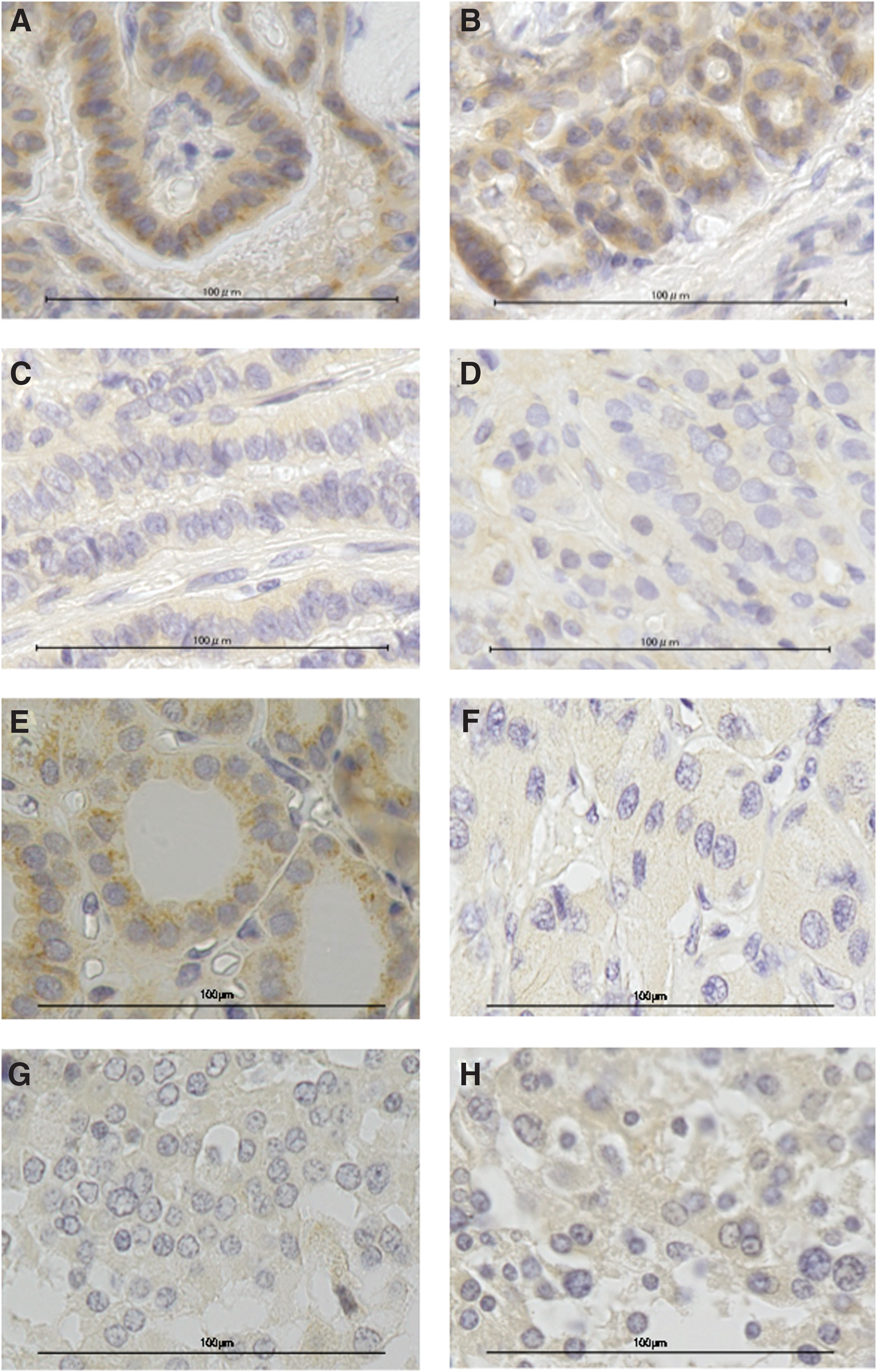
Expression of GalNAc-T3 in thyroid carcinomas. Representative findings of immunohistochemical analyses in the papillary carcinoma: positive staining in
GalNAc-T3 expression in papillary thyroid carcinomas
As the expression of GalNAc-T3 was detected most frequently in papillary carcinomas, we further evaluated the association between GalNAc-T3 expression and its clinicopathological features. The clinicopathological features of 134 papillary carcinomas are summarized in Table 3. As differentiated papillary carcinoma often comprises diverse histological components, we examined the expression of GalNAc-T3 in individual histological components of papillary carcinoma classified pathologically (Fig. 2). The well-differentiated components of papillary thyroid carcinomas were mainly papillary (Fig. 2A) and/or follicular (Fig. 2B). In contrast, poorly differentiated components were mainly trabecular (Fig. 2C) and/or solid (Fig. 2D). Two hundred fifteen well-differentiated components (102 papillary and 113 follicular) and 89 poorly differentiated components (49 trabecular and 40 solid) were detected in 134 tumors. We next evaluated GalNAc-T3 expression in individual components. Positive staining for GalNAc-T3 was detected in 152 (70.7%) of 215 well-differentiated components, whereas only 13 (14.6%) of 89 poorly differentiated components were positive for GalNAc-T3. Thus, the well-differentiated components of papillary carcinomas showed GalNAc-T3 expression significantly more frequently compared with poorly differentiated components (p<0.001). With regard to other clinicopathological factors, a statistically significant association between the expression of GalNac-T3 and the extent of extraglandular tumor invasion was observed (p<0.05).
GalNAc-T3 expression in follicular thyroid carcinomas
With regard to follicular thyroid carcinomas, 5 (45.5%) of 11 tumors expressed GalNAc-T3. Follicular carcinomas consist of both well-differentiated (follicular) and poorly differentiated components (trabecular or solid). We evaluated the expression of GalNAc-T3 in individual components (Table 4; Fig. 2). Ten well-differentiated and 10 poorly differentiated components were detected in 11 follicular carcinomas (Fig. 2E, F). The expression of GalNAc-T3 was more frequently observed in the well-differentiated components compared with the poorly differentiated components (40.0% vs. 20.0%, respectively); however, no significant difference was observed.
Expression of thyroglobulin and GalNAc-T3 in differentiated thyroid carcinomas
Thyroglobulin is a protein produced in the follicular cells of the normal thyroid gland and in differentiated carcinoma cells. Consequently, thyroglobulin is used as a marker for the differentiation of both normal and malignant thyroid tissue. To evaluate any correlation between GalNAc-T3 expression and the production of thyroglobulin in thyroid carcinomas, the expression of both GalNAc-T3 and thyroglobulin was assessed by immunostaining (Table 5; Fig. 3A, B). In papillary carcinomas, thyroglobulin was detected in 208 of 215 (96.7%) well-differentiated components and in 72 of 89 (80.9%) poorly differentiated components. However, no significant association was observed between the expression of thyroglobulin and GalNAc-T3. With regard to follicular carcinomas, thyroglobulin was detected in all tumors except one, and no correlation between the expression of thyroglobulin and GalNAc-T3 was observed. With regard to medullary carcinomas, thyroglobulin and GalNAc-T3 were not detected in any of the studied tumors (Fig. 2G, H).
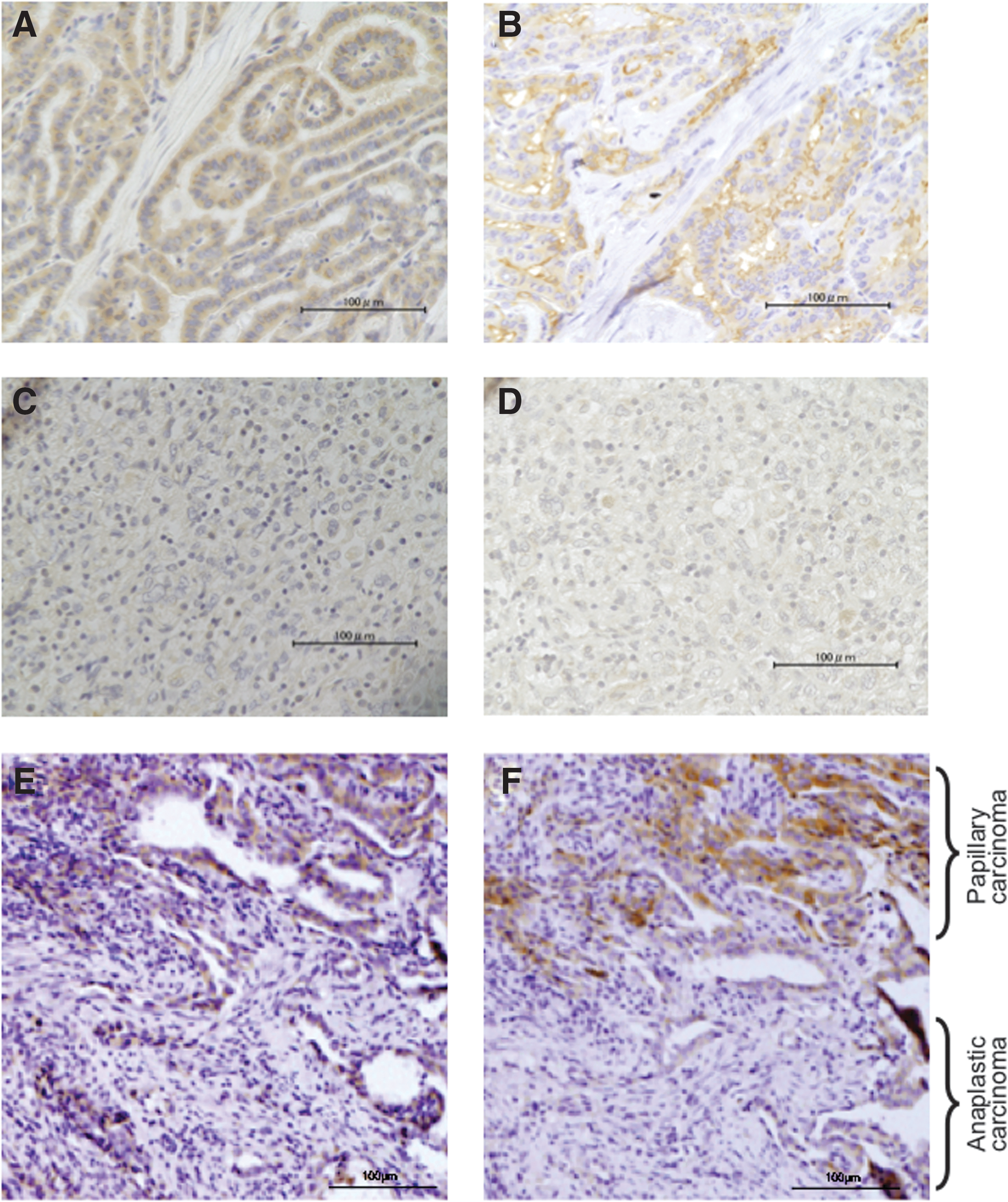
Expression of GalNAc-T3 and thyroglobulin in papillary and anaplastic thyroid carcinomas. Representative findings of immunohistochemical analyses of GalNAc-T3 and thyroglobulin expression in serial sections of the tumor. Both GalNAc-T3
NA, not available.
Expression of thyroglobulin and GalNAc-T3 in anaplastic thyroid carcinomas
In anaplastic thyroid carcinomas, the expression of GalNAc-T3 was not detected in 13 of 15 tumors, and the expression of thyroglobulin was detected only in tumors positive for GalNAc-T3 expression (Table 6; Fig. 3C, D). Furthermore, in seven cases that underwent surgical resection, a transition from papillary to anaplastic carcinoma was observed in the resected specimens. In these cases, the papillary carcinoma components were positive for GalNAc-T3 and thyroglobulin in all tumors, whereas the expression of neither GalNac-T3 nor thyroglobulin was detected in the anaplastic carcinoma component in the same tumors, except in one case (Fig. 3E, F).
NA, not available.
Discussion
It is well known that the biological behavior and aggressiveness of thyroid carcinomas depend on histological type, but the mechanisms attributed to variation in biological behavior remain to be fully elucidated (33,34). Many studies have demonstrated that GalNAc-T3 expression is associated with the differentiation or biological behavior of adenocarcinomas. In the present study, we demonstrated for the first time that GalNAc-T3 expression is associated with both the histological type of thyroid carcinomas and the structural difference in differentiated carcinomas: the expression of GalNAc-T3 was higher in the well-differentiated components and lower in the poorly differentiated components. Furthermore, the expression of GalNAc-T3 was rarely detected in anaplastic carcinomas.
In the present study, the expression of GalNAc-T3 was detected in the majority of follicular epithelial cells in normal thyroid. Previous studies demonstrated strong positivity in normal colorectal epithelium (27), whereas weak expression of GalNAc-T3 was reported in normal breast glandular tissue and normal bile duct epithelium (29,32). Thus, the expression of GalNAc-T3 in normal epithelial cells depends on the organ involved.
With regard to the association of GalNAc-T3 expression and thyroid cancer phenotype, the expression of GalNAc-T3 was detected more frequently in the well-differentiated components compared with the poorly differentiated components, and interestingly, GalNAc-T3 was rarely detected in anaplastic thyroid carcinomas. Consistent with the association of GalNAc-T3 expression and differentiation of thyroid carcinomas, GalNAc-T3 was not detected in anaplastic lesions from specimens showing a transition from papillary to anaplastic carcinoma, except in one case. Thus, the expression of GalNAc-T3 in thyroid carcinomas was decreased in parallel with tumor transition to a more aggressive phenotype.
Although the expression of GalNAc-T3 was inversely correlated with tumor aggressiveness in thyroid carcinomas in the present study, previous studies have demonstrated that positive GalNAc-T3 expression is correlated with tumor aggressiveness in gastric and esophageal carcinomas (25,31). With regard to the expression of GalNAc-T3 in other adenocarcinomas, colorectal carcinoma has been reported to demonstrate a lower intensity than normal colorectal cells (27). In contrast, weak expression of GalNAc-T3 was correlated with an aggressive phenotype in pancreatic and gallbladder carcinomas (28,30). Moreover, in gallbladder and extrahepatic bile duct carcinomas, high-intensity GalNAc-T3 expression was detected in noninvasive or minimally invasive carcinomas, and the proportion of low-intensity GalNAc-T3 expression was increased at the invasive border of advanced carcinomas (29). Thus, our data, together with those of other studies, suggest that the expression pattern of GalNAc-T3 in endocrine and exocrine organs is different from that in epithelial cells in other organs. In the present study, the expression of GalNAc-T3 was not detected in medullary carcinomas. Considering that medullary thyroid carcinoma derives from neuroendocrine parafollicular C cells of the thyroid gland, which is different from the origin of papillary and follicular thyroid cancer, the negative expression of GalNAc-T3 in medullary carcinoma may reflect difference in the origin of the cancer cells.
In the present study, a significantly positive association between the expression of GalNAc-T3 and that of thyroglobulin was observed in tumor lesions showing a transition from papillary to anaplastic carcinoma. Human thyroglobulin is a large molecule containing 2750 amino acids, with a molecular weight of 330 kDa (35). Although 20 putative N-linked glycosylation sites have been proposed, only 16 are glycosylated in the mature protein (36). Immunohistochemical detection of thyroglobulin in surgical specimens is useful in the differential diagnosis of tumors of unknown origin, and measurement of serum thyroglobulin is primarily used as a tumor marker in the postoperative management of patients with differentiated thyroid cancer. Thus, thyroglobulin measurements in tissue and serum play an integral role in the evaluation of patients with thyroid cancer (37). It is known that during malignant transformation, epithelial cells modulate the glycosylation profile of their secretion products and that these post-translational modifications may represent exploitable diagnostic or prognostic markers, which may provide important tools for elucidating the molecular mechanisms responsible for the progression of these tumors (38). In thyroid malignancies, heterogeneity in the carbohydrate chains has been reported (30,39); moreover, promising results of studies using lentil (Lens culinaris) agglutinin–reactive serum thyroglobulin ratios to distinguish differentiated thyroid cancer from benign thyroid lesions have been reported (40,41). Thus, differences in post-translational modifications of the glycosylation sites of thyroglobulin occurring in differentiated thyroid cancer may have potential as a tumor-specific marker, but the alterations in the glycosylation of thyroglobulin occurring in the process of malignant transformation have not been precisely elucidated. The expression of thyroglobulin is known to be detectable in less than 10% of anaplastic thyroid cancers, whereas thyroglobulin is considered to be a marker in differentiated thyroid carcinomas (42 –44), indicating that thyroid cancer cells lose the ability to produce thyroglobulin in the process of dedifferentiation to a more aggressive phenotype. As no direct association of GalNAc-T3 and thyroglobulin has been demonstrated in the thyroid gland, our data suggest that absent expression of GalNAc-T3 is attributed to the post-translational modification of glycosylation induced along with the anaplastic transformation of differentiated thyroid carcinoma.
In conclusion, further studies are required to elucidate the function of GalNAc-T3 in thyroid carcinomas, and GalNAc-T3 is not useful in distinguishing a malignant lesion from a benign lesion. However, the present study suggests that lower expression of GalNAc-T3 could be a useful marker for identifying differentiated thyroid carcinomas with a more aggressive phenotype.
Footnotes
Author Disclosure Statement
The authors declare that there is no conflict of interest that could be perceived as prejudicing the impartiality of the research reported.