
Abstract
Background:
Differentiated thyroid carcinomas originating from thyroid follicular cells are frequent tumors of the thyroid with relatively good prognosis due to improved surgical techniques and follow-up procedures. Poorly differentiated thyroid cancers, which lose iodine uptake ability, in most cases still express thyrotropin (TSH) receptors (TSHR). Therefore, the aim of this study was to radiolabel a superagonist recombinant human TSH (rhTSH) analogue for imaging poorly differentiated thyroid cancer.
Methods:
The TSHR superagonist, TR1401, was labeled with 99mTc using an indirect method via succinimidyl-6-hydrazinonicotinate hydrochloride conjugation. In vitro quality controls included SDS-PAGE, cysteine challenge, and cell-binding assay on TSHR positive cell lines (JP09 and ML-1). In vivo studies included tumor targeting experiments in athymic nude CD-1 mice xenografted with several different TSHR positive cells (JP09, K1, and ML-1) and TSHR negative cells (JP02) as control.
Results:
The superagonist rhTSH analogue TR1401 was labeled with high labeling efficiency (>95%) and high specific activity (9250 MBq/mg). The labeled molecule retained its biologic activity and structural integrity. In tumor targeting experiments, a focal uptake of radiolabeled TR1401 was observed in TSHR positive cells but not in TSHR negative cells. The same observation was made in a dog with spontaneous intraglandular thyroid cancer.
Conclusions:
We were able to radiolabel the rhTSH superagonist analogue TR1401 with 99mTc efficiently with retention of in vitro and in vivo binding capacity to TSHR. The relative role of such novel radiopharmaceutical versus 131I scanning of thyroid cancer will require future histopathologic and clinical studies, but it may open new perspectives for presurgical staging of thyroid cancer, and diagnosis of radioiodine negative local relapses and/or distant metastases.
Introduction
P
In patients with poorly differentiated thyroid cancer with low or no iodine uptake (either 124I or 131I) but with high Tg values, a PET scan with [18F]-FDG can be performed for staging (12,13). This often allows the detection of residual disease and enables surgery to be planned in selected cases. In most cases of poorly differentiated thyroid cancer, however, there is no satisfactory therapy to date, despite recent attempts using tyrosine kinase inhibitors (14).
PET scanning with [18F]-FDG has also shown a higher diagnostic accuracy when performed in hypothyroidism or after rhTSH administration, indicating that poorly differentiated thyroid cancers still express TSHR despite loss of NIS (15). The expression of other receptors has been correlated with the degree of cell differentiation, such as, for example, the somatostatin and VEGF receptors (VEGFR) (16 –19). Therefore, it appears that a wide range of differentiation can occur in thyroid cancer cells from well-differentiated ones, expressing NIS, TSHR, making Tg, and expressing low levels of glucose transporters (GLUTs), to non-differentiated cancer cells with complete loss of NIS and low production of Tg but high expression of glucose transporters, TSHR, and VEGFR (5,20).
Given this wide range of de-differentiation characteristics, it is relevant to develop new diagnostic tools for the in vivo characterization of thyroid-derived lesions, with the aim of performing better staging and follow-up evaluations in order to select the most appropriate therapy.
In the present study, we describe the radiolabeling with 99mTechnetium (99mTc) of a new superagonist rhTSH analogue to produce a new radiopharmaceutical for single photon emission computed tomography (SPECT) imaging of differentiated and poorly differentiated thyroid cancer.
We selected the analogue TR1401 (21,22) whose sequence mainly differs from hTSH by four additional positively charged amino acids located in close spatial proximity at the α-L1 loop, of which three are located at identical positions in bovine TSH (bTSH). Such positive charges are involved in receptor binding, increasing the affinity of TR1401 (and bTSH) for the TSHR, as revealed by increased cAMP signaling. Specific negatively charged amino acids in the hinge region of the TSHR have been implicated as mediators of the superagonistic activity of TR1401 as compared to the wild-type rhTSH (21).
Materials and Methods
Labeling of superagonist rhTSH analogue TR1401 with 99mTc
The human rhTSH analogue TR1401 was produced at Trophogen, Inc., by site-directed mutagenesis techniques in stably transfected Chinese Hamster Ovary (CHO) cells, and purified by a combination of dye, ion exchange, and gel filtration high-performance liquid chromatography (HPLC). The TR1401 molecule contains four lysine substitutions (Q13K+E14K+P16K+Q20K) in the α subunit of the glycoprotein in comparison to the wild-type molecule (21 –23).
Indirect labeling of the highly purified TR1401 molecule was performed by conjugation with the bifunctional chelator succinimidyl-6-hydrazinonicotinate hydrochloride (HYNIC), which is able to bind both proteins and 99mTc. The analogue was modified by conjugation with SHNH (SoluLink) in dimethylformamide (DMF; Sigma-Aldrich Chemical) and purified from free SHNH by G-25 Sephadex PD10 column (GE Healthcare) using nitrogen-purged phosphate buffer saline (pH 7.4) as an eluant. In order to identify the best labeling conditions, 40 μg TR1401 (in 100 μL phosphate-buffered saline (PBS)) was labeled with 370 MBq of freshly eluted 99mTcO4 − (100 μL) in the presence of 200 μL tricine (Sigma-Aldrich; from 0.9 mg/mL to 200 mg/mL PBS) and 5 μL SnCl2 (Sigma-Aldrich; from 2 mg/mL to 20 mg/mL 0.1 M HCl). The reaction volume was always 405 μL. Labeling efficiency (LE) and colloid percentages were measured after 10, 30, and 60 min. After labeling, an additional purification by size exclusion chromatography was performed using a PD10 column (GE Healthcare). Free pertechnetate, tricine, and SnCl2 were eluted with the flow-through.
The number of HYNIC groups bound per molecule of TR1401 was determined by molar substitution ratio (MSR) assay. Briefly, 2 μL of conjugated TR1401 was added to 18 μL of a 0.5 mM solution of 2-sulfobenzaldehyde in 0.1 M 2-(N-morpholino)ethanesulfonic acid (MES) buffer, pH 5.0, and incubated at room temperature for 2 h. PBS alone was used as blank, and duplicates were prepared. After 2 h, the absorbance at 345 nm (A345) of each reaction was measured with a spectrophotometer, and the number of HYNIC groups per molecule was calculated as indicated in the SoluLink data sheet.
In vitro quality controls
Quality controls were performed using both instant thin-layer chromatography-silica gel (ITLC-SG) strip (Pall Life Sciences) and thin-layer chromatography silica gel (TLC-SG) plates (J.T. Baker). Results were analyzed by a radio-scanner (Bioscan, Inc.) to calculate the LE of 99mTc-HYNIC-TR1401. The mobile phase for LE determination was a 0.9% NaCl solution, whereas for colloid determination it was a NH3:H2O:EtOH (1:5:3) solution. Quality controls were performed before and after an additional purification step with a PD10 column. Stability assays were performed adding 100 μL of 99mTc-HYNIC-TR1401 to 900 μL of fresh human blood serum or to 900 μL of 0.9% NaCl solution. The vials were incubated for up to 24 h at 37°C in a water bath. The radiochemical purity was measured at 1, 3, 6, and 24 h by ITLC analysis. In addition, a cysteine challenge assay was performed, incubating the labeled TR1401 at 37°C for 60 min with different cysteine concentration, ranging from 1000:1 (cysteine:TR1401) to 0.1:1 molar ratio. For each time point, radiochemical purity was evaluated by ITLC as described above. All known chemical forms of 99mTc-cysteine have R f values between 0.5 and 1, when normal saline was used as mobile phase. Integrity of the labeled TR1401 molecule was also checked by SDS-PAGE electrophoresis carried out according to Laemmli's method (24) on a 15% polyacrylamide gel under nonreducing conditions. Proteins were visualized by staining the gels with Coomassie Brilliant Blue (Pierce). Radioactivity associated with each band was determined scanning the gel with a radio scanner.
Cell lines and FACS analysis
Chinese hamster ovary (CHO) JP09 and JP02 cell lines (25,26) were cultured in complete Ham's F12 medium (Eurobio Laboratories) with 10% fetal calf serum (FCS; Gibco), 100 IU/mL penicillin, 100 μg/mL streptomycin, and 2 mM
TSHR expression was analyzed in JP09, JP02, ML-1, and K1 cell lines by immunofluorescence staining and cytofluorimetric analysis. Experiments were repeated twice. Briefly, one million cells were seeded in Eppendorf vials and incubated for 15 min with 10% inactivated murine serum in PBS at 4°C. At the end of the incubation, 10 μL of a PE-conjugated anti-TSHR antibody (Santa-Cruz) was added. Vials were incubated at 4°C for 30 min before the cells were analyzed by cytofluorimeter.
In vitro binding studies
Measurements of cell uptake and retention of labeled TR1401 was performed in vitro using a semi-automatic system: LigandTracer™ (Ridgeview Instruments AB) (29). Briefly, 106 mL−1 cells were seeded in a tilted Petri dish and incubated in a humidified incubator at 37°C and 5% CO2 for 24 h. The dish was then placed in the LigandTracer and allowed to rotate continuously for 15 min to allow weakly attached cells to detach. After one gentle wash, 2 mL of 30 nM radiolabeled TR1401 was added to the cell culture medium, and the dish rotated for 1 h. The device was then stopped, and the liquid was removed and replaced with culture medium without labeled TR1401 for calculating the release of radioactivity from the cells. Additionally, the medium was counted over time to evaluate the duration of the signal from internalized 99mTc-HYNIC-TR1401. A binding/release curve was obtained, and data were analyzed with GraphPad Prism (GraphPad Software, Inc.) for calculating k on, k off, and K d values.
For binding assay with JP09 cells that do not stably adhere to Petri dishes (30), a standard saturation binding assay was performed, as described elsewhere (31). Briefly, 3×x106 cells were placed in triplicate in Eppendorf vials and incubated with decreasing concentrations of labeled TR1401 for 1 h at 4°C to calculate the total binding curve. The same experiment was performed adding a 100-fold molar excess of unlabeled TR1401 to each vial, to calculate nonspecific binding. At the end of the incubation time, the cells were washed twice with 0.5 mL of PBS. Cell pellets and the supernatants were counted separately in a single-well gamma counter (Gammatom). Nonlinear regression analysis was performed using GraphPad Prism.
In vivo high-resolution imaging
A high-resolution portable mini-gamma camera (HRC), IP-Guardian (Li-tech S.r.l.) was used as previously described (32,33). It is composed of a crystal-collimator structure coupled to a Hamamatsu H8500 Position Sensitive Photo Multiplier Tube (PSPMT), a charge readout electronics, and a data-acquisition system. The system allows real-time acquisitions to be performed with a refresh time of 0.5 s. The HRC energy resolution is about 20% at 140 keV (99mTc), the sensitivity is 210 cps/MBq, and the uniformity is±5%, while it provides 2.2 mm intrinsic resolution suitable for imaging experiments in vivo in small animals.
For in vivo biodistribution studies, 5.5 MBq (100 μL) of labeled TR1401 was injected in the tail vein of 12 nude CD-1 mice, and static planar posterior images were acquired using the HRC at 1, 3, 6, and 24 h, under light ether anesthesia. At the end of each imaging point, three mice were sacrificed, and major organs were collected and counted in a single-well gamma counter. Time–activity curves in organs were created for both in vitro and ex vivo data.
For in vivo cell-targeting studies, we studied 12 nude CD-1 mice divided into four groups (three mice per group). Each group was injected subcutaneously with a different cell line. Cells—8×10⁶ JP09, K1, ML-1, or JP02—were mixed with BD Matrigel (BD Biosciences; 1:1) and injected subcutaneously into the right thigh; the same volume of BD Matrigel alone was injected into the left thigh as control. Approximately 1 h after cell injection, 5.5 MBq of labeled TR1401 were administered intravenously (i.v.), and static planar posterior HRC images were acquired at 1, 3, 6, and 24 h under light ether anesthesia.
In another set of experiments, three groups of three nude CD-1 mice were injected in the right thigh with 2×10⁶ JP09, ML-1, or K1 cells. In the first group, 2×10⁶ TSHR negative JP02 cells were also injected in the left thigh. After consistent tumor growth (approximately 0.6–1 cm3 in 20 days), mice were injected i.v. with 5.5 MBq of labeled TR1401, and static HRC images were acquired at 1, 3, 6, and 24 h under light ether anesthesia. All images were acquired positioning the HRC over the back of the mouse, in contact with it, for 60 s per image.
Case study in a Dachshund dog with spontaneous thyroid cancer
A seven-year-old castrated Dachshund with a visible, palpable cervical mass (about 3 cm in diameter) was selected for the study. Blood tests and fine-needle aspiration biopsy were performed to assess the thyroidal origin of the lesion, suspected to be a papillary thyroid carcinoma. A whole body SPECT/CT was performed 3 h after injection of 222 MBq of 99mTc-HYNIC-TR1401 under deep anesthesia. The dog underwent surgery, the lesion was removed, and immunohistochemistry was performed using a rabbit anti-dog TSHR (LifeSpan Biosciences).
Results
Labeling of TR1401 with 99mTc and quality controls
The highest labeling efficiency was obtained when the analogue was conjugated with a HYNIC:TR1401 ratio higher than 8:1. Determination of the molar substitution ratio of HYNIC-conjugated TR1401 demonstrated that an average of 1.9, 4.7, and 6.6 molecules of SHNH were bound per molecule of analogue, using a 4:1, 8:1, and a 12:1 HYNIC:TR1401 ratio respectively. The 8:1 ratio was selected for further experiments to avoid overconjugation of the hormone and possible structural modification (Fig. 1). Optimization of the labeling procedure of the HYNIC-TR1401 conjugate (40 μg) with 99mTc showed that the use of 200 μl of tricine (1.1 mM) and 5 μl of SnCl2 (50 nM), SnCl2:tricine ratio of 1:800, gave the highest LE (94±2%) and the lowest amount of colloids (<5%) after just 10 min of incubation (Fig. 1A). Specific activity of resulting 99mTc-TR1401 was 7400 MBq/mg. Radiolabeled TR1401 was stable up to 24 h both in human serum and in a 0.9% NaCl solution at 37°C (Fig. 1B), as well as in solutions containing increasing cysteine concentrations (Fig. 1C). A slight decrease in the radiochemical purity was observed only at high cysteine concentrations (>1000:1). SDS-PAGE analysis of radiolabeled, conjugated, and unconjugated analogue showed no significant differences and the absence of significant degradation or aggregation resulting from conjugation and/or labeling (Fig. 1D).
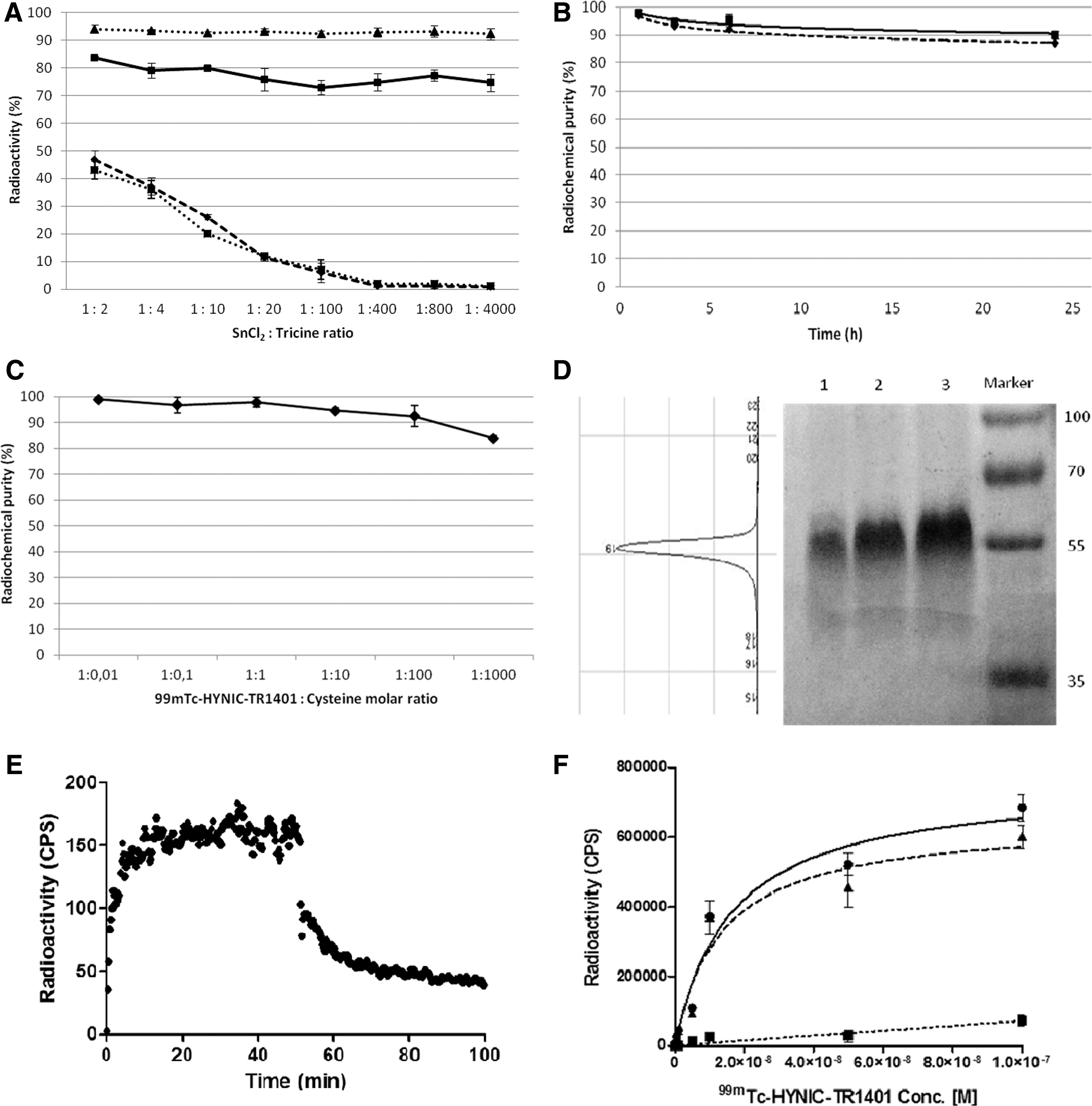
In vitro quality controls of 99mTc-HYNIC-TR1401.
Cell lines and FACS analysis
JP09, JP02, ML-1, and K1 showed excellent growth in vitro, although a progressive decrease of TSHR expression was noticed by JP09 cells in following passages. ML-1 cells had a very slow growing rate when injected in mice. Mean fluorescence intensity (range of MFI) was higher for K-1 cells (14.64–23.34), followed by ML-1 (6.93–7.99) and JP09 (5.59–8.93). JP02 cells showed a MFI<3, as expected, due to the absence of TSHR expression.
In vitro binding studies
ML-1 cells showed very fast radiopharmaceutical uptake, by LigandTracer, reaching a plateau within 20 min and a slow dissociation from the TSHR with time. The calculated K d was 2.51 nM (Fig. 1E), and a retention experiment allowed us to determine that a third of the bound radioactivity was internalized, decreasing over time, with a half-life of 13.6±2 h. The binding assay performed with JP09 cells showed a Kd of 8.79 nM (Fig. 1F ) by nonlinear regression analysis.
In vivo high-resolution imaging
Visual analysis of images related to the biodistribution of 99mTc-HYNIC-TR1401 in mice showed a rapid and persistent uptake by the kidneys and a moderate uptake by the liver with almost no signal from other organs and blood pool, as shown in Figure 2. Single organ counting revealed that thyroid uptake, even if low in absolute amounts compared to other major organs, is very high per gram of tissue and increases over time up to 6 h, with a decrease at 24 h (Fig. 3).
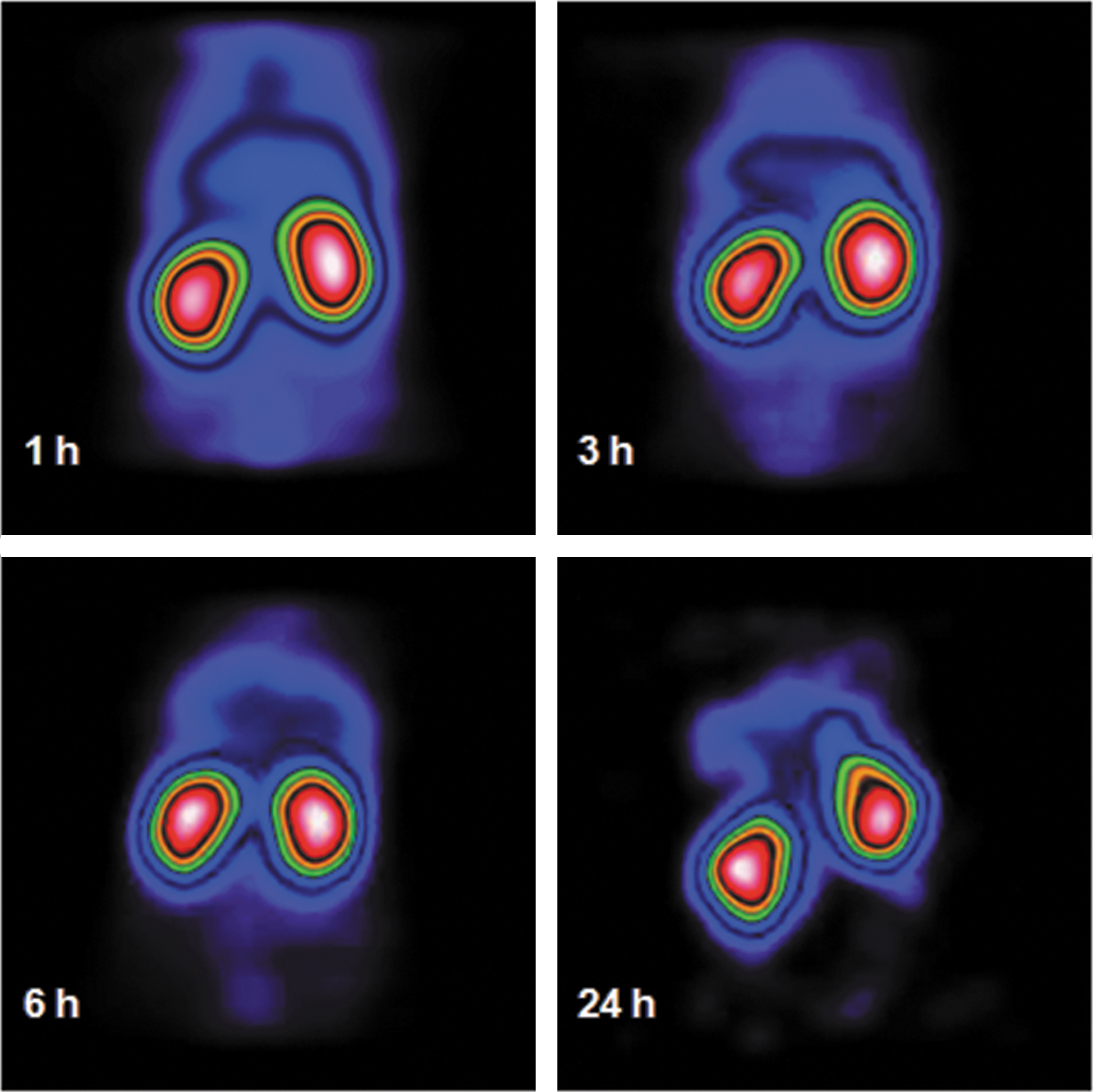
Planar posterior whole body images, at different time points, of nude CD1 mice after the injection of 370 Bq of TR1401. Images were acquired with a portable high-resolution mini-gamma camera and show the rapid and persistent kidney activity.
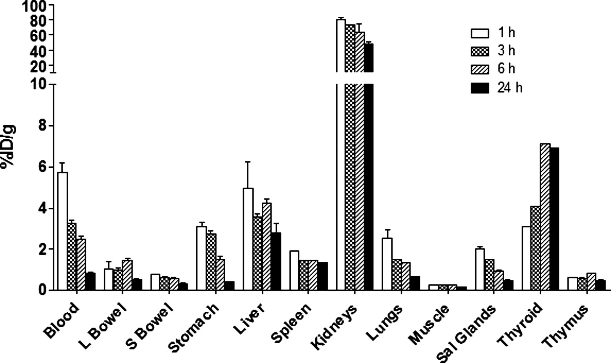
Single organ counts at 1 h (white bar), 3 h (squared bar), 6 h (dashed bar), and 24 h (black bar). Data are shown as % ID/g over time. Each point is the average of three mice. An evident uptake was found in the thyroid, kidneys, and liver.
Figure 4 shows the images acquired in mice injected with different cell lines in the right thigh. In animals injected with TSHR-positive cells, there is a focal uptake of the radiopharmaceutical where cells were injected. Regions of interest (ROIs) were drawn over the right and left thigh, and tumor/background (T/B) ratios were calculated at each time point (Fig. 5). All cell lines showed the highest T/B ratio at 3 h, and a slight decrease was observed at 6 h up to 24 h.
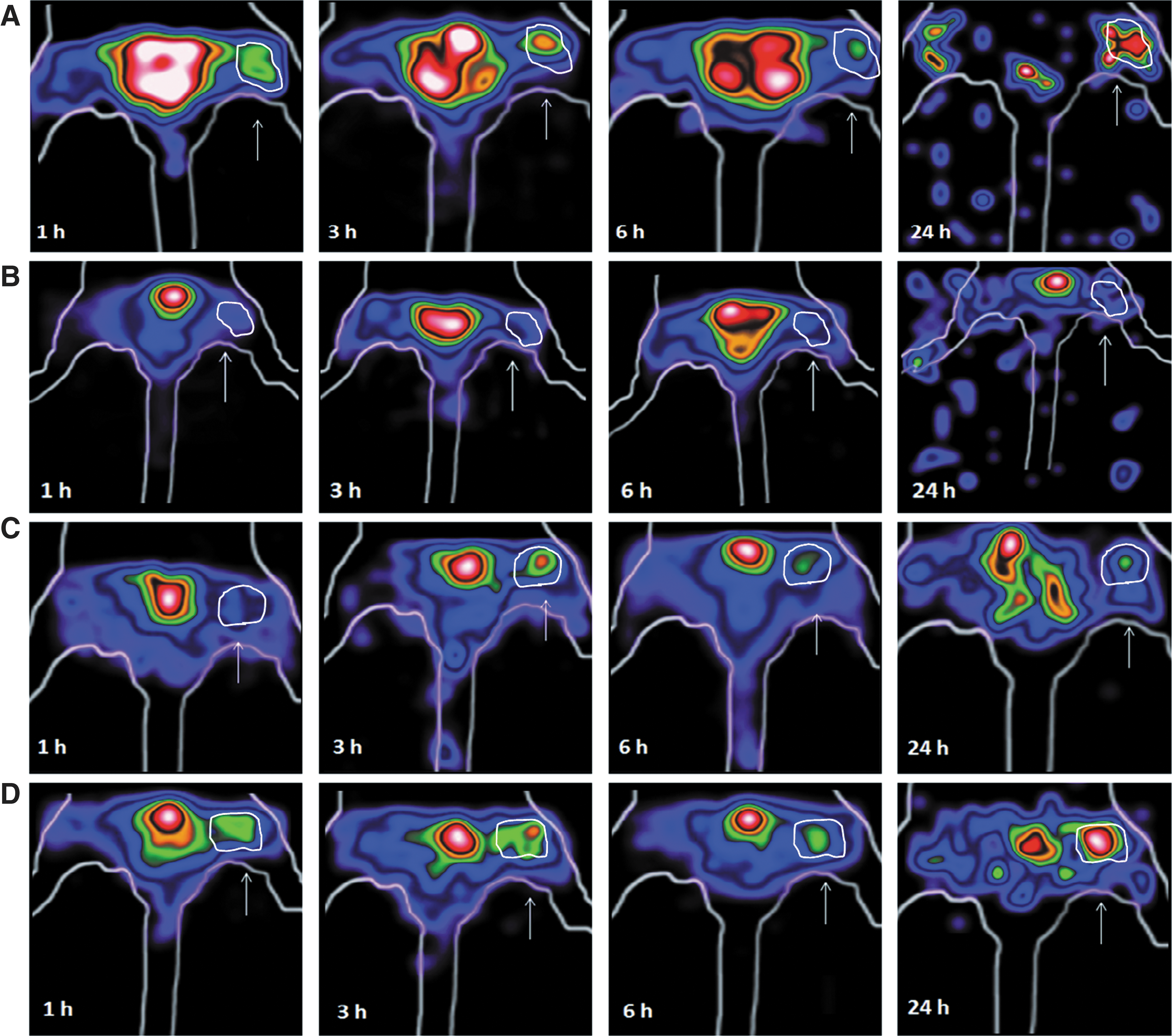
Planar posterior high-resolution camera (HRC) images of the legs of mice injected in the right thigh with TSHR-positive JP09
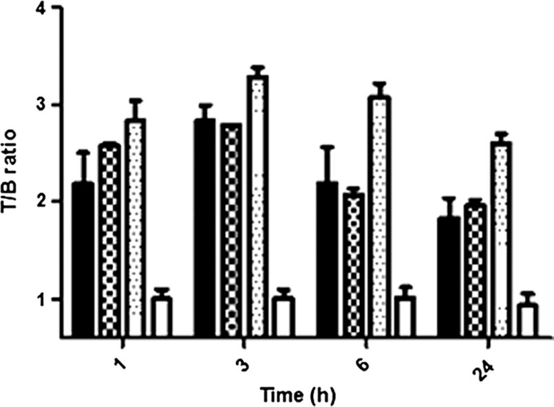
T/B ratios calculated on planar posterior images obtained at 1, 3, 6, and 24 h after i.v. injection of 99mTc-HYNIC-TR1401, by drawing a region of interest (ROI) over the injected cells and over the contralateral leg. JP09 (black bar), ML-1 (squared bar), K1 (dotted bar), and JP02 cells (white bar). The best T/B ratios were obtained at 3 h for all cell lines, confirming results of visual analysis of images and demonstrating the rapid uptake of this radiopharmaceutical with very fast plasma clearance.
Tumor xenografts in mice injected with JP09 and K1 cells were detectable after 20 days, and radiolabeled TR1401 was able to visualize the lesion with a high T/B ratio (6.8 and 2.9 respectively) as early as at 3 h after injection. No uptake was observed in tumors derived from TSHR negative cells (Fig. 6). ML-1 tumors were smaller and less vascularized than other tumors at autopsy. Images with 99mTc-HYNIC-TR1401 showed the tumors best at 3 h after injection with a calculated T/B of 1.7.
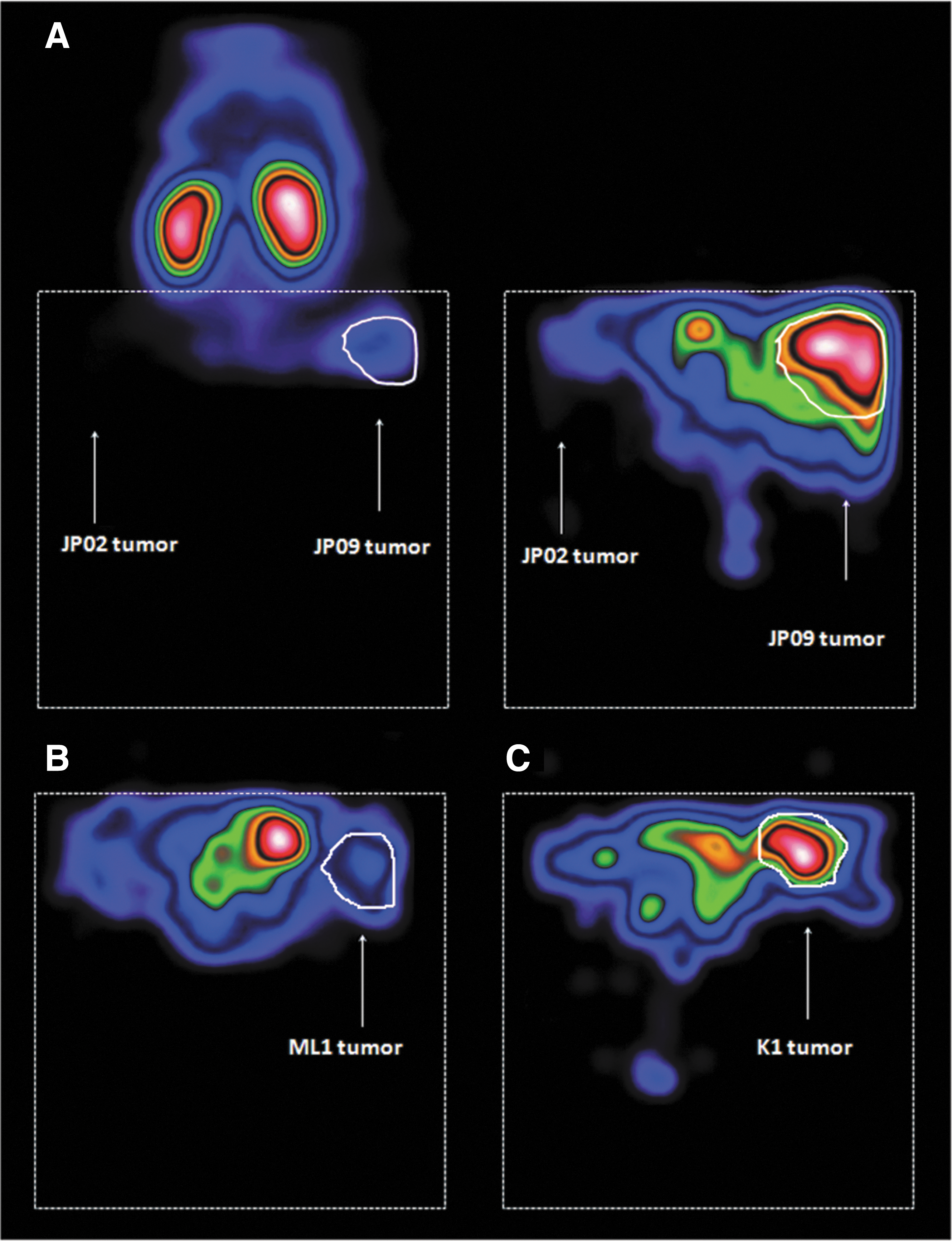
Planar posterior HRC images of nude mice bearing a JP09 tumor in the right thigh and a JP02 tumor in the left thigh
Case study in a Dachshund dog with spontaneous thyroid cancer
The Dachshund dog was clinically symptom free, and routine hematologic and biochemical parameters were all within the reference ranges, including thyroxine and TSH levels. SPECT/CT images showed high uptake in the left thyroid lobe corresponding to the thyroid nodule with no significant uptake in the contralateral lobe (Fig. 7). No focal uptake was found in regional lymph nodes nor in other tissues, but it was found in the liver and kidneys as a consequence of 99mTc-TR1401 metabolism. Despite the initial diagnosis, histologic examinations revealed that the excised lesion was a poorly differentiated thyroid cancer with high TSHR expression (Fig. 8).

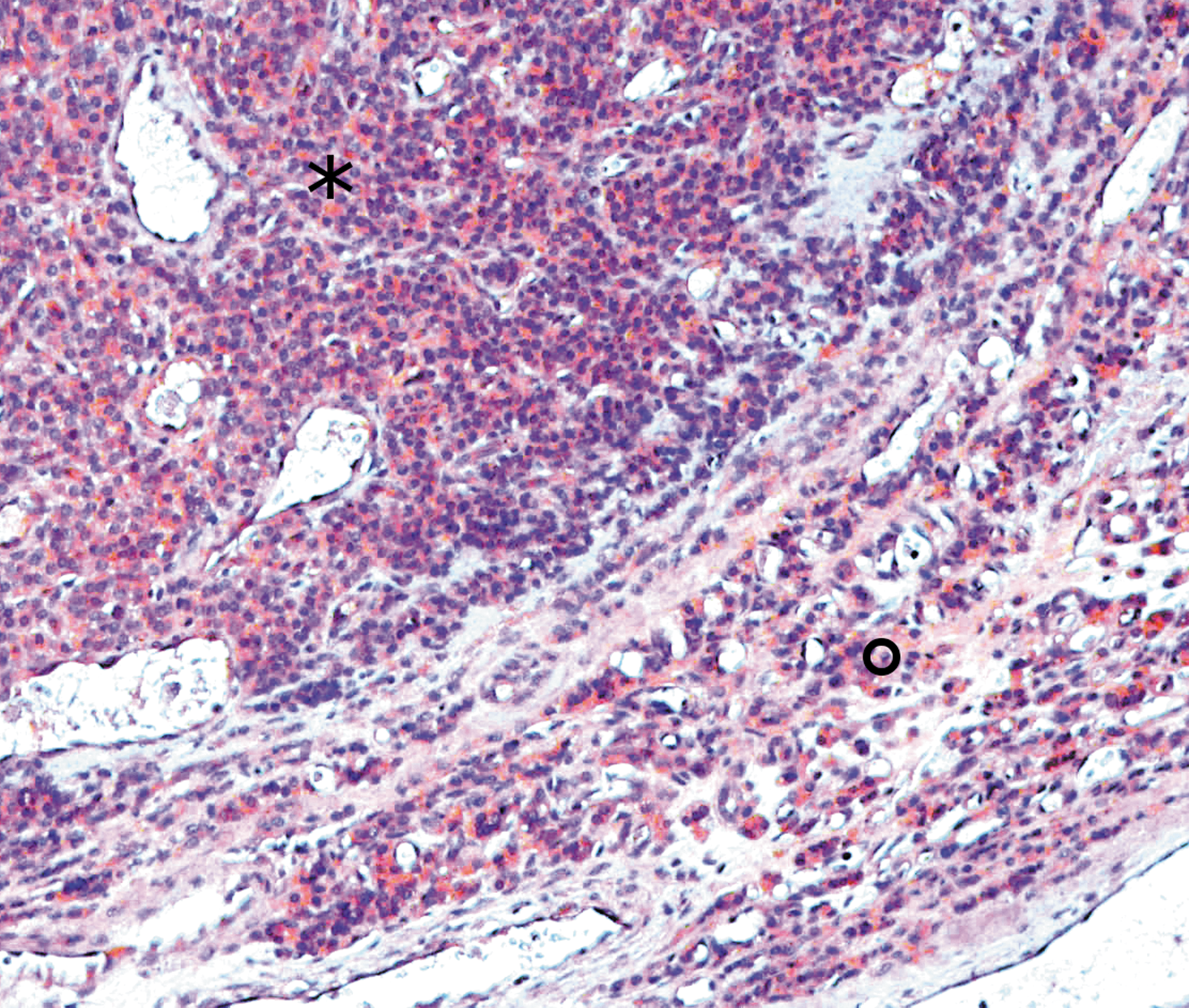
Histologic section of the excised lesion showing a poorly differentiated thyroid carcinoma (*) and the surrounding normal thyroid tissue (o). The section is stained with hematoxylin and eosin and with an anti-TSHR antibody specific for canine TSHR. The malignant tissue shows much higher density of TSHR expression compared to the normal tissue.
Discussion
Although 99mTc-methoxyisobutylisonitrile (MIBI) scintigraphy (34), [18F]-FDG PET (13), radiolabeled somatostatin analogues (35), Doppler-US (36), contrast-enhanced magnetic resonance imaging (37), and others (38) have shown to be useful for the in vivo characterization of thyroid and extra-thyroid lesions, more accurate imaging techniques are needed.
We previously published an alternative approach for imaging differentiated thyroid cancers based on the use of a radiolabeled anti-Galectin-3 mAb (39). Despite the excellent results in preclinical studies, we had difficulties in translating this methodology to humans due to the murine nature of the antibody and the long plasma half-life of the labeled antibody that reduces T/B ratios and the possibility of detecting small lesions. A humanized antibody fragment radiolabeled with a positron-emitting isotope would have been more successful in humans. In the following years, we investigated, like others, the use of radiolabeled peptides and receptor agonist analogues.
Along this line, Sager et al. (40) investigated the role of scintigraphy with radiolabeled somatostatin analogues that showed a sensitivity ranging between 25% and 75% depending on patient selection criteria. They compared 18[F]-FDG and 99mTc-MIBI with 99mTc-HYNIC-TOC, preferring it to 111In-octreotide due to its lower cost, higher resolution, and lower radiation dose to the patient. They found that the sensitivity of somatostatin receptor scintigraphy was higher than MIBI, but lower than 18[F]-FDG-PET. Similarly, Rodrigues et al. (41) proposed the use of 99mTc-depreotide that showed, in many cases, a higher sensitivity than 18[F]-FDG-PET, but also nonspecific uptake in cells that play a role in granulomatous diseases, such as activated lymphocytes, leading to false-positive results.
Finally, Zhao et al. (42) proposed the use of the Arginine-Glycine-Aspartic acid (RGD) molecule 3PRGD2 labeled with 99mTc that has a high affinity and specificity for integrin αvβ3, present in various malignant tumors. In 10 patients with negative 131I-WBS and high Tg, 99mTc-3PRGD2 scintigraphy was able to visualize thyroid cancer metastases with a high T/B ratio that also correlated with growth rate. Moreover, the low background in the lungs facilitated the detection of metastases in the chest.
As an alternative to the above-mentioned imaging approaches, we have investigated the use of radiolabeled human TSH (43). Since the TSHR is overexpressed in some de-differentiated thyroid cancer, the relevance of this imaging approach depends on the potential of using this radiopharmaceutical not only for follow-up but also for preoperative staging of differentiated thyroid cancer. A further advantage of this technique could be the possibility of avoiding the induction of hypothyroidism either by thyroxine withdrawal or rhTSH administration.
We therefore radiolabeled rhTSH with 123I and obtained interesting preclinical results. However, images obtained with 123I-rhTSH were of relatively poor quality for three possible reasons: (a) the low specific activity of the radiopharmaceutical as a consequence of the low labeling efficiency of rhTSH with 123I; (b) the low affinity binding of rhTSH to the TSHR; (c) the possible saturation of the TSHR by endogenous TSH. We therefore attempted to label rhTSH with 99mTc with high specific activity and performed preliminary studies in animals and patients with metastatic thyroid cancer (unpublished data). However, tumor lesions were poorly distinguishable from background, and we explained these results by the low binding affinity of rhTSH to the TSHR.
This highlighted the need of searching for a new TSH analogue (TSH receptor agonist) with higher receptor binding affinity than rhTSH (44,45). The relatively low affinity of rhTSH, which is much lower than bovine or rodent TSH, is the result of an evolutionary change from an acute thermogenic hormone to one whose primary role in humans is to promote a gradual and chronic increase of NIS during periods of fasting or low iodine diet, as an adaptation to nomadic life (22,45). Recently, a novel second-generation superagonist rhTSH analogue named TR1401 has been produced and purified to homogeneity (>99%). It has very high in vitro receptor binding affinity and in vivo bioactivity (Trophogen, Inc., unpublished data). This new analogue has higher biopotency and maximal efficacy compared to standard wild-type hormone and first generation rhTSH analogues, and previous studies demonstrated its capacity to increase radioiodine and 18[F]-FDG uptake in FRTL-5 cells (22,46,47). Since the heterodimeric TSH molecule has crucial disulfide bonds, involved in the formation of a cysteine-knot in each subunit, we focused on the labeling of TR1401 with 99mTc using an indirect method based on SHNH conjugation, obtaining a reliable and reproducible protocol that allowed us to radiolabel the analogue without affecting its biologic activity. SHNH is bound to lysine residues and then chelates the reduced technetium, thus avoiding opening the S–S bonds to insert Technetium, as this is done with the so-called direct methods (48).
Using the indirect method, tricine is used as a co-ligand, and its quantity, as well as that of SnCl2, must be carefully selected in order to reduce 99mTc-colloid formation and increase LE. We found that a SnCl2:tricine ratio of 1:800 gave a 94% LE with negligible amounts of colloids (SA of 7400 MBq/mg). Labeled TR1401 was stable in both saline and human serum for up to 24 h and the binding on TSHR-positive cells was specific because in saturation binding assays performed on JP09 cells, an excess of unlabeled TR1401 was able to saturate the receptors and prevent further binding of labeled molecules. With ML-1 cells, a K d of 2.51 nM was calculated.
This high affinity of TR1401 and the high specific activity of 99mTc-TR1401 allowed us to inject relatively low doses of the radiopharmaceutical into the animals and obtain excellent tumor localization by gamma camera imaging. This could also be achieved in humans, thus reducing the radiation dose and any biologic effect on TSHR positive tumors.
In mice, the radiolabeled analogue showed early targeting to tumors and the thyroid, as well as fast renal metabolism with very low blood pool activity at late time points.
The thyroid showed, ex vivo, the highest % ID/g, even if it could not be clearly visualized with the mini-gamma camera because of its small size, which was below its resolution. JP09 tumors were visualized in vivo with a high T/B ratio (6.8) at 3 h, whereas K1 and ML-1 tumors showed a lesser uptake (2.9 and 1.7 respectively, both at 3 h). It should be mentioned that T/B values did not quantitatively correlate with the TSHR density calculated by FACS analysis. Our interpretation of these results is based on the consideration that cells in xenografts behave very differently from cultures, and the vascularization of xenografts may also vary and affect the uptake of radiopharmaceutical. Thus, ML-1 tumors were slow growing, small, and poorly vascularised. K1 tumors grew well but differ from ML-1 and JP09 because of their origin. The case study in a Dachshund dog with a spontaneous poorly differentiated thyroid cancer, although not fully conclusive, is of interest because it highlights the potential of this new radiopharmaceutical for detection of intrathyroidal lesions. SPECT/CT images performed 3 h after i.v. injection confirmed that TR1401 rapidly binds in vivo to tumor cells, with a very low blood pool activity and predominant kidney accumulation observed previously for highly sialylated rhTSH preparations (47). The clear visualization of the tumor within the thyroid gland in euthyroid conditions suggests the possibility of using this radiopharmaceutical for preoperative TSHR-based staging in addition to patient follow-up and for imaging de-differentiated thyroid cancer metastases. Indeed, we can assume that the lower the level of endogenous TSH, the better the condition for using radiolabeled TR1401 because of a decreased likelihood of competition at the TSHR. However, radiolabeled TR1401 affinity for the human TSHR is much higher than that of wild-type hTSH, and suppression of endogenous TSH will most likely not be necessary in patients.
Because of this favorable biodistribution, high binding affinity, and retention in the cytoplasm, radiolabeled-TR1401 could also be suitable for therapeutic purposes, if radiolabeled with a beta-emitting isotope such as 177Lu, providing the ability to perform a renal protection by amino acid infusion, as for other radiolabeled peptides (49). Moreover, novel long-acting superagonist rhTSH analogues are under development and may replace the high renal uptake of TR1401. From a therapeutic point of view, 99mTc-TR1401 could be useful to predict which radioiodine-negative thyroid cancers might respond to therapies with 177Lu-labeled TSH analogues, which are currently under development in our laboratory and at Trophogen.
The relative role of 99mTc-TR1401 versus 131I scanning of thyroid cancer will require future histopathologic and clinical studies in patients receiving both scanning modalities, since the in vivo functional status of the TSHR versus NIS has not yet been clarified. Therefore, it would be important to understand which patients may benefit from such novel radiopharmaceuticals, and whether 99mTc-TR1401 scintigraphy could become complementary to radioiodine scanning in the management of differentiated thyroid cancer patients. As a minimum, 99mTc-TR1401 scanning will provide a major new unmet clinical need to image thyroid cancers that have lost the expression or membrane insertion of NIS while retaining expression of functional TSHRs. These thyroid cancers are typically associated with finding a positive serum Tg and a negative iodine scan, thus posing a major diagnostic and therapeutic challenge. The fact that most of these patients respond with an increase in serum Tg to endogenous or rhTSH proves that they retain an appreciable numbers of TSHRs that allow cancer imaging with the new high affinity TSH analogue. In addition, even in cancers with a functional NIS and TSHR overexpression, 99mTc-TR1401 may have advantages for imaging of intrathyroidal cancers, or local nodes in presurgical staging and distant metastases, allowing physicians to perform the scintigraphy before thyroidectomy without interference of the thyroid. This would allow the surgeon to remove local non-iodine uptaking nodes at the moment of the thyroidectomy. Our study, as a proof of concept, opens new diagnostic and therapeutic strategies.
Conclusions
The highly pure rhTSH superagonist TR1401 was efficiently labeled with 99mTc with high specific activity and used for imaging TSHR-positive cell lines in vitro and TSHR-positive thyroid tumor xenografts in mice. A spontaneous poorly differentiated thyroid cancer within the thyroid was also imaged in a dog. In the light of these results, we will investigate the possibility of using this radiopharmaceutical for imaging intrathyroidal differentiated thyroid cancer and, particularly, radioiodine-negative metastases.
Due to fast in vivo binding of 99mTc-HYNIC-TR1401, a possible use of short half-life positron emitting isotopes can also be considered (such as 18F or 68Ga) for PET/CT imaging. More studies are necessary to evaluate the possibility of labeling TR1401 with a beta-emitting isotope for therapeutic purposes.
Footnotes
Acknowledgments
We wish to acknowledge Drs. Meng Zhang and Vladimir Wolf for their contributions at Trophogen, Inc., to the development, production, purification, and initial characterization of purified TR1401 analogue glycoprotein. Dr. Armando Bartolazzi performed the immunohistochemical staining of dog tissues. Funding for this project was provided in part by Trophogen, Inc., NIH-SBIR Grant R43DK076302, AIRC IG Grant 2010 10359, AIRC IG-13234 and “Sapienza” University research project C26A10H3AN. We also wish to acknowledge the nonprofit association Nuclear Medicine Discovery for support.
Author Disclosure Statement
B.W., M.S., and V.F. are employed by Trophogen, Inc. F.G., I.M., G.P., L.B., R.D., and A.S. have no competing financial interests.