
Abstract

Multiple endocrine neoplasia 2B (MEN-2B) is caused by the M918T and A883F germline mutations in the rearranged during transfection (RET) proto-oncogene for >95% and 2–3% of cases, respectively (1). The figures refer to the original RET consortium study in which 75 of 79 MEN-2B families had the M918T mutation and 4 families had unknown mutations, 2 of which were subsequently reported with the A883F mutation. Very few cases of the A883F mutation have been reported (2 –4). We report of a MEN-2B case with aggressive medullary thyroid carcinoma (MTC) due to the A883F mutation.
A 10-year-old boy with approximately 20 lingual nodules, prominent lips, and thickening of the corneal nerves was referred to the Department of Endocrinology and Internal Medicine, Aarhus University Hospital, THG. Six years earlier the patient had been examined for toe-walking and lumbar hyperlordosis leading to the suspicion of mild cerebral palsy. Previous examination for von Recklinghausen's disease was negative. The lingual nodules had earlier been histologically verified as neuromas. No evidence of marfanoid stature was noted.
The patient was the first-born of fraternal twins and had poorer developed motor and linguistic skills compared to his brother. There was no clinical family history of thyroid cancer or other MEN-2B features. Initial biochemical measurements showed normal plasma metanephrines and a calcitonin level of 100 pmol/L (<3.8 pmol/L).
Chest X-ray and ultrasonography of the liver and kidneys were normal. Ultrasonography of the neck revealed two thyroid cystadenomas measuring 7.2 mm and 4.3 mm in the left and right lobe, respectively. Furthermore, two enlarged lymph nodes on the left side and one on the right side of the neck were visualized. Fine-needle aspiration cytology from the left lobe suggested MTC. Calcitonin staining was not performed. Mutational analysis was positive for an A883F germline mutation. The mutation was confirmed to be de novo mutation as RET gene analysis of the parents and brother was normal.
One and a half months after referral, the patient underwent total thyroidectomy with bilateral central neck dissection and modified radical left sided neck dissection (levels 3–5). Surgical pathology revealed metastases in 3 of 5 lymph nodes from the central neck and in 1 of 34 from the left side of the neck. Calcitonin levels decreased from 100 to 5 pmol/L.
On subsequent follow-up, 5 months post-thyroidectomy, the patient presented with multiple ultrasonographic suspicious lymph nodes in the neck. He underwent a modified radical right-sided neck dissection (level 2–5) and radical dissection of left-sided lymph nodes (level 2). One of 25 left-sided lymph nodes was positive and none of the 39 right-sided lymph nodes were positive. Thus, in total metastases were found in 5 of 103 lymph nodes.
Three months later, 18F-dihydroxyphenylalanine positron emission tomography (18F-DOPA PET) was performed due to a calcitonin doubling time of 0.49 years (American Thyroid Association [ATA] Web-based calculator). The scan showed no signs of residual MTC but instead both adrenal glands incidentally showed increased activity suggestive for pheochromocytoma (PHEO) (Fig. 1). Prior to the scan, plasma metanephrines were slightly elevated and a previously performed ultrasonography for detecting PHEOs was negative. The patient underwent bilateral total adrenalectomy. Cortical-sparing surgery was considered but ultimately not performed after discussions between surgeons and endocrinologists because of the risk of recurrent PHEOs in this very young patient. Histology confirmed PHEOs and cortical hyperplasia in both adrenal glands.
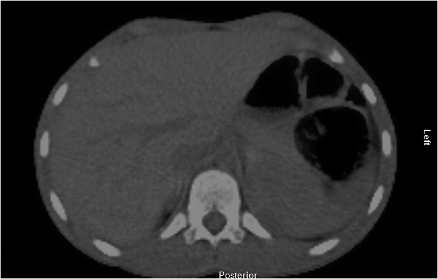
18F-dihydroxyphenylalanine positron emission tomography (18F-DOPA PET) indicative of pheochromocytoma.
Three years after the second neck surgery further palpable lymph nodes on the left side of the neck were found and the calcitonin doubling time was 1.02 years. Thus another 18F-DOPA PET was performed and showed a PET-positive lymph node in the left side of the neck. Ultrasonography was not used regularly in the surveillance. Two lymph nodes were excised and pathology revealed metastasis in both. Hereafter, calcitonin levels decreased from 92 to 44 pmol/L.
Six years later the patient had no symptoms, but slowly increasing calcitonin levels, currently at 120 pmol/L. The patient was considered to have residual disease without further surgical treatment options at that moment. Future screening will consist of regular measurements of calcitonin and plasma metanephrines.
According to the latest recommendations from the ATA regarding RET mutations and MTC aggressiveness, the A883F mutation is classified among the highest risk mutations, ATA-D (1). However, in a single observation made subsequent to publication of the recommendations, it has been suggested that the mutation could be a lower risk mutation than previously thought (3). However, the aggressive MTC of our patient corresponds well with the current ATA-D classification of the A883f mutation.
Footnotes
Acknowledgment
Special thanks to senior consultant Karin Hjorthaug, Department of Nuclear Medicine, Aarhus University Hospital, Brendstrupgaardsvej 100, 8200 Aarhus N, Denmark, for providing the figure.
Author Disclosure Statement
The authors declare that no competing financial interests exist.