
Abstract
Background:
No standard chemotherapy is available for anaplastic thyroid cancer (ATC). Drug-loaded nanobubbles (NBs) are a promising innovative anticancer drug formulation, and combining them with an externally applied trigger may further control drug release at the target region. Extracorporeal shock waves (ESWs) are acoustic waves widely used in urology and orthopedics, with no side effects. The aim of the present work was to combine ESWs and new doxorubicin-loaded glycol chitosan NBs in order to target doxorubicin and enhance its antitumor effect in ATC cell lines.
Methods:
CAL-62 and 8305C cells were treated with empty NBs, fluorescent NBs, free doxorubicin, and doxorubicin-loaded NBs in the presence or in the absence of ESWs. NB entrance was evaluated by fluorescence microscopy and flow cytofluorimetry. Cell viability was assessed by Trypan Blue exclusion and WST-1 proliferation assays. Doxorubicin intracellular content was measured by high-performance liquid chromatography.
Results:
Treatment with empty NBs and ESWs, even in combination, was safe, as cell viability and growth were not affected. Loading NBs with doxorubicin and combining them with ESWs generated the highest cytotoxic effect, resulting in drug GI50 reduction of about 40%. Mechanistically, ESWs triggered intracellular drug release from NBs, resulting in the highest nuclear drug content.
Conclusions:
Combined treatment with doxorubicin-loaded NBs and ESWs is a promising drug delivery tool for ATC treatment with the possibility of using lower doxorubicin doses and thus limiting its systemic side effects.
Introduction
A
In order to find new therapeutic tools that allow the cumulative dose to be reduced, as well as to target the drug to the tumor site, nanoparticles encapsulating anticancer drugs appear to be promising delivery systems. In fact, they can carry loaded drugs to the tumor site through the bloodstream, taking advantage of the enhanced permeability and retention effect (EPR), due to the defective vascular architecture of the tumor tissue (17).
Recently, growing attention in the field of nanomedicine has been given to micro- and nanobubbles (NBs). NBs, composed of an external shell and a gas core, can deliver diverse molecules, such as DNA and drugs, to target tissues in response to physical triggers, such as ultrasound (US). US-induced drug delivery at specific sites is the result of bubble cavitation and increased cell permeability (18 –23).
Extracorporeal shock waves (ESWs)—acoustic waves widely used in urology for lithotripsy (24) since the 1980s—produce cavitation without heat induction. ESWs can be focused with high precision in depth and determine permeabilization of plasma membranes (25 –27). The authors have already reported elsewhere that ESWs increase paclitaxel-induced apoptosis in breast-cancer cell lines (25), in anaplastic thyroid cancer cells (28), and in a Mat B-III rat syngeneic model of breast cancer (29). Furthermore, ESWs enhance the cytotoxic effect of doxorubicin and methotrexate in human osteosarcoma cell lines (30). Finally, ESW exposure has been shown to mediate DNA uptake into cells supporting in vitro transfection (27), and DNA-loaded microbubbles are associated with an increase in transgene expression in cultured cells exposed to ESWs (31). All these features make ESWs an ideal alternative to US in combination with drug-loaded NBs in delivery strategies.
The aim of this in vitro study was to combine the new doxorubicin-loaded glycol chitosan NBs and ESWs in order to enhance doxorubicin antitumor activity, increasing intracellular drug release in ATC cells. The main goal was to provide the basis for future in vivo studies aiming at a new therapeutic approach for ATC patients.
Materials and Methods
Materials
Ethanol (96%) was sourced from Carlo Erba (Milan, Italy). Epikuron 200® (soy lecithin containing 95% of dipalmitoyl phosphatidylcholine) was kindly gifted by Cargill (Hamburg, Germany). Doxorubicin hydrochloride was a kind gift from Pharmacia & Upjohn, while 6-coumarin was purchased from Acros Organics (Geel, Belgium). Ultrapure water was obtained using a 1-800 Millipore system (Molsheim, France). Penicillin and streptomycin were purchased from Gibco (Life Technologies Corp., Grand Island, NY). Dulbecco's modified Eagle's medium combined with Ham's F12 (DMEM-F12) was purchased from Invitrogen (Groningen, The Netherlands). RPMI 1640 and fetal calf serum (FCS) were purchased from Euroclone (Wetherby, United Kingdom). Phenylmethylsulfonyl fluoride (PMSF) and WST-1 cell proliferation reagent were purchased from Roche Diagnostics Corporation (Indianopolis, IN). Penicillin, streptomycin, non-essential amino acids (NEAA), palmitic acid, perfluoropentane, glycol chitosan (Mw = 68 kDa), fluorescein isothiocyanate (FITC), Nonidet 40 (NP40), desoxycholate sodium, sodium dodecyl sulphate (SDS), phosphate-buffered saline (PBS), aprotinin, sodium orthovanadate, 4-(2-Hydroxyethyl) piperazine-1-ethanesulfonic acid (Hepes), sodium carbonate (Na2CO3), magnesium chloride (MgCl2), potassium chloride (KCl), sodium chloride (NaCl), 1,4-dithiothreitol (DTT), Triton X-100, glycerol, and ethylenediaminetetraacetic acid (EDTA) were all purchased from Sigma-Aldrich (St Louis, MO).
Preparation of NBs
NBs were formulated by purposely tuning the method previously reported (18) using perfluoropentane as the inner core component and glycol chitosan for the shell. First, an ethanolic solution containing palmitic acid and Epikuron 200® (1% w/v) was added to perfluoropentane to form a pre-emulsion. After the addition of ultrapure water, the system was homogenized using a high shear homogenizer (Ultraturrax®; IKA, Königswinter, Germany) in an ice bath. Then, to obtain the polymeric NBs, an aqueous solution of the glycol chitosan polymer (2.7% w/v, pH 5) was dropwise added under mild magnetic stirring.
Doxorubicin as a base was obtained by adding a saturated solution of Na2CO3 (1.4 g/mL) to a doxorubicin HCl solution in deionized water (200 mg/mL) drop by drop. To obtain the drug-loaded NBs, a pre-emulsion was obtained by adding an Epikuron® 200 and palmitic acid ethanolic solution (1% w/v) containing doxorubicin base to perfluoropentane under magnetic stirring. After the addition of ultrapure water, the system was homogenized using a high shear homogenizer. To obtain the NBs, an aqueous glycol chitosan solution (2.7% w/v, pH 5) was dropwise added under magnetic stirring. A purification step by dialysis was then carried out to eliminate the potential free drug.
Two types of fluorescent glycol chitosan NBs were prepared. Empty fluorescent NBs were obtained by adding 6-coumarin to the perfluoropentane core. FITC-labeled doxorubicin-loaded NBs were prepared by inserting the green fluorescent FITC in the NB shell, in order to have a double labeling, exploiting the intrinsic fluorescence of doxorubicin. For this purpose, an amount of FITC (0.02% w/v) was added to the preformed doxorubicin-loaded NB suspension and incubated under stirring for 24 hours in the dark at room temperature.
Characterization and stability of NBs
The average diameter and polydispersity index of NB formulations were determined by photon correlation spectroscopy; the zeta potential was determined by electrophoretic mobility using a 90 Plus instrument (Brookhaven, NY). The analyses were performed at a scattering angle of 90° at a temperature of 25°C, using NB suspension diluted with deionized distilled water. For zeta potential determination, samples of diluted NB formulations were placed in the electrophoretic cell, where an electric field of approximately 15 V/cm was applied.
The morphology of formulations was evaluated by transmission electron microscopy (TEM), using a Philips CM10 instrument (Eindhoven, NL).
In vitro doxorubicin release kinetics
In vitro release studies were carried out using a multi-compartment rotating cell. The donor compartment containing 1 mL of doxorubicin-loaded glycol chitosan NB suspension or doxorubicin hydrochloride aqueous solution as control was separated by a dialysis membrane (Spectrapore, cutoff 12,000–14,000 Da) from a receiving compartment, containing saline phosphate buffer at pH 7.4. At fixed times, the receiving phase was withdrawn and replaced with fresh receiving medium. The doxorubicin content was determined by a high-performance liquid chromatography (HPLC) system consisting of a pump (LC-9A PUMP C, Shimadzu, Kyoto, Japan) equipped with a fluorescence detector (Chrompack, Tokyo, Japan). Analyses were performed using an Agilent TC C18 column (250 mm ×4.6 mm, 5 μm). The external standard method was used to calculate the drug concentration. For this purpose, 1 mg of doxorubicin was weighed, placed in a volumetric flask, and dissolved in water to obtain a stock standard solution. This solution was then diluted using the mobile phase, providing a series of calibration solutions, subsequently injected into the HPLC system.
Moreover, in vitro release kinetics of doxorubicin from doxorubicin-loaded glycol chitosan NBs were investigated after ESW treatment, whereby 1 mL of doxorubicin-loaded NBs was placed in 20 mm polypropylene tubes (Nunc, Wiesbaden, Germany) and treated with ESWs (0.59 mJ/mm2, 500 pulses).
Cell lines and culture conditions
ATC cell lines, CAL-62 and 8305C, were purchased from Deutsche Sammlung von Mikroorganismen and Zellkulturen (Braunschweig, Germany), which certifies the origin and identity of the cells. The rat embryonic cardiomyocyte cell line, H9C2, was purchased from ATCC (Manassas, VA). Cells were routinely maintained in 75 cm2 flasks at 37°C, in 5% CO2 and 95% humidity, with 100 IU/mL penicillin and 100 μg/mL streptomycin added in DMEM-F12 for CAL-62 and H9C2 cells, or RPMI 1640 plus 1% NEAA for 8305C cells, supplemented with 10% FCS.
ESW treatment
The shock-wave generator utilized for the in vitro experiments is a piezoelectric device (Piezoson 100; Richard Wolf, Knittlingen, Germany) designed for clinical use in orthopedics and traumatology. The experimental setup has been reported elsewhere (25). Aliquots of 1 mL of cell suspension adjusted to 1 × 10⁶ cells/mL were placed in 20 mm polypropylene tubes, completely filled with culture medium. Subsequently, cells were gently pelleted by centrifugation at 250 g in order to minimize motion during shock-wave treatment. Each cell-containing tube was placed in vertical alignment with the focal area and was adjusted so that the central point of the focal area corresponded to the center of the tube bottom. The shock-wave unit was kept in contact with the cell-containing tube by means of a water-filled cushion. Common ultrasound gel was used as a contact medium between cushion and tube. ESW treatment was as follows: energy flux density (EFD) = 0.59 mJ/mm2, 500 pulses (frequency = 4 shocks/sec), peak positive pressure 64 MPa, peak negative pressure 12 MPa. After treatment, cell viability was evaluated in a hemocytometer chamber by a Trypan Blue dye exclusion assay.
Cell viability assay
CAL-62 and 8305C cells were treated with ESWs, empty NBs (0.9–5.4 × 10⁶ NBs/mL), free doxorubicin (0.25 –3 μM), doxorubicin-loaded NBs (0.25 – 3 μM), empty NBs + ESWs, doxorubicin + ESWs, doxorubicin-loaded NBs + ESWs, and empty NBs + free doxorubicin + ESWs. H9C2 cells were treated with empty NBs (0.9 – 5.4 × 10⁶ NBs/mL), free doxorubicin (0.25 – 3 μM), and doxorubicin-loaded NBs (0.25–3 μM). Cells in maintenance medium plus 1% FCS were used as controls (Basal). After different treatments, cells were seeded at 3 × 10³ cells/well in 96-well plates (Corning, New York, NY). After 24 h, drug-containing medium was replaced with maintenance medium plus 1% FCS. Viable cells were determined at different times using the cell proliferation reagent WST-1, following the manufacturer's instructions. This is a colorimetric assay for the quantification of cell viability and proliferation based on the cleavage of the tetrazolium salt WST-1 by mitochondrial dehydrogenases. Briefly, 10 μL of WST-1 was added to each well. After 1 h incubation, absorbance at 450 nm was measured using a plate reader (Model 680 Microplate Reader; Bio-Rad, Hercules, CA). Four replicate wells were used to determine each data point. Growth inhibition fifty (GI50), corresponding to the concentration of the compound that inhibits 50% cell growth, was calculated.
NB internalization
Cells were seeded at 5 × 10⁵ cells/well in six-well plates (Corning) and treated with 6-coumarin-labeled glycol chitosan NBs (0.9 – 5.4 × 10⁶ NBs/mL). After 24 h, green fluorescence in the cells was assessed by flow cytometer (EPICS XL, Coulter Corp., Hialeah, FL). Ten thousand events were acquired for each sample.
To evaluate NB entrance, cells were seeded at 3 × 10³ cells/well in 96-well plates and treated with 6-coumarin-labeled glycol chitosan NBs at 0.9 and 1.8 × 10⁶ NBs/mL. After 24 h, cells were observed with an inverted fluorescence microscope Leica DMI 4000 B (Leica Microsystems, Wetzlar, Germany), and photos were taken with a Leica DCF340 FX digital camera system (Leica Microsystems) at 200× and 400× magnifications.
To study the ESW effects on NBs, cells were treated with 1 μM doxorubicin-loaded FITC-NBs in the presence or absence of ESWs. One hour after ESW treatment, cells were cytospun on slices and observed with an inverted fluorescence microscope Leica DMI 4000 B. Photos of single channels and overlayers were taken with a Leica DCF340 FX digital camera system at 200× final magnification.
Cell drug uptake
Cell drug uptake was investigated by the quantitative evaluation of intracellular doxorubicin accumulation. CAL-62 and 8305C cells were treated with 1 μM free doxorubicin or doxorubicin-loaded glycol chitosan NBs in the presence or in the absence of ESW treatment and seeded at 5 × 10⁶ cells/well in six-well plates (Corning).
After treatment for 24 h, whole cell lysates were prepared in RIPA buffer (1% NP40, 0.5% desoxycholate sodium, 0.1% SDS in PBS pH 7.4, with 10 mg/mL PMSF, 30 μL/mL aprotinin, and 100 mM sodium orthovanadate) and incubated on ice for 30 min. Cells were then centrifuged for 20 min at 15,000 g at 4°C, and clear supernatants were used.
Nuclear extracts were obtained in hypotonic buffer (10 mM Hepes, 1.5 mM MgCl2, 10 mM KCl, 0.5 mM DTT, 1 mM PMSF, pH 7.9), added with Triton X-100 (0.1% final concentration) and incubated for 15 min on ice. Cells were then disrupted by repeated passages into a syringe. Nuclear pellets, obtained by centrifugation at 11,000 g for 20 min, were rinsed once in hypotonic buffer, resuspended in hypertonic buffer (20 mM Hepes, 1.5 mM MgCl2, 25% (v/v) glycerol, 420 mM NaCl, 0.5 mM DTT, 1 mM PSMF, 0.2 mM EDTA, pH 7.9), gently shaken for 30 min at 4°C, and then centrifuged at 15,000 g for 10 min. The final supernatants were used. After dilution with the mobile phase, the samples were stirred for 2 min and injected into the HPLC system for the quantitative determination of doxorubicin. Cell uptake was expressed as nanograms of doxorubicin/106 cells.
Statistical analysis
Data are expressed throughout the text as means ± standard deviation, calculated from at least three different experiments. Comparison between groups was performed with analysis of variance (two-way ANOVA) and the threshold of significance was calculated with the Bonferroni test. Comparison between ESW-treated cells and non-ESW-treated cells was performed with a t-test. Statistical significance was set at p < 0.05.
Results
Characterization of new doxorubicin-loaded glycol chitosan NBs
The average diameter, polydispersity index (PDI), and zeta potential values of the NBs, before and after loading with doxorubicin, determined at 25°C, are reported in Table 1. Empty NBs showed an average diameter <500 nm and a positive surface charge. Doxorubicin loading did not significantly alter these values, and neither did fluorescent labeling (data not shown). A TEM image of doxorubicin-loaded glycol chitosan NBs is shown in Figure 1A. The NBs showed a spherical shape, smooth surface, and a well-defined core-shell structure, with a polymeric shell thickness of about 40 nm.
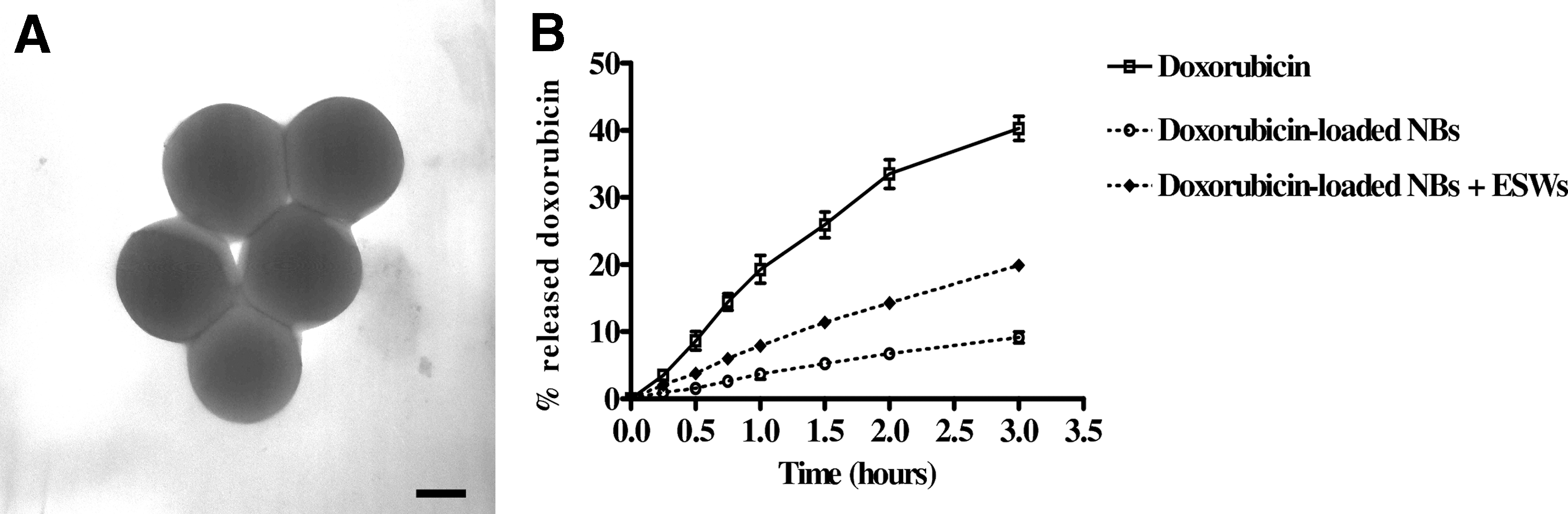
Characterization of nanobubbles (NBs). Transmission electron microscopy (TEM) image of glycol chitosan NBs (scale bar 250 nm) (
NB, nanobubble; PDI, polydispersity index; SD, standard deviation.
Figure 1B shows the in vitro release profile of doxorubicin from NBs in phosphate buffer (pH 7.4) in the presence or absence of ESW treatment compared with a doxorubicin hydrochloride aqueous solution used in therapy. After 3 h, only 9.2% of the drug was released in the absence of ESW with respect to 40.3% of free doxorubicin (p < 0.001). Moreover, no initial burst effect was observed, showing NB stability. Interestingly, doxorubicin was released to a larger extent following ESW treatment with 500 pulses at 0.59 mJ/mm2. In particular, after 3 h, ESWs generated an increase of about 70% of released doxorubicin with respect to NBs without ESW treatment (p < 0.001).
Safety of ESW treatment and empty NBs
In a preliminary series of experiments, cells were treated with different numbers of pulses (from 100 to 2000 at EFD = 0.59 mJ/mm2) in order to find the best treatment in terms of cell viability and growth. The used energy was within the range of clinical use; in ESW therapy, shock waves are applied ranging from 0.01 to 0.6 mJ/mm2 (32). Soon after treatment with 500 pulses, cell viability in both CAL-62 and 8305C cell lines was >87% (data not shown). Moreover, the same treatment did not affect cell growth of both cell lines up to 72 h (Fig. 2A and B). Therefore, this treatment schedule was chosen for further experiments.
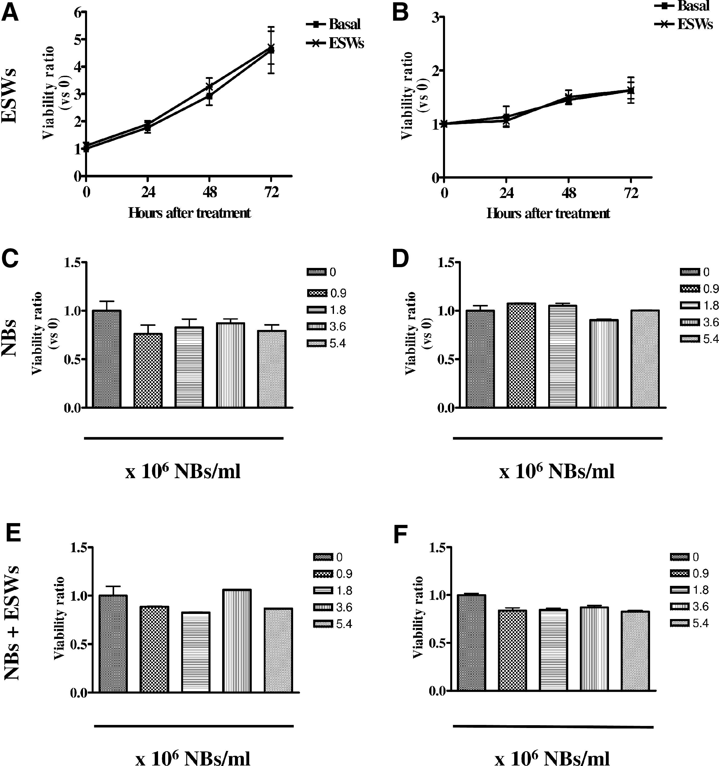
Effect of ESWs and empty NBs on cell viability. Growth curve of CAL-62 (
As the shell influences NB characteristics and behavior, the study tested whether empty NBs affected the cell viability of both ATC cell lines. Glycol chitosan NBs had no significant effect on cell viability of both cell lines (Fig. 2C and D). Moreover, as shown in Figure 2E and F, empty NBs had no significant effect on cell growth, even when used in combination with ESWs, and this allowed any non-specific effect of the combined treatment on cell viability to be excluded. In both ATC cell lines, glycol chitosan NBs entered the cells in a dose-dependent manner, as shown in Figure 3A and B, and were visible inside the cells (Fig. 3C and D).

Glycol chitosan NB entrance. Cytofluorimetric analysis of CAL-62 (
Cytotoxicity of doxorubicin-loaded glycol chitosan NBs combined with ESWs
As shown in Figure 4A and B, free doxorubicin had cytotoxic effect in both ATC cell lines only at levels hardly obtainable in patients receiving the doxorubicin dose of 60 mg/m2 (33) with a GI50 at 48 h of 1.57 μM in CAL-62 and 3.93 μM in 8305C cells, respectively (Table 2). ESW treatment increased the cytotoxic effect of the free drug (Fig. 4C and D). In fact, the GI50 decreased to 1.33 μM in CAL-62 (p < 0.05) and to 3.17 μM in 8305C cells (p < 0.05; Table 2). Drug-loaded NBs, even without ESWs, were more effective in inducing cytotoxicity in both cell lines with respect to free doxorubicin (Fig. 4E and F). In fact, as reported in Table 2, the GI50 decreased to 1.13 μM in CAL-62 (p < 0.001) and to 2.98 μM in 8305C cells (p < 0.05). The combination of ESWs with doxorubicin-loaded NBs had the greatest cytotoxic effect (Fig. 4G and H) with a GI50 of 0.95 μM in CAL-62 (p < 0.001) and 2.12 μM in 8305C (p < 0.001), respectively (Table 2). In a second series of experiments, the cytotoxicity of CAL-62 treated with empty NBs in combination with free doxorubicin and ESWs was evaluated. This treatment was more cytotoxic with respect to the free drug alone. In fact, the GI50 decreased to 1.26 μM (p < 0.01; Table 2). However, combining ESWs with free doxorubicin plus empty NBs was less effective than using ESWs with doxorubicin loaded into NBs.
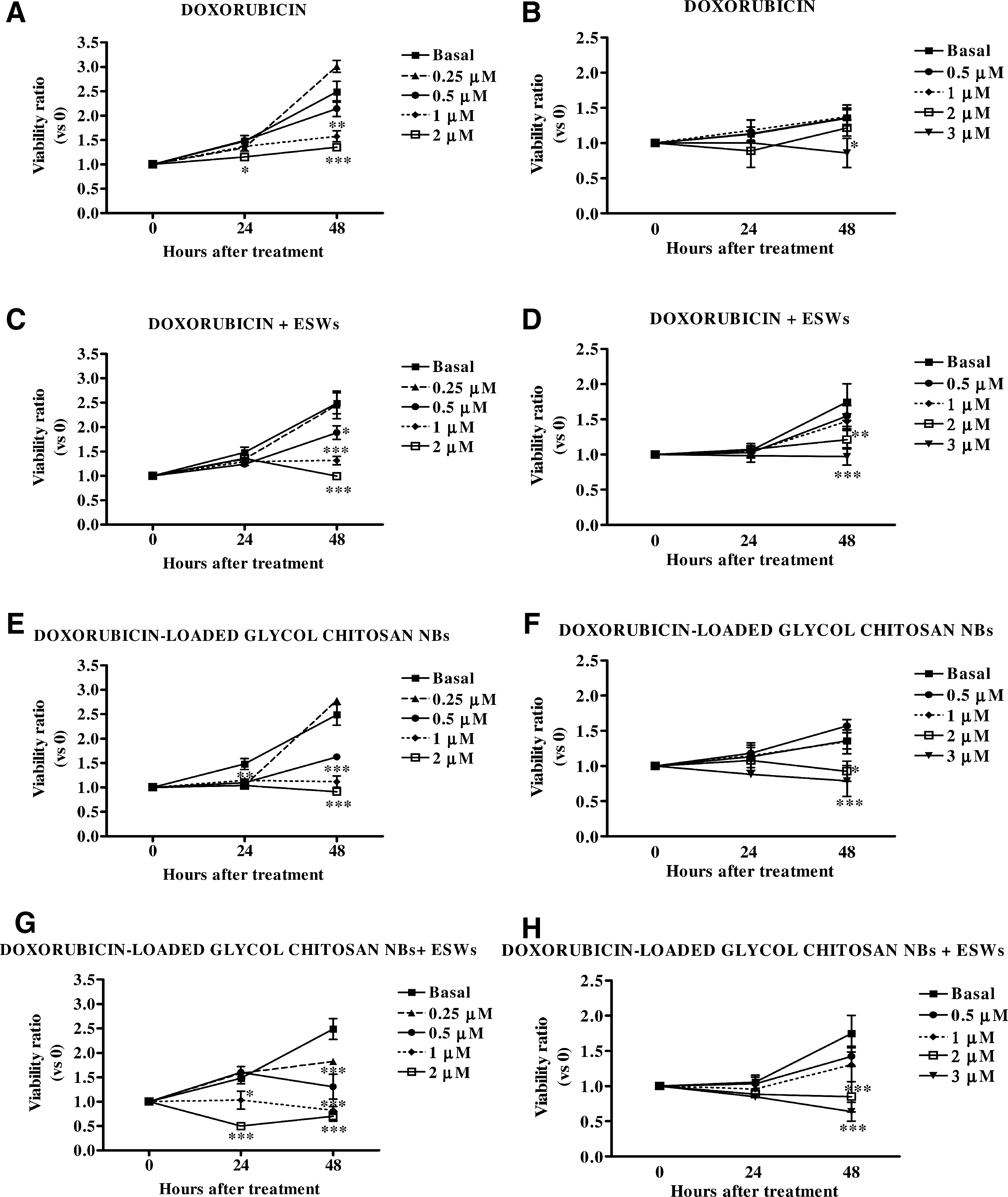
Cytotoxicity of doxorubicin-loaded NBs. Cytotoxic effects of doxorubicin: (
Significance: doxorubicin + ESWs versus doxorubicin, * p < 0.05 in both cell lines; doxorubicin-loaded NBs versus doxorubicin, *** p < 0.001 in CAL-62 and * p < 0.05 in 8305C; doxorubicin-loaded NBs + ESWs versus doxorubicin, *** p < 0.001 in both cell lines; doxorubicin + empty NBs + ESWs versus doxorubicin, ** p < 0.01 in CAL-62; doxorubicin-loaded NBs + ESWs versus doxorubicin-loaded NBs, °p < 0.05 in CAL-62 and °°p < 0.01 in 8305C.
ESWs enhanced doxorubicin-loaded NB cytotoxicity, decreasing the drug GI50 of 40% in CAL-62 and 46% in 8305C cells, respectively.
ATC, anaplastic thyroid carcinoma; GI50, growth inhibition fifty.
Effect of empty NBs and doxorubicin-loaded NBs on rat cardiomyocytes
Empty NBs were found to be safe in rat cardiomyocytes H9C2 cells (Fig. 5A), and they entered the cells at a level comparable to that observed for cancer cells (Fig. 5B). In these cells, drug-loaded NBs did not increase the cytotoxic effects of doxorubicin (Fig. 5C and D), with a GI50 at 48 h of 1.75 μM and 1.68 μM for doxorubicin-loaded NBs and free doxorubicin, respectively.

Effect of NBs on rat H9C2 cardiomyocytes. Viability of H9C2 cells after 72 h of treatment with empty NBs (0.9–5.4 × 10⁶ NBs/mL) (
ESW effects on intracellular doxorubicin release and content
To obtain insight into the mechanisms of ESW action on NB drug delivery, cells were treated with FITC-labeled doxorubicin-loaded glycol chitosan NBs, in either the presence or the absence of ESWs. In the cells treated with NBs alone, the co-localization of the two fluorescent molecules (FITC and doxorubicin) was observed, indicating no perturbation of the NB structure after internalization into the cells (Fig. 6, panels c and c′). In contrast, in the cells that received the combined treatment (NBs plus ESWs), there was a loss of co-localization of the two molecules (Fig. 6, panels f and f′).
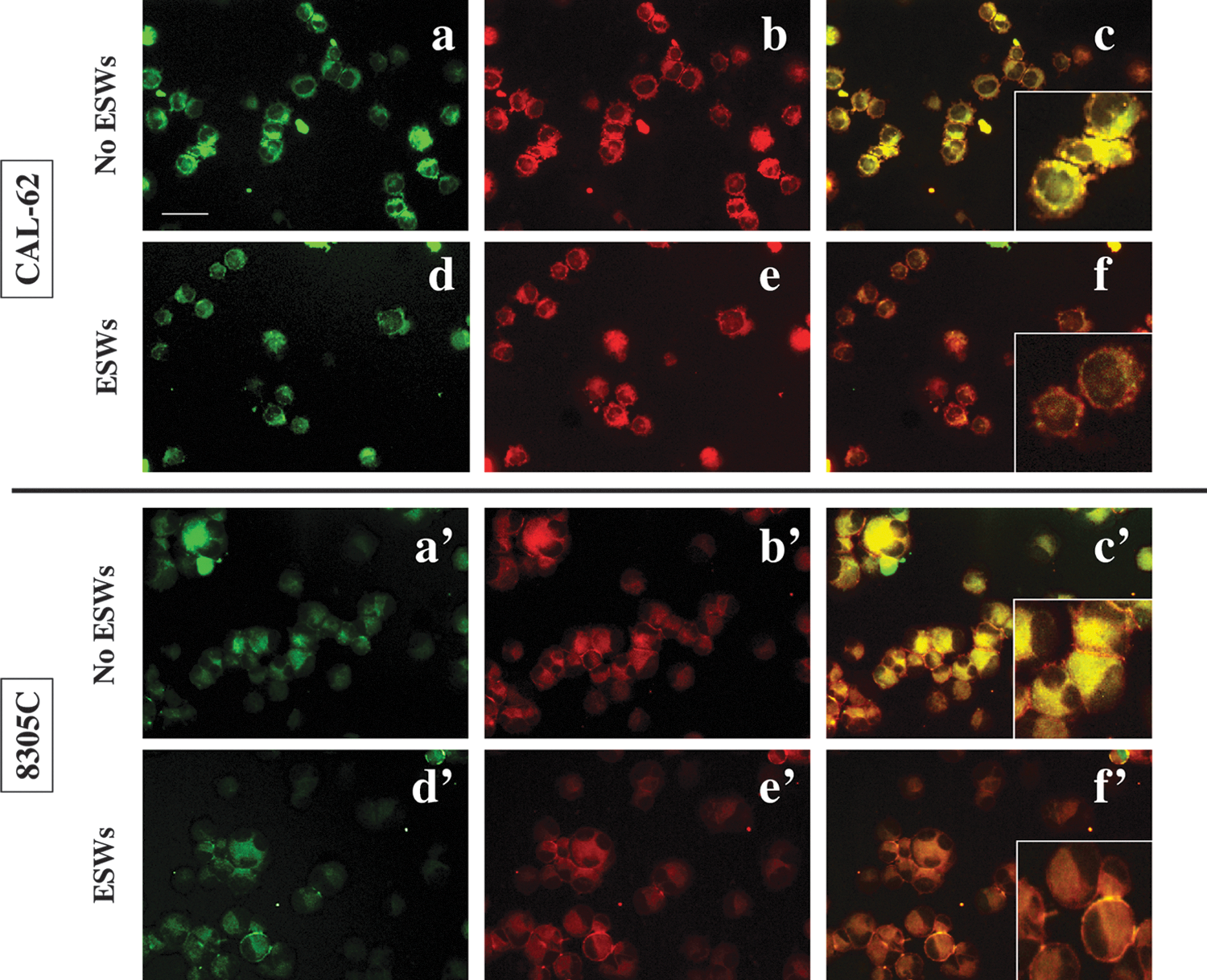
Effects of ESWs on NBs. Pictures of CAL-62 (upper panels) and 8305C (lower panels) cells treated with FITC-labeled doxorubicin-loaded glycol chitosan NBs, in the presence (d–f; d′–f′) or absence (a–c; a′–c′) of ESWs (500 pulses, 0.59 mJ/mm2). Green fluorescence of FITC (a, d, a′, d′), red fluorescence of doxorubicin (b, e, b′, e′), and merge (c, f, c′, f′) photos were taken at 200× final magnification (scale bar: 100 μm). In the right inset, an enlarged detail of the overlaid pictures is shown. The images are representative of three independent experiments. Color images are available online at
Finally, the intracellular doxorubicin content was investigated after ESW treatment. As shown in Figure 7A, ESWs increased the concentration of intracellular free doxorubicin from 0.3 to 1.8 ng/106 cells (p < 0.01). A further increase to 17.2 ng/106 cells (p < 0.001) was obtained when the drug was loaded within NBs, but it was the combined treatment that showed the best effect with a doxorubicin concentration in the cell lysate of about 30 ng/106 cells (p < 0.001).

Intracellular content of doxorubicin. Doxorubicin content was measured in whole cell lysate (
As shown in Figure 7B, NB formulations greatly increased the nuclear amount of doxorubicin to 15.2 ng/106 cells with respect to 0.2 ng/106 cells of the free drug (p < 0.001). However, again, the highest nuclear drug concentration was obtained after the combined treatment with ESWs and doxorubicin-loaded NBs (26.2 ng/106 cells, p < 0.001). Consequently, doxorubicin accumulated into the nuclei, its site of action, resulting in cell death.
Discussion
The present study demonstrates for the first time that the combined treatment with doxorubicin-loaded glycol chitosan NBs and ESWs enhances cytotoxicity of doxorubicin in ATC cells.
Since a curative therapy for ATC has not yet been identified (5,6), it is mandatory to characterize the mutational landscape of ATC better in order to identify new possible candidates for targeted therapy (34,35), and to search novel therapies against this fatal disease. Unfortunately, to date, different targeted therapies used in ATC have not shown any success, and others that have demonstrated a potential effectiveness have not reached a clinical setting. Thyroidectomy with neoadjuvant PLX4720, an inhibitor of BRAFV600E , was effective in an ATC BRAFV600E orthotopic mouse model (36). The treatment of an ATC mouse model overexpressing FOXM1 (Forkhead Box Protein M1) with thiostrepton, a compound that reduces FOXM1 activity (37,38), strongly reduced tumor burden and abolished metastasis (39). LBH589, a potent pan-deacetylase inhibitor with anticancer activity showed potent cytotoxic and anti-invasive activities in both in vitro and in vivo models of ATC (40,41) and conferred radioiodine sensitivity to ATC cell lines and primary cultures (42).
Doxorubicin is the only chemotherapeutical drug approved by the FDA for ATC treatment in monotherapy (12), but it has a poor clinical response (13,14), and extremely severe side effects (15,16,43). Therefore, targeting doxorubicin to the tumor cells and increasing its cytotoxicity while reducing side effects could be important goals in ATC therapy.
In this study, new glycol chitosan-shelled NBs were designed for the delivery of doxorubicin. Chitosan derivatives are attractive excipients for their excellent biocompatibility, biodegradability, and low immunogenicity. Glycol chitosan has already been used with promising results in many studies for drug targeting and delivery. Previous in vivo studies reported very low systemic toxicity for different glycol chitosan nanoparticles and their preferential tumor localization in tumor-bearing animals (44 –48).
NBs filled with perfluorocarbon have already been used for contrast agent preparation because of their stability and biological inertness. Moreover, the very low water solubility, which reduces the gas dissolution rate from the NB inner core into the bloodstream, stabilizes the system and increases the in vivo lifetime of the bubbles. As described elsewhere, perfluoropentane cored and chitosan-shelled NBs have recently been designed as theranostic agents (49).
Safety testing of unloaded NBs was fundamental in order to exclude any undesired effect of the shell. In the present study, empty glycol chitosan NBs entered CAL-62 and 8305C cells and had no effect on cell viability of either of the ATC cell lines. Thus, they appear to be safe, even when used in combination with ESWs. Once glycol chitosan NB safety was proven, it was demonstrated that doxorubicin-loaded NBs generated a higher cytotoxic effect compared with the free drug. Even if increased doxorubicin cytotoxic effects have been reported for drug combinations with either PXD101 (50) or valproic acid (51) histone deacetylase inhibitors, to date, no other study has used delivery systems to target doxorubicin in ATC. Nevertheless, the cytotoxic efficacy of other types of nanoparticles to deliver doxorubicin has been reported in other tumor models. For instance, polylactide-graft-doxorubicin nanoparticles with pH-triggered drug delivery exhibited higher efficacy in killing MCF-7 breast cancer cells (52), and doxorubicin-loaded glucan nanoparticles decorated with trastuzumab antibodies (against HER2) were more efficient in reducing tumor volume in an in vivo murine breast cancer HER2-positive model (53) compared with free doxorubicin. Yu et al. (54) reported that folic acid-conjugated glycol chitosan nanoparticles have a great cytotoxicity against folate receptor-positive HeLa cervical cancer cells, and other authors (55) have demonstrated that doxorubicin-loaded apotransferrin and lactoferrin nanoparticles were more cytotoxic than the free drug in an in vivo rat hepatocellular carcinoma model.
The novelty of the present work is the use of ESWs as physical strategy to trigger drug release from NBs specifically at the tumor site. This study reports that ESWs enhanced in vitro doxorubicin-loaded NB cytotoxicity, decreasing the drug GI50 of 40% in CAL-62 and 46% in 8305C cells, respectively. The reduction of the GI50 is notable, since the use of lower doses of doxorubicin is essential to reduce its side effects (56). Moreover, an increased cytotoxic effect was observed, even when the ESWs were used in combination with the free doxorubicin formulation, in either the absence (free doxorubicin plus ESWs) or the presence of empty NBs (free doxorubicin plus empty NBs and ESWs). All these effects may be the consequences of cavitation, which occurs when acoustic waves propagate into fluids. In fact, cavitation might determine both the perturbation of the NBs, resulting in the drug release, as well as the transient permeabilization of both plasma and nuclear membranes, thus allowing drug penetration into the cell. It has already been reported that ESWs increase cell permeability (26 –28), affecting drug efficacy in breast cancer cells (25), in anaplastic thyroid cancer cells (28), and in an in vivo breast cancer model (29). However, the greatest effect was obtained with doxorubicin-loaded NBs used in combination with ESWs.
As far as the mechanism of action of the combined treatment (doxorubicin-loaded NBs and ESWs) is concerned, ESWs increased the intracellular doxorubicin release from NBs, and, as a consequence, the drug accumulated in the nuclei, where doxorubicin intercalates into DNA and inhibits topoisomerase II, resulting in DNA damage and cell death (57). As nuclear localization is considered a mechanism to overcome drug resistance (58,59), combining ESWs and drug-loaded NBs may be also viewed as a new therapeutic approach to circumvent cellular drug resistance.
All these observations explain the enhanced cytotoxic effects of the combined treatment allowing insights into its mechanism of action. It is suggested that (i) NBs act as stable doxorubicin reservoir in the thyroid cancer cells with prolonged drug release; and (ii) ESWs further increase doxorubicin release from NBs leading to a higher intracellular drug content and favoring its nuclear accumulation.
The cytotoxic effects of doxorubicin-loaded NBs and ESWs, which will allow lower doses to be used, together with the EPR effect, will reduce the exposure of non-target organs to doxorubicin. However, as expected, the in vitro experiments on rat cardiomyocytes H9C2 did not show any significant difference between free doxorubicin and doxorubicin loaded into NBs, but they cannot completely predict the in vivo toxicity on off-target tissues. Only further in vivo experiments will ultimately define the ability of this combined treatment in limiting the systemic side effects of the drug and in the enhancement of its efficacy. Many authors have already reported a higher concentration of doxorubicin in tumors of animals treated with doxorubicin-loaded nanocarriers compared with those treated with the free drug (60 –62). However, to date, nobody has used ESWs to trigger drug release. The ESW generator can be easily placed in contact with a water-based gel on the skin, and ESWs can be focused at the tumor site, thus potentially allowing specific ATC lesions to be targeted. Finally, unlike US, ESWs do not have heating effects. This could be an advantage for in vivo application, since temperature elevation is difficult to control spatially and temporally, especially in large tumors with heterogeneous vascularization, such as ATC (63).
In conclusion, this first in vitro study on the use of ESWs and doxorubicin-loaded NBs, suggests that this combined treatment may be a promising drug delivery tool for targeted ATC treatment, but this approach now needs further characterization in vivo in preclinical models.
Footnotes
Acknowledgments
We thank Sebastiano Colombatto, Department of Oncology, University of Turin, Turin, Italy. We also thank Med & Sport 2000 S.r.l., Turin, Italy, for providing the shock-wave generator. The study was supported by Fondazione CRT Turin, Italy, to Maria Graziella Catalano; by “Progetto Ateneo 2011,” University of Turin, Turin, to Roberto Frairia; and by “Research Fund ex-60%,” University of Turin, Turin, to Roberta Cavalli.
Author Disclosure Statement
No competing financial interests exist.