
Abstract
Background and Objective:
A cell-based bioassay for the measurement of thyroid blocking autoantibodies (TBAb) has been recently reported. The analytical performance and validation of this bioassay is assessed and described.
Methods:
Chinese hamster ovary cells expressing a chimeric thyrotropin receptor were treated with bovine (b) TSH and different concentrations of an immunoglobulin G (IgG) monoclonal human TBAb (K1-70). TBAb was measured as a function of luciferase activity relative to bTSH alone and expressed as percent inhibition. Results obtained in the chimeric cell line were compared with those of a wild-type cell line. Analytical performance studies were subsequently performed with the chimeric cell line only.
Results:
Immunodepletion of K1-70 IgG by using a protein G-Sepharose column showed that positive percent inhibition in the TBAb bioassay was detectable from K1-70 IgG only. The limit of blank was determined to be 12.2%. The limit of detection was 14% inhibition, equivalent to 0.4 ng/mL K1-70, while the limit of quantitation was 22% (coefficient of variation [CV] 12%) equivalent to 0.625 ng/mL K1-70. The dynamic range was between 14 ± 3.7 (mean % inhibition ± standard deviation) and 101 ± 2.6, equivalent to 0.4–10 ng/mL K1-70. The linear range was between 22 ± 2.6 and 93 ± 0.6 inhibition, equivalent to 0.625–5 ng/mL K1-70. The upper limit of the 99th percent reference range was 34% inhibition. In two laboratories, CV values for the intra- and inter-assay precisions for K1-70 ranged from 2% to 12% and from 1.7% to 14.5%, respectively. For patient sera, the CV values for the intra- and inter-assay precisions ranged from 3% to 9% and from 3% to 11%, respectively. No interference was found when follicle-stimulating hormone, luteinizing hormone, and human chorionic gonadotrophin were tested in the TBAb bioassay. The median of % inhibition values in 40 TBAb positive sera from patients with autoimmune thyroid disease were 93.5 (range 25–103) and 92 (range 64–107) for the wild type and chimeric cell lines, respectively. Further, all 40 samples of patients with various non-thyroidal autoimmune diseases were TBAb negative.
Conclusions:
This TBAb bioassay exhibits excellent analytical performance and high level of reproducibility.
Introduction
T
Isolation of human TSHR monoclonal antibodies (mAb) with either blocking (K1-70, 5C9) or stimulating (M22, K1-18) activities has been a major advance to study the interactions of TSHR-Ab (5,21 –27). The well-characterized pure blocking mAb K1-70 was isolated from a patient with hypothyroidism and high levels of TSHR-Ab. Peripheral blood lymphocytes were immortalized with the Epstein–Barr virus (EBV) and fused with the K6H6/B5 heterohybridoma cells line (5). K1-70 has a high affinity to the TSHR (4 × 10¹⁰ L/mol) and has been shown to block the stimulation of cyclic AMP (cAMP) in Chinese hamster ovary (CHO) cells expressing the human TSHR in a dose-dependent manner, with complete inhibition at 100 ng/ml K1-70 IgG (5). The inhibiting effect of K1-70 on thyroid function was also tested in vivo in rats (28).
In comparison, M22 has been recently used to assess the analytical performance of both a Food and Drug Administration–cleared TSAb bioassay and an automated binding assay (29,30). The M22 mAb is used in a thyrotropin-binding inhibiting immunoglobulin (TBII) assay that has been shown to be more sensitive than a bTSH-based binding assay (31). The present study aimed to establish and evaluate the analytical performance characteristics of a functional bioassay for the detection of TBAb (32) by using K1-70. Previous work has shown that K1-70 has no stimulatory activity in a TSAb assay and demonstrated blocking activity in our TBAb bioassay (32).
Materials and Methods
TBAb bioassay procedure
CHO cells expressing either a chimeric or the wild-type human TSHR and a cAMP response element (CRE)-dependent luciferase were used to measure blocking activity, as previously described (32). In the chimeric construct, the amino acid (AA) residues 262–368 of the human TSHR are replaced with the AA residues 262–334 from the rat luteinizing and chorionic gonadotrophin hormone receptor. In the plasmid, the TSHR cDNA is driven by the SV40 promoter/enhancer (SV40 pro) and a preceding beta globin intron. The firefly luciferase reporter gene is driven by a glycoprotein hormone alpha subunit promoter, which contains two CREB (cAMP response element-binding protein) binding sites. Briefly, before seeding the cells, the wells were treated with cell attachment solution (Quidel, Athens, OH). The frozen cells were thawed and immediately seeded at 6.7 × 10⁴ cells/well and then incubated in growth medium (Quidel) at 37°C, 5% CO2 for 15–18 h before addition of the samples. The cells were plated in the inner 48 wells of a 96-well plate with black-wall, clear-bottom wells. The final passage number used for the chimeric CHO cells in the Thyretain® kit is 13. For the sample preparation, 180 μL of reaction buffer (Quidel), 220 μL of bTSH (Sigma Aldrich, St. Louis, MO; product number T8931, Lot number SLBN3854V), and 40 μL of the human blocking mAb K1-70 (10 μg K1-70 IgG/vial; RSR Ltd, Cardiff, United Kingdom) were mixed. The final dilution was 1:11, and 100 μL of each sample was added to each well. K1-70 samples and three controls, consisting of a reference standard bTSH, normal serum, and positive TBAb control were tested in duplicate. After 3 h induction time, the luciferase expression levels of cell lysates were measured directly in the wells following addition of substrate and lysis reagent (Promega, Madison, WI) using a multi-well plate luminometer (Tecan Infinite M200, Tecan GmbH, Crailsheim, Germany; or VERITAS Micro plate Luminometer, Turner Bio Systems, Sunnyvale, CA). Blocking activity was defined as percent inhibition of luciferase expression relative to induction by bTSH alone (100× (1 – sample + bTSH/bTSH alone). The concentration of bTSH (reference) in the well after dilution for the chimeric CHO cells is 100 mIU/L, and for the wild type CHO cells 25 mIU/L. Prior to calculating the percent inhibition; the background relative light units (RLU) were subtracted from the sample RLU and the bTSH RLU. Background RLU was determined using normal serum diluted 1:11 in reaction buffer. The reference control comprises bovine thyrotropin (TSH) 1:11 diluted in normal serum and reaction buffer. The assay cutoff for the chimeric CHO cells is at 40% inhibition. All data were analyzed with the GraphPad Prism software (v5.04).
Sample handling and sharing
All measured samples, including patient sera, K1-70, and glycoprotein hormone samples, were prepared and aliquoted in the Quidel Research and Development Laboratory and subsequently forwarded to the Johannes Gutenberg University laboratory, which was blinded to the findings of the first laboratory. All measurements were performed at both sites.
Comparison of CHO cells expressing either a chimeric or a wild-type TSHR
A total of 80 samples—40 TBAb-positive sera and 40 specimens of non-thyroidal autoimmune diseases (type 1 diabetes, rheumatoid arthritis, systematic lupus erythematosus, and autoimmune gastritis)—were measured in both CHO cell lines to confirm that some TBAb were not missed when the chimeric TSHR cell line was used.
Protein G-Sepharose chromatography
A protein G spin kit (Cat #89949; Thermo Scientific, Waltham, MA) was used to confirm the TBAb assay's specificity for detecting the thyroid blocking antibody K1-70. Serum containing 10 ng of K1-70 was directly tested in the TBAb bioassay as positive control. Each K1-70 concentration was measured three times. Briefly, the K1-70 antibody was applied to the protein G spin column with “end-over-end mixing” for 10 min at room temperature. After mixing, the column was centrifuged. The mixture passing through the column and eluted fractions from the column were collected separately. The bound material was eluted with the elution buffer. Both unbound and eluted fractions were tested in the TBAb assay.
Analytical performance
Determination of the limits of blank, detection, and quantitation
The limits of blank (LoB), detection (LoD), and quantitation (LoQ) were determined according to the Clinical and Laboratory Standards Institute (CLSI)-approved guideline for protocols determining LoD and LoQ (33). LoB and LoD were calculated according to the following formulas: LoB = M blank + 1.64 × standard deviation (SD)blank and LoD = LoB +1.64 × M SD low concentration sample. LoQ was defined as the lowest value of TBAb that can be quantitatively determined with stated acceptable imprecision below a coefficient of variation (CV) of 20% (34,35). A total of 120 replicate measurements of a negative serum sample diluted 1:11 in reaction buffer were performed to determine the LoB. The serum sample was obtained from a healthy euthyroid control subject: baseline serum TSH 0.95 mIU/L (reference range 0.27–4.20 mIU/L); free triiodothyronine 4.5 pmol/L (reference range 3.1–6.8 pmol/L); and free thyroxine 16 pmol/L (reference range 12–22 pmol/L). The subject was negative for all three thyroid antibodies: thyroperoxidase antibodies 7.54 IU/mL (normal <34 IU/mL); thyroglobulin antibodies 21.83 IU/mL (normal <115 IU/mL), and TSHR antibodies 0.92 IU/L (normal <1.75 IU/L). Concentrations of total IgG and IgG subtypes were IgG 12.53 g/L (5.4–18.2 g/L), Ig1 4.6 g/L (2.8–8 g/L), Ig2 3.46 g/L (1.15–5.7 g/L), Ig3 0.86 g/L (0.24–1.25 g/L), and Ig4 0.71 (0.052–1.25 g/L), respectively. Prior to the establishment of the LoD, multiple replicates of six positive K1-70 concentrations 0.3125, 0.625, 1.25, 2.5, 5, and 10 ng/mL diluted in normal serum by using twofold serial dilutions were measured in the TBAb bioassay in order to demonstrate that the luminescence readings (% inhibition) at each K1-70 concentration are normally distributed. This is required for the subsequent calculation of LoD.
Determination of the TBAb bioassay reference range
Serum samples from 282 healthy euthyroid non-smokers (151 females; aged 18–75 years) were measured in the TBAb assay to determine the reference range. All healthy controls were devoid of thyroid disorders and autoimmune diseases, had normal baseline serum thyroid-related hormones, and were negative for thyroperoxidase, thyroglobulin, and TSHR antibodies. The reference range was established by using the 99th percentile for the normal population.
Intra- and inter-assay precision
To determine the precision of the TBAb bioassay, four concentrations of K1-70 (1.25, 2.5, 5, and 10 ng/mL diluted in normal serum only) and one low, one moderate, and one high TBAb positive serum sample of patients with Hashimoto's thyroiditis (HT) and a euthyroid healthy control were measured in accordance with the CLSI Precision Performance Guideline EP05-A2 (36) in two laboratories (two users per laboratory measured one plate for 20 consecutive days). The tested K1-70 concentrations are the final concentrations in the well after dilution in reaction buffer. The CV% was calculated as follows: CV% = (standard deviation RLU specimen/mean RLU specimen) × 100. To evaluate the inter-assay precision, the CV% values of the same K1-70 concentrations were measured multiple times by different individuals in two different laboratories.
Interference testing with the glycoprotein hormone subfamily members
Glycoprotein hormones that share the identical alpha chain structure with TSH were tested for interference in the TBAb bioassay. In the chimeric TSHR CHO cells, luciferase induction with the glycoprotein hormones follicle-stimulating hormone (FSH), luteinizing hormone (LH), and human chorionic gonadotrophin (hCG) were tested with various concentrations and in two conditions consisting of 1:11 diluted normal serum with 5 ng/mL K1-70 mAb or in reaction buffer both in the presence of bTSH. The concentrations of the stock solutions were 8000 IU/L for FSH, 3500 IU/L for LH, and 650,000 IU/L for hCG. All hormones were reconstituted in buffer solution. The concentrations of the glycoprotein hormones were prepared twofold higher because the stock solution is applied to an equal volume of reaction buffer in each well. Each concentration represented the average from triplicate measurements. All hormones were from the National Institute for Biological Standards and Control (FSH code 83/575, LH code 80/552, and HCG code 75/589).
Results
Comparison of CHO cells expressing either a chimeric or a wild-type TSHR
The comparison of the CHO cells expressing either the chimeric or the wild-type TSHR is shown in Figure 1. The median of percent inhibition values in 40 TBAb positive sera from patients with AITD were 93.5 (range 25–103) and 92 (range 64–107) for the wild type and chimeric cell lines, respectively. In comparison, all samples in 40 patients with various non-thyroidal autoimmune diseases were TBAb negative (wild type: 21, –11 to 39; chimeric: 14.5, –52 to 39).
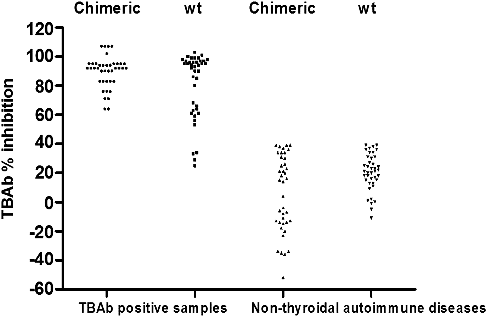
Comparison of the thyroid blocking autoantibodies (TBAb) bioassay measurements in the Chinese hamster ovary cell lines expressing either the chimeric or the wild-type thyrotropin receptor (TSHR). On the y-axis, the TBAb percent inhibition values are shown obtained with the 40 TBAb-positive samples of patients with autoimmune thyroid disease (AITD) and the 40 patient samples with various non-thyroidal autoimmune diseases (x-axis).
Use of a protein G-Sepharose column to deplete TBAb activity of K1-70
Dose–response curves showed that K1-70 had 100% inhibitory activity in the TBAb assay at a concentration of 10 ng/mL K1-70 (32). The ability of protein G to remove the blocking activity of K1-70 was tested by passing a sample containing K1-70 at 10 ng/mL over a protein G column (n = 3). The blocking activity prior to adding to the column was 113%. Mean, median, SD, min, and max of the RLU raw data were 2005, 2005, 3.5, 2002, and 2009, respectively. After binding to the column overnight (ON) the activity of the eluent was 37% (8787, 8786, 2.6, 8785, and 8790, respectively) and was therefore negative for blocking activity. The eluted fraction exhibited 114% inhibition (1871, 1870, 2.6, 1869, and 1874, respectively). Several different times of incubation and other K1-70 IgG concentrations were tested and gave similar results (data not shown).
Analytical performance
Determination of the limits of blank, detection, and quantitation
The results of 120 replicate measurements of a single serum showed a Gaussian distribution with a calculated mean (±SD) inhibition of 4 ± 5%. For the single serum, LoB was 4% +1.64 × 5% = 12.2% inhibition. The measurement of various K1-70 concentrations (from 0.3125 to 10 ng/mL) showed for the K1-70 concentration of 1.25 ng/mL a normal distribution. The LoD was calculated at 14% inhibition, corresponding to 0.4 ng/mL K1-70, while the LoQ was 22% (CV 12%), equivalent to 0.625 ng/mL K1-70. The mean (±SD) dynamic range was between 14 ± 3.7 and 101 ± 2.6, equivalent to 0.4–10 ng/mL K1-70. The linear range was between 22 ± 2.6 and 93 ± 0.6 inhibition, equivalent to 0.625–5 ng/mL K1-70.
Determination of the TBAb bioassay reference range
The results of 846 replicate measurements of 282 control serum samples showed that a total of 20.9% samples had inhibitory activities above the LoD of 14%. The 99th percentile of these samples was determined at 34% inhibition. Mean percent inhibition was –0.8% inhibition, and the confidence interval (CI) of the mean ranged from –2.9% to 1.3% according to the CLSI, EP28-A3C (35).
Precision with the blocking mAb K1-70
The intra- and inter-assay precision data of the TBAb bioassay are shown in Tables 1 and 2. The CV values for the intra- and inter-assay precision ranged from 2% to 12% and from 1.7% to 14.5%, respectively.
CV, coefficient of variation; mAb, monoclonal antibodies; SD, standard deviation.
Patient samples
One low, one moderate, and one high TBAb-positive serum sample from three patients with HT and one sample from a euthyroid healthy control were measured over three consecutive days (Table 3). Two plates were run on each day, and all samples were measured three times in duplicate on every plate. The CV values for the intra- and inter-assay precision ranged from 3% to 9% and from 3% to 11%, respectively.
Interference testing with hormones of the glycoprotein hormone subfamily
No interference was found when FSH, LH, and HCG were tested in the TBAb bioassay at concentrations within and above the physiological range (Table 4).
The luteinizing hormone (LH), follicle-stimulating hormone (FSH), and human choriongonadotropin (hCG) hormone were tested at various levels. Testing was performed in reaction buffer alone (negative control) and with K1-70 at 5 ng/mL.
Discussion
The present study describes in detail and for the first time the analytical performance and validation of a recently introduced chimeric cell-based bioassay for the measurement of TSHR autoantibodies with functional blocking activity or TBAb (32). The measurement of TSHR blocking autoantibodies may be clinically useful in patients with AITD, for monitoring patients during pregnancy and the postpartum period, and during treatment with antithyroid medication, since the appearance of such antibodies may impact the course of the disease. In other words, the development and validation of such a reproducible qualitative and sensitive bioassay may offer a useful laboratory marker for the management of selected patients with AITD. In comparison, and although easier to handle in large laboratories, the measurement of binding TSH receptor antibodies with widely available commercial binding assays does not differentiate between neutral, blocking, and stimulating antibodies, and does not offer an accurate mirror of a possible switch and/or variation of ratios between these antibodies (6). Furthermore and as previously published, this TBAb bioassay was approximately 20-fold more sensitive than a standard TSHR binding assay (32). In contrast to this latter automated binding assay, sera with high levels of TBAb activity were able to be diluted several 100-fold and still exhibit blocking activity above the cutoff level. Thus, this TBAb bioassay may provide a useful tool for measuring the activity of anti-TSHR antibodies and may help clinicians to characterize the diverse clinical presentations of certain patients with AITD. Indeed, there are a handful of good clinical international studies focused on the clinical value of TBAb in patients with AITD, and most studies used TBAb assays that were not fully characterized. This is in contrast to significant experience with TSAb bioassays, which are widely used both in research as well as in clinical laboratories worldwide (2,29,37,38).
A comprehensive comparison between two CHO cell lines expressing either the human chimeric or the wild-type TSHR was performed. In the TBAb bioassay performed with the CHO cells expressing the chimeric TSHR, all samples were blocking antibody positive (>40% inhibition). However, 10% of the CHO cells expressing the wild-type TSHR had similar percent inhibition values to the non-thyroidal autoimmune disease samples. This is why, and as previously shown in detail (32), the CHO cell line expressing the chimeric TSHR was subsequently used for the measurement of TBAb.
For this investigation, a human monoclonal antibody K1-70 IgG with strong antagonist activity was used. The protein G-mediated depletion of K1-70 IgG 1 lambda demonstrated clearly that positive percent inhibition seen in the TBAb assay with K1-70 entirely originates from the IgG and not any contaminating inhibitory substance. The present study indicates that the results obtained in the TBAb bioassay reliably detected the TBAb and demonstrates that the bioassay has a high level of reproducibility. The measured TBAb percent inhibition values were consistent throughout the study as shown by low CV% values in two different laboratories.
The blank samples (LoB) were measured in a single serum of a healthy euthyroid subject devoid of thyroid and autoimmune diseases and for the determination of the TBAb bioassay reference range in a large control collective. However, most inhibitory activities for normal serum were below the LoD of the assay. Both the low LoD and the dynamic range of the TBAb bioassay show that very low levels of serum TBAb can be detected. Assuming that blocking autoantibodies have similar specific inhibitory activity as K1-70, this would represent approximately 0.01% of total IgG. This underlines the high analytical sensitivity of this TBAb bioassay.
Structurally related members of the heterodimeric glycoprotein family other than TSH should be tested for interference. This is a relevant parameter for the evaluation in the TBAb bioassay. TSH shares a common alpha subunit with FSH, LH, and hCG, but the beta subunit differs between the four hormones, hence resulting in distinct specificities. The results showed no interference by other glycoprotein members, and demonstrate that the blocking activity of K1-70 is very specific to bovine TSH. However, since serum levels of hCG can peak to extremely high values in some pregnancies, monitoring patients during pregnancy may require excluding an interference with very high hCG concentrations.
Looking for similar reports, a literature search found no studies reporting the analytical performance measured in other cell-based bioassays for TBAb. RSR Ltd offers an assay service for research use only to measure blocking serum activity (39). In this assay, K1-70 IgG is also used to determine the lower detection limit, which is at 21% inhibition (M ± 3 SD in assay of pool of healthy blood donor sera in the presence of TSH, n = 25). The detection range is 1.2–10 μg/mL K1-70 IgG, and the assay cutoff is at 30% inhibition. No further information regarding the analytical performance is available on the Web site.
Finally, since no international standards for the calibration of TBAb assays are available, standardization of this TBAb bioassay is planned, as previously done with the TSAb assay (30). Furthermore, and subsequent to the evaluation of the analytical performance, a prospective multicenter clinical trial is foreseen to investigate the clinical relevance of TBAb in patients with AITD, that is, Graves' disease and HT, prior to as well as during specific treatment.
Footnotes
Acknowledgments
We are grateful to Liping Yu for data collection and to Jeffrey Houtz for his careful evaluation of the manuscript.
Author Disclosure Statement
P.D.O. and G.J.K. consult for and Y.L. and H.K. are employees of Quidel, CA. T.D., M.K., and K.J.L. have nothing to disclose.