
Abstract
Background:
Previous studies reported significant differences in the clinical presentation and outcomes of papillary thyroid carcinoma (PTC) in pediatric patients compared with adults. Previous studies have suggested that the clinicopathological differences observed between pediatric and adult PTCs may be due the existence of distinct genetic alterations. However, the knowledge of genetic events in pediatric PTCs is based primarily on studies in radiation-exposed PTCs or in the few studies that enrolled predominantly adolescent patients. The aim of this study was to characterize the known oncogenic alterations of the MAPK pathway found in adult and radiation-exposed PTCs in a cohort of predominantly sporadic pediatric PTC patients.
Methods:
Thirty-five pediatric PTCs were screened for the most prevalent fusions (RET/PTC1, RET/PTC2, RET/PTC3, ETV6-NTRK3, and AGK-BRAF) and point mutations (BRAFV600E and NRASQ61 ) described in sporadic pediatric PTCs. The mutational status was correlated with clinicopathological data.
Results:
Mutations were found in 20 out of 35 (57%) PTC cases. Fusion oncogenes were the main genetic alterations found. RET/PTC1-3 rearrangements were found in 13 (37%), ETV6-NTRK3 in 3 (9%), AGK-BRAF in 4 (11%), and BRAFV600E in 3 (9%). No mutation was found in NRASQ61 . BRAFV600E was associated with older age and larger tumor size (p < 0.05), and RET/PTC3 was associated with a larger tumor size and multifocality (p < 0.05).
Conclusions:
The genetic signature in this cohort was remarkably different than that observed in adults. Although observed at a lower prevalence, the spectrum of mutations was quite similar to that described in radiation-exposed pediatric PTCs. As mutations were unidentifiable in over 40% of the PTC cases, more comprehensive studies conducted in these patients will help to decipher the genetic landscape of sporadic pediatric PTCs.
Introduction
T
Previous studies reported significant differences in the clinical presentation and outcomes of PTCs in pediatric patients compared with adults. Although pediatric PTC patients usually present at a more advanced stage of disease at diagnosis and have a higher risk of recurrence, the overall mortality is lower (8,9). Nevertheless, a marked heterogeneity within the pediatric group has been reported. Children (<10 years) present with more aggressive local disease and are more likely to have lymph node metastases at diagnosis and are probably more prone to develop subsequent metastases than adolescents (≥10–18 years) (4,10 –13). They also experience recurrence more frequently and earlier than adolescents (14).
There has been an ongoing debate about whether the clinicopathological differences observed between pediatric and adult PTCs may be due to the existence of distinct genetic alterations (15). Efforts have been made to identify these acquired genetic abnormalities in tumors from pediatric patients that will determine the tumors' biological behavior and that will ultimately allow molecular prognostication. However, most studies were performed in pediatric PTCs after radiation exposure such as the Chernobyl disaster.
The prevalence of the BRAFV600E mutation, the most common genetic event found in adult PTCs (16,17), is significantly lower in sporadic and radiation-exposed pediatric PTCs (18 –20). RET/PTC rearrangement, the second most common event in adult PTCs, is the major genetic alteration found in sporadic (21 –24) and radiation-exposed pediatric PTC (20,24 –26). Moreover, RAS mutations are a rare genetic event in pediatric PTCs. The incidence rates range from 0% to 7% in sporadic cases (15,20,27,28), while no mutation was reported in radiation-exposed tumors (15,20,29). In the few pediatric patients in whom RAS point mutations were detected, only the NRASQ61R was described.
Recently, the ETV6-NTRK3 fusion oncogene was identified as the second most common rearrangement found in radiation-exposed pediatric PTCs (20,30). A similar prevalence was described in pediatric PTCs with no known radiation exposure from the same geographic area (20). Remarkably, the authors also reported AGK-BRAF rearrangements in 4% (1/26) of radiation-exposed pediatric PTC patients, but this genetic alteration was not found in sporadic cases (20). However, our group reported AGK-BRAF fusions in 10% (3/30) of sporadic pediatric PTC patients (31), suggesting the possibility of genetic and environmental differences between ethnicities and geographic regions.
Given that the prevalence and specificity of each genetic event varies geographically in pediatric populations, additional studies—performed across different populations—are required to gain insight into disease biology and to develop strategies for molecular diagnosis and prognosis as well as treatments for pediatric thyroid cancer.
In the present study, we screened a cohort of 35 Brazilian patients with sporadic pediatric PTCs for the most common genetic alterations found in adult and pediatric PTCs diagnosed in other countries and correlated these findings with clinicopathological features.
Materials and Methods
Patients and samples
The series consists of 35 formalin-fixed paraffin embedded (FFPE) sections from primary tumors from patients who underwent thyroid surgery at Hospital São Paulo (Universidade Federal de São Paulo) and Hospital da Santa Casa de São Paulo. The samples were reviewed by two pathologists (R.D. and M.T.S.A.). The study included 16 classical PTCs (CPTCs), 13 follicular variants (FVPTCs), 4 diffuse sclerosing variants (DSVPTCs), and 2 other variants of PTCs.
All of the patients were ≤18 years of age at the time of diagnosis, as recommended by the American Thyroid Association guidelines for children with thyroid nodules and DTCs (32). The pediatric cases were further separated into two age groups: children (<10 years old) and adolescents (≥10–18 years old). Because information about pubertal development was not available for all of the patients, the age of 10 years was used as the cutoff. This cutoff point was recommended by the World Health Organization and used to determine the effect of age on time to recurrence and mortality rates in pediatric DTCs (14,33,34).
The clinical and pathological features evaluated are summarized in Supplementary Table S1 (Supplementary Data are available online at
DNA isolation and BRAFV600E and NRASQ61R genotyping
DNA was isolated from 10-μm-thick formalin-fixed paraffin-embedded (FFPE) sections using the NucleoSpin Tissue kit (Macherey-Nagel, Duren, Germany). The DNA was quantified using a NanoDrop 2000c spectrophotometer (NanoDrop Technologies, Wilmington, DE). Somatic mutations in BRAF (BRAFV600E ) and NRASQ61R were screened by PCR. Exon 15 of the BRAF gene was amplified by PCR as previously reported (35). Exon 3 of the NRAS was amplified as follows: 94°C for 5 minutes, followed by 40 cycles of 94°C for 30 seconds, 56°C for 30 seconds, and 72°C for 30 seconds. The PCR products were resolved by electrophoresis, purified, and submitted to cycle sequencing using a BigDye Terminator v3.1 cycle sequencing kit (Applied Biosystems, Foster City, CA). The samples were sequenced at least twice and in both directions. PCR primers are summarized in Supplementary Table S2.
RNA isolation and cDNA synthesis
Total RNA was isolated from 10 μm thick FFPE sections using the Recover All Total Nucleic Acid isolation kit (Ambion Inc., Austin, TX). Total RNA (500 ng) was treated with DNAse and reverse transcribed into cDNA with oligo-dT12–18 (50 μM) and random hexamers (50 ng) using a Superscript III reverse transcriptase kit (Invitrogen Corp., Carlsbad, CA), following manufacturer's instructions.
Expression of fusion oncogenes in thyroid cell lines
PCCL3 cells (normal follicular thyroid cells derived from Fischer rats) were cultured in Ham's F12 medium (Life Technologies, Carlsbad, CA) supplemented with 5% fetal bovine serum, thyrotropin (1 mU/mL), hydrocortisone (10 ng/mL), transferrin (5 μg/mL), and insulin (10 μg/mL) (Sigma-Aldrich, Saint Louis, MO). Transient transfections were performed using 10 μg of plasmids encoding RET/PTC1, RET/PTC2, RET/PTC3, ETV6-NTRK3, and AGK-BRAF. The oncogene-transfected cells were harvested, and the total RNA isolated using TRIzol Reagents (Invitrogen Corp.) and reverse transcribed into cDNA using oligo(dT)12–18 and Superscript III reverse transcriptase, as previously described (36). The cDNA generated from cells expressing the fusion transcripts was used as positive control.
RET/PTC2, RET/PTC3, ETV6-NTRK3, and AGK-BRAF plasmids were kindly donated by Dr. James Fagin (Memorial Sloan-Kettering Cancer Center), and the RET/PTC1 construct was donated by Massimo Santoro (Università di Napoli Federico II).
Detection of ETV6-NTRK3 fusion transcript
All of the samples were screened for the presence of the ETV6-NTRK3 fusion transcript by RT-PCR, as previously described (20). Briefly, cDNA (2 μL) was subjected to PCR amplification using 2 pmol of each specific primer and Platinum Taq DNA Polymerase (Invitrogen Corp.). The PCR products were resolved on a 2.5% agarose gel and visualized on a Bio-Rad Gel Doc EZ System (Bio-Rad, Hercules, CA). The presence of the fusion transcripts was confirmed by direct sequencing. The primers used were previously described and validated (20) (Supplementary Table S2).
Detection of AGK-BRAF fusion transcript
The AGK-BRAF fusion was previously investigated in 30 cases of PTC (31). In this study, a total of 35 patients were screened for the presence of the AGK-BRAF fusion transcript by RT-PCR, as previously described (31). The presence of AGK-BRAF was confirmed by fluorescence in situ hybridization analysis.
Detection of RET/PTC rearrangements by quantitative RT-PCR
The thyroid samples were screened for RET/PTC1, RET/PTC2, and RET/PTC3 rearrangements according to standard quantitative RT-PCR. Briefly, cDNA (2 μL) was subjected to PCR amplification using 1 × SYBR Green PCR Master Mix (PE Applied Biosystems) and predesigned primers that span the known breakpoints specific for each of the RET/PTC transcripts (23) or an internal control (RPS8). After an incubation at 50°C for 10 minutes and 95°C for 10 minutes, the PCR reaction was subjected to 40 cycles at 95°C for 15 seconds and 60°C for 1 minute. The qPCR reaction was performed in triplicate, and the threshold cycle was obtained using Applied Biosystem software. Post-amplification fluorescent melting curve analysis was performed. The PCR products were analyzed by electrophoresis on a 2% agarose gel and visualized on a Bio-Rad Gel Doc EZ system (Bio-Rad). The samples were considered positive when at least two replicates had amplification curves that crossed the threshold before cycle 38, the melt curve aligned with the positive control, and the gel analysis revealed a band of the expected product size. To confirm the identity of the amplified products, randomly selected samples positive for RET/PTC isoforms were sequenced using the BigDye Terminator Cycle Sequencing Kit (PE Applied Biosystems). The primers used to detect the RET/PTC fusion transcripts were previously described and validated (23) (Supplementary Table S2).
Statistical analysis
Statistical analysis was performed using GraphPad Prism 6.0 (GraphPad Software, La Jolla, CA) software. Shapiro-Wilk test was used to verify the normality of distribution. Mann-Whitney U test or the Student's t-test was used to compare continuous variables, and Fisher's exact test was used for dichotomous variables. Results with p < 0.05 were considered to be statistically significant and a trend with borderline significance was considered when p was ≥0.05 and <0.10.
Results
Clinical and pathological features
The mean age at diagnosis was 11.8 years (range, 4 to 18 years). The female to male ratio was 26:9, with female predominance mainly in the adolescent group. The pathological findings included multifocality in 17 (49%), extrathyroidal (ETE) extension in 17 (49%), lymph node metastasis (LNM) in 28 (80%), and lung metastasis in 11 (31%) patients. There was no death from PTC in patients enrolled in this study. Two of the patients had family history for PTCs, and three cases had a history of previous radiation exposure during childhood to treat another cancer. Demographic data were previously reported (37) and are summarized in Supplementary Table S3.
BRAFV600E and NRASQ61 mutation in sporadic pediatric PTCs
We systematically investigated the prevalence of the BRAFV600E mutation in 35 pediatric PTCs. The BRAF mutation was found in 3 (9%) patients (Fig. 1). The age of the patients ranged from 16 to 18 years old (mean 17.33). None of the patients had a mutation in exon 3 of NRAS.
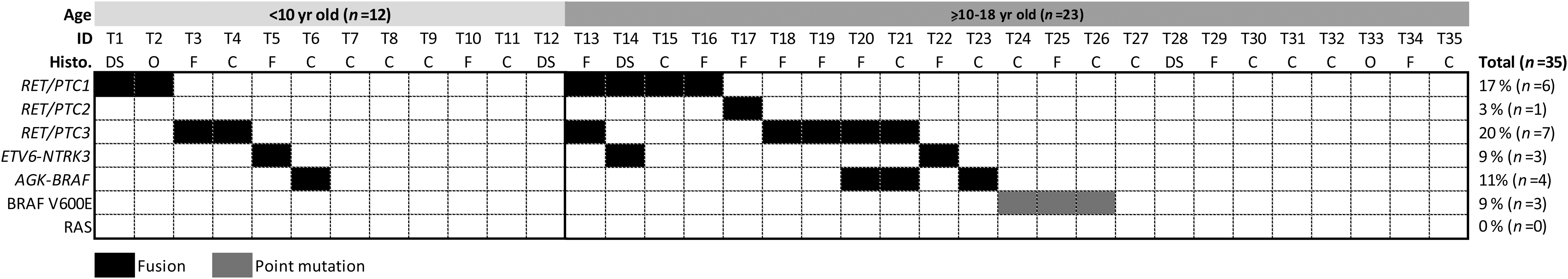
Prevalence of RET/PTC1, RET/PTC2, RET/PTC3, BRAFV600E , ETV6-NTRK3, AGK-BRAF, and NRAS in all of the pediatric papillary thyroid carcinomas (PTCs) (n = 35). The prevalence of genetic alterations in pediatric PTC was classified according to age: children (<10 years old) or adolescents (≥10–18 years old) and histological variants. C, classical PTC; DS, diffuse sclerosing variant of PTCs; F, follicular variant of PTCs; O, other variants of PTCs (solid and encapsulated variants of PTCs).
ETV6-NTRK3 in sporadic PTCs
ETV6-NTRK3 was identified in 3 (9%) patients with PTC (Fig. 1). Although different isoforms of ETV6-NTRK3 rearrangements were previously identified in PTCs, in this study, the fusion transcript that juxtaposes exons 1–4 of ETV6 to exons 12–18 of NTRK3 was observed in all ETV6-NTRK3–positive samples. Sequencing analysis confirmed the identity of the ETV6-NTRK3 rearrangements identified on gel analysis. None of the cases positive for ETV6-NTRK3 had a radiation exposure history.
AGK-BRAF was identified in both sporadic and radiation-exposed pediatric PTCs
The AGK-BRAF fusion was detected in 4 (11%) patients; one case had a radiation exposure history for the treatment of Hodgkin's lymphoma. The remaining cases were sporadic pediatric PTC patients (31).
RET/PTC rearrangements in sporadic pediatric PTCs
Overall, RET/PTC was identified in 13 (37%) patients. RET/PTC1 alone was identified in 4 (11%), RET/PTC2 in 1 (3%), and RET/PTC3 in 4 (11%). The co-occurrence of RET/PTC1 and RET/PTC3 was identified in 1 (3%), RET/PTC1 and ETV6-NTRK3 was detected in 1 (3%), and RET/PTC3 and AGK-BRAF was found in 2 (6%) patients (Fig. 1). Only one case with a RET/PTC3 and AGK-BRAF rearrangement had a radiation exposure history. Sequencing analysis confirmed the identity of the RET/PTC rearrangements identified on gel analysis.
Correlation of mutational status with clinicopathological features
Of the 35 PTCs screened using a candidate gene approach, 20 (57%) were positive for BRAFV600E , RET/PTC1, RET/PTC2, RET/PTC3, ETV6-NTRK3, or AGK-BRAF (Fig. 1 and Supplementary Table S1).
When the tumors harboring fusion oncogenes and BRAF point mutations were grouped and compared with tumors negative for genetic alterations, the mutation-positive cases were of larger size than the mutation-negative cases (p < 0.05). When each genetic event was independently analyzed, tumors harboring RET/PTC3 or BRAFV600E were of a larger size than the mutation-negative cases (p < 0.05).
When each mutation was considered as an independent variable, RET/PTC3 was correlated with multifocality (p = 0.0325) and a tendency to ETE (p = 0.0867), and BRAFV600E was significantly associated with older age of the patients (p = 0.0221).
The presence of genetic event was not significantly correlated with cervical or distant metastases. Eighteen out of twenty (90%) tumors positive for mutations had LNM at diagnosis, whereas 10/15 (67%) tumors without mutations had LNM at diagnosis (p = 0.1122). Distant metastases were identified in 7/20 (35%) tumors with an identified genetic alteration and in 4/15 (27%) tumors negative for mutations. Moreover, when each mutation was considered individually, none of the genetic alterations were correlated with the presence of cervical or distant metastases.
When tumors were classified according histological subtypes, a trend to a higher prevalence of rearrangement in FVPTCs (70%) than in CVPTCs (31%) (9/13 vs. 5/16; p = 0.0618) was identified. Of note, a trend to an increased incidence of RET/PTC fusion transcripts was identified in FVPTCs (46%) compared with CVPTCs (19%) (6/13 vs. 2/16; p = 0.0648). Therefore, there was an apparent correlation with the nature of the causal mutation. Consistent with previously reported findings (38), RET/PTC1 was a common event found in DSVPTCs.
Interestingly, the prevalence of genetic alterations did not differ significantly between the two age groups: 6/12 (50%) and 14/23 (61%) of alterations occurred in the <10 years and in the ≥10 to 18 years age groups respectively (Fig. 1). RET/PTC, the most prevalent genetic alteration, was equally prevalent in both age groups, which may explain the lack of correlation between genetic mutation and clinicopathological findings in children and adolescents. Of note, BRAFV600E mutation was found only in the ≥10 to 18 years age group.
Discussion
In this cohort, the lack of the BRAFV600E mutation in children and a lower prevalence (9%) of this mutation in pediatric PTCs compared with adult PTCs is in agreement with previous reports (18 –20,39). Our findings suggest that the prevalence of BRAFV600E is not only lower in pediatric sporadic PTCs than in adult PTCs, but that its prevalence also increases with age. Although similar findings were previously reported (27,39), a higher prevalence of BRAFV600E was recently described in sporadic pediatric PTCs (28,40,41). It is worth mentioning that these last reports enrolled predominantly adolescent patients. Moreover, the study with the highest prevalence of BRAFV600E considered pediatric patients up to 21 years of age (41).
RET/PTC fusion transcripts were previously identified as the central genetic event in both radiation-exposed and sporadic pediatric PTCs (24). Indeed, a RET/PTC rearrangement was the most prevalent mutation found in this patient cohort. Although the prevalence (37%) was lower than the one reported in radiation-exposed PTCs (24,38,42), our data are consistent with those observed in sporadic pediatric PTCs from different geographical areas (27,43). RET/PTC1 and RET/PTC3 were the most prevalent isoforms identified in our cohort. Although RET/PTC3 was mainly found in the patients who were exposed to 131I from the Chernobyl accident (24,42), only one patient with the RET/PTC3 rearrangement had a previous radiation history in the cohort presented here. To the best of our knowledge, this is the first study to report the prevalence of RET/PTC fusions in pediatric patients from Brazil.
Because both the prevalence and distribution of isoforms diverge widely among different countries, the difference observed in the distributions of the RET/PTC isoforms in our series could be, at least in part, due to the exposure to different environmental factors and genetic background. Another potential explanation is that a large proportion of the tumors were follicular variants of PTC in our series. Previous studies reported such an association between RET/PTC3 and solid and follicular variant PTCs (24,42).
ETV6-NTRK3 was reported as the second most common rearrangement found in radiation-exposed PTC diagnosed among individuals who were <18 years of age at the time of the Chernobyl accident (20,30). In this cohort, the prevalence of ETV6-NTRK3 (9%) was consistent with that reported in adult (5%) and sporadic pediatric (7%) PTC (20,30).
While the mutations described above occur in a mutually exclusive pattern in most tumors, we identified one tumor with concomitant presence of a RET/PTC1 and RET/PTC3 rearrangement, another tumor with RET/PTC1 and ETV6-NTRK3, and two tumors with RET/PTC3 and AGK-BRAF fusions. Although ETV6-NTRK3 was initially identified in radiation-exposed pediatric PTCs lacking known common driver mutations (20), further validation analyses showed the co-occurrence of ETV6-NTRK3 with RET/PTC1 or with BRAFV600E (30).
Although the studied mutations were equally prevalent in children and adolescents, the spectrum of mutations notably diverges between these age groups. The occurrence of BRAFV600E mutation and concomitant rearrangements were found only in adolescents. These findings suggest that there are differences in tumor biology according to age.
Although the BRAFV600E mutation was previously reported to be correlated with a more aggressive tumor phenotype in adults (17,44,45), we did not find such a correlation in sporadic pediatric PTCs. Other groups have previously reported an absent association between biological behavior of pediatric PTCs and the presence of the BRAFV600E mutation (15,41). These findings reflect the fact that in this age group, BRAFV600E is uncommon, and the aggressiveness of PTC is likely associated with other genetic events. Whether additional changes in tumor microenvironment, metabolism, microRNA pattern, or other genetic events that activate the mitogen-activated protein kinase (MAPK) pathway such as the ETV6-NTRK3 or AGK-BRAF fusion oncogenes (20) are associated with a more aggressive phenotype, is still unclear.
Although ETV6-NTRK3 reportedly activates both MAPK and phosphoinositide 3-kinase signaling pathways and AGK-BRAF activates MAPK (20), the ability of these fusions proteins to activate these pathways and the biological effects, compared with BRAFV600E and RET/PTC, are still uncertain.
Remarkably the RET/PTC3 isoform was associated with pathological features related with tumor aggressiveness, such as larger tumor size and multifocality, and there was a trend towards a higher prevalence of ETE. RET/PTC3 was previously associated with solid-follicular growth pattern in radiation-exposed pediatric PTCs and more aggressive disease, whereas RET/PTC1 was mainly associated with noninvasive disease (23,24,42,43).
There was no statistically significant difference between the presence of a genetic mutation and other features related to a worse prognosis, such as local or distant metastasis, in this study. Nevertheless, further studies—ideally involving a multicenter approach—are needed to confirm the absence of such an association.
There are some limitations to our study. First, our study is limited by the inherent biases of a retrospective analysis, and therefore, the lack of a proper follow-up with all of the patients. Second, it is limited by a small sample size. However, most of the previous studies usually enrolled a similar or even smaller number of sporadic pediatric PTC patients (15,19,20,27,28,43). The much lower incidence of sporadic pediatric PTCs, especially in patients under 10 years old, compared with adult PTCs makes it difficult to collect a larger number of patients.
The investigation of the most prevalent mutations found in adult PTCs, including the recently identified ETV6-NTRK3 and AGK-BRAF fusion transcripts in a cohort of pediatric sporadic PTCs, contributes to better understanding of the pathogenesis the pediatric population. We observed that the prevalence of mutations in this cohort was remarkably different compared with adult PTCs, even in the same geographical area (17,46). Nearly 9% of the patients had activating mutations, while 49% had rearrangements. Although the prevalence of fusion oncogenes was lower than in radiation exposed PTCs (20), they were the most prevalent genetic event found in this cohort. Importantly, about 43% of the pediatric patients were negative for any of the studied mutations, suggesting that the most prevalent mutations found in adults and radiation-exposed PTCs are not the major events in sporadic pediatric PTCs. Because the tissue samples used in this study were particularly enriched in tumors cells, we believe that the false negative rate is very low.
In summary, the clinical features of PTCs are markedly different according to age, and this fact is probably associated with the differences in molecular profiles. The high number of patients in whom no genetic event was identified suggests that other genetic/epigenetic factors may be associated with the pathogenesis and biological behavior of sporadic pediatric PTCs. More comprehensive studies of the PTCs negative for mutations are needed in order to decipher their genetic signature and to reduce the so-called “dark matter” in the genome of sporadic pediatric PTCs.
Footnotes
Acknowledgments
The study was supported by research grants 2013/03867-5 and 2015/60330-8 from the São Paulo State Research Foundation and grant 470441/2013-5 from the Brazilian Research Council. L.M. and A.U.B. are recipients of fellowships from the São Paulo State Research Foundation. J.M.C. is a recipient of a scholarship of Research Productivity from the Brazilian Research Council.
Author Disclosure Statement
The authors have no conflicts of interest to declare.
References
Supplementary Material
Please find the following supplemental material available below.
For Open Access articles published under a Creative Commons License, all supplemental material carries the same license as the article it is associated with.
For non-Open Access articles published, all supplemental material carries a non-exclusive license, and permission requests for re-use of supplemental material or any part of supplemental material shall be sent directly to the copyright owner as specified in the copyright notice associated with the article.