
Abstract
Background:
The aim of this study was to evaluate whether 18F-fluorodeoxyglucose positron emission tomography/computed tomography (FDG-PET/CT) is useful in the further characterization of sonographically suspicious and scintigraphically hypofunctional thyroid nodules.
Methods:
Sixty-five patients with sonographically suspicious thyroid nodules that were hypofunctional on 99m-Tc-pertechnetate scintigraphy (diameter >1 cm) were retrospectively analyzed. All patients underwent evaluation with FDG-PET/CT. Thyroid nodules were sonographically categorized by Thyroid Image Reporting and Data System (TIRADS) criteria. FDG uptake in the thyroid nodules was visually compared to the remainder of the thyroid tissue and categorized as pathological or non-pathological. In cases of pathologically increased uptake, maximum standardized uptake values (SUVmax) of the suspicious nodule and the perinodular thyroid tissue were determined. Depending on the results of the FDG-PET/CT, patients underwent thyroid surgery (pathological FDG uptake) or follow-up examinations (non-pathological FDG uptake). The endpoints for comparison with the FDG uptake were either histological results or sonographic follow-up examinations of at least five years.
Results:
In 18/65 (28%) patients, PET/CT showed visually pathological FDG uptake in the suspicious thyroid nodules (SUVmax 7.1 ± 4.6). Of these nodules, 3/18 (17%) were sonographically categorized as TIRADS 4a, 11/18 (61%) nodules as TIRADS 4b, 3/18 (17%) nodules as TIRADS 4c, and 1/18 (6%) nodule as TIRADS 5. The other nodules without pathological FDG uptake were categorized as TIRADS 4a in 24/47 (51%) patients, as TIRADS 4b in 18/47 (38%), and as TIRADS 4c in 5/47 (11%) patients. Twenty-three patients (18 FDG positive, 5 FDG negative) underwent surgery. The other patients underwent follow-up examinations with stability on observation over at least five years as a surrogate endpoint. Taking into consideration that FDG-PET/CT was rated as true negative in 42/47 patients with stability on sonographic follow-up, sensitivity, specificity, positive predictive value, and negative predictive value of FDG-PET/CT in detecting malignancy in the suspicious thyroid nodules were 100%, 87%, 61%, and 100%, respectively.
Conclusion:
FDG-PET/CT allows stratification of patients with sonographically suspicious and scintigraphically hypofunctional thyroid nodules with a positive predictive value of 61% and negative predictive value of 100%. The absence of visually pathological FDG uptake in suspicious thyroid nodules may be useful for avoiding unnecessary thyroid surgery.
Introduction
T
Another recommended option is the sonographic surveillance of suspicious nodules. Additional diagnostic tests such as thyroid scintigraphy is only recommended by the American Thyroid Association for patients with a low thyrotropin level (1), and is not recommended at all by the British Thyroid Association (2). Measurement of serum thyroglobulin is not informative in the evaluation of thyroid nodules (8) and is not recommended by international guidelines (1,2,9).
Several studies have shown a high risk of malignancy in thyroid incidentalomas with a high glucose uptake in 18F-fluorodeoxyglucose positron emission tomography-computed tomography (FDG-PET/CT) (10 –15). The guidelines recommend further characterization of nodules with focal FDG uptake with sonography and FNA (1,2). Moreover, FDG-PET/CT can be useful in the initial staging of patients with thyroid cancer and the re-staging of patients with recurrent thyroid cancer (16 –25). An emerging field of application of FDG-PET/CT is the preoperative characterization of thyroid nodules in case of an indeterminate cytology. Some studies have demonstrated that FDG-PET/CT has a high sensitivity and high NPV in suspicious thyroid nodules (26 –31).
In the present study, patients with sonographically suspicious thyroid nodules that were hypofunctional on 99m-Tc-pertechnetate scintigraphy were analyzed with FDG-PET/CT. The nodules of these patients were not evaluated with FNA. Depending on the results, patients underwent diagnostic surgery or periodical ultrasound follow-up for at least five years. The aim was to evaluate whether FDG-PET/CT can help in the patient selection for either surgery or observation.
Materials and Methods
Patients
The data of 65 patients (36 females and 29 males; M age ± standard deviation = 61 ± 10.8 years) with a sonographically suspicious and scintigraphically hypofunctional thyroid nodule (diameter >1 cm) were retrospectively analyzed. In these patients, no FNA was performed due to anticoagulation with vitamin K antagonist or because patients refused FNA. Instead of FNA, patients underwent FDG-PET/CT as further preoperative diagnostic evaluation. Imaging findings were validated by histopathology or follow-up examinations of at least five years.
Thyroid ultrasound and scintigraphy
Each nodule was sonographically examined by an experienced physician in nuclear medicine using an LOGIQ P6 ultrasound (GE Healthcare GmbH, Solingen, Germany). Thyroid scintigraphies were performed using a Nucline TH-22 Gamma Camera (Mediso GmbH, Münster, Germany). About 20 minutes after intravenous injection of approximately 60 MBq of 99mTc pertechnetate, images were acquired with a scan duration of 10 minutes or until 100,000 counts were recorded. In principle, other tracers such as 123I or 131I could also be used for this purpose. However, 99mTc is more readily available (technetium generator for in-house production of 99mTc on demand), cheaper, and associated with a lower radiation exposure. All images were digitally stored.
FDG-PET/CT
Whole-body FDG-PET/CT was performed using a Biograph 16 LSO PET/CT scanner (Siemens AG, Healthcare Sector, Erlangen, Germany). Patients fasted for at least 6 h before the scan, and a blood glucose level <150 mg/dL at the time of tracer injection was assured. Whole-body emission data were acquired 60 ± 5 min after intravenous tracer application (weight-dependent FDG dose, mean activity 321 ± 20.7 MBq) by scanning from the upper thigh to the head in three-dimensional mode using six to eight bed positions of 2 min each. A non-diagnostic CT scan was performed (non-contrast-enhanced, 120 kV tube voltage, 80 mA/s, 5 mm slice thickness, 0.5 s per rotation). The CT data were used for attenuation correction of the PET data. For image reconstruction, attenuation-weighted ordered subset expectation maximization iterative algorithm with two iterations and eight subsets, Gaussian filter with 5.0 mm full width at half maximum, and attenuation image segmentation were used.
Image analysis
Two physicians experienced in nuclear medicine analyzed the thyroid sonography and thyroid scintigraphy in one session and the FDG-PET/CT studies in another session; the imaging studies were de-identified. In cases of differences in scoring, a consensus was obtained. Based on sonography, thyroid nodules were categorized according to the modified Thyroid Image Reporting and Data System (TIRADS) criteria by Kwak et al. (32), which are listed in Table 1.
Suspicious US features: solid without cystic component, hypoechogenicity or marked hypoechogenicity, microlobulated or irregular margins, microcalcifications, and taller-than-wide shape.
TIRADS, Thyroid Image Reporting and Data System; FDG-PET/CT, 18F-fluorodeoxyglucose positron emission tomography/computed tomography; US, ultrasound.
FDG-PET/CT images were visually evaluated regarding FDG uptake in the suspicious thyroid nodules and were compared with the remainder of the thyroid tissue. Imaging findings were categorized as pathological (focal FDG uptake higher than perinodular tissue) or non-pathological (FDG uptake not increased). If the uptake was pathologically increased, the maximum standardized uptake value (SUVmax) of the thyroid nodule and the perinodular tissue were determined in a semi-quantitative analysis. A ratio of thyroid nodule SUVmax divided by the perinodular SUVmax was calculated (SUVmax ratio). These findings were compared with the histological results or follow-up examinations.
Surgery and follow-up examinations
Every patient with pathologically increased FDG uptake in a thyroid nodule underwent diagnostic surgery (subtotal lobectomy) of the suspicious thyroid nodule. In case of malignant findings in the intra-surgical frozen section, a total thyroidectomy was performed. Surgical pathology was available for all specimens. A few patients without pathological findings in the FDG-PET/CT requested a pathological clarification and underwent surgery for this reason. Patients without pathological findings on FDG-PET/CT did not undergo surgery but underwent periodical follow-up examinations, including at least semi-annual ultrasound of the thyroid and neck for lymph node characterization. Additional FDG-PET/CT studies were not performed. A follow-up examination was rated as non-pathological if the thyroid nodule did not significantly grow (measuring inaccuracy of 10% was allowed) and the sonographic pattern did not change (scored as “stable”). The results of the latest available follow-up examination (at least five years) served as endpoint for comparison.
Statistics
The descriptive analysis included mean values, standard deviations, sensitivity, specificity, positive predictive value (PPV), and NPV. Statistical analysis of differences between SUVmax values and SUVmax ratio was performed with a two-tailed, unpaired Mann–Whitney test and was considered statistically significant if p < 0.05 (GraphPad Prism v6.0; GraphPad Software, Inc., La Jolla, CA). This study was conducted according to the local ethical rules and regulations. Since this was a retrospective analysis of clinically indicated examinations, no approval by the Institutional Review Board was required.
Results
Sonography and FDG-PET/CT of the thyroid nodules
According to the TIRADS criteria by Kwak et al. (32), 27/65 (42%) thyroid nodules were categorized as TIRADS 4a, 29/65 (45%) nodules as TIRADS 4b, 8/65 (12%) nodules as TIRADS 4c, and 1 (2%) nodule as TIRADS 5.
FDG-PET/CT detected pathologically increased FDG-uptake in 18/65 (28%) nodules. Among them, 3/18 (17%) nodules were categorized as TIRADS 4a, 11/18 (61%) nodules as TIRADS 4b, 3/18 (17%) nodules as TIRADS 4c, and 1/18 (6%) nodule as TIRADS 5. The mean SUVmax value of the suspicious nodule, the perinodular tissue, and the SUVmax ratio were 7.1 ± 4.6 (median 5.5; range 2.7–20.2), 2.1 ± 0.7 (median 1.9; range 1.3–3.9), and 3.5 ± 2.0 (median 2.8; range 2.1–10.6), respectively. The other 47/65 (72%) nodules without pathological increased FDG uptake were rated as follows: TIRADS 4a in 24/47 (51%) nodules, TIRADS 4b in 18/47 (38%) nodules, and TIRADS 4c in 5/47 (11%) nodules.
Thyroid surgery
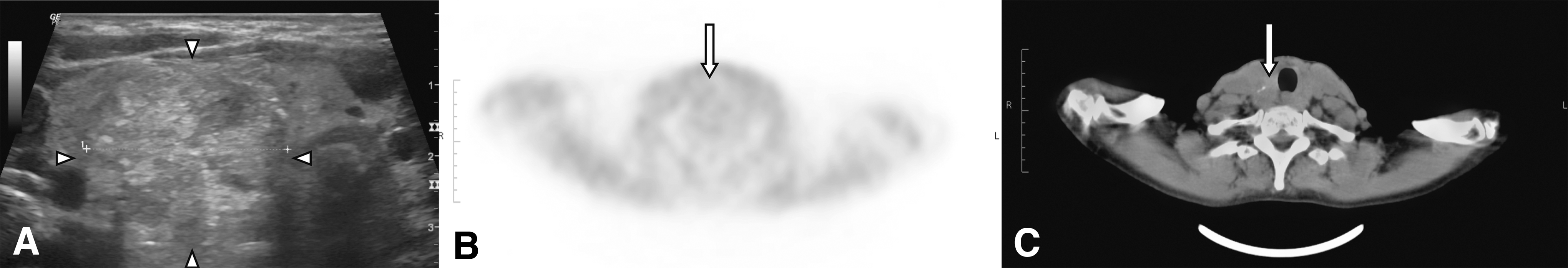
The 18 patients with a sonographically suspicious, scintigraphically hypofunctional, and FDG-PET/CT-positive nodule underwent surgery. Pathological examination revealed thyroid carcinoma in 11/18 (61%) nodules (FDG-PET/CT true positive): 10 papillary thyroid carcinomas (n = 4 pT1b, n = 5 pT2, and n = 1 pT3a; AJCC/TNM 7th edition) and one anaplastic thyroid carcinoma. An illustrative example is shown in Figure 1. In 7/18 (39%) thyroid nodules, no carcinoma was found (FDG-PET/CT false positive). The TIRADS classification of the FDG true-positive nodules was: TIRADS 4a, n = 3; 4b, n = 5; 4c, n = 2; and 5, n = 1. The mean SUVmax value in the FDG true-positive thyroid nodules was 7.9 ± 5.3 (median 5.6; range 3.7–20.2) versus. 5.9 ± 3.2 (median 4.3; range 2.7–10.1) in the FDG false-positive nodules. The SUVmax ratio was 3.5 ± 2.4 (median 2.8; range 2.2–10.6) versus 3.4 ± 1.4 (median 3.0; range 2.1–5.6). The SUVmax and SUVmax ratio were not statistically different (p = 0.36 and p = 0.93, respectively).

Nodule in the right thyroid lobe with marked hypoechogenicity and irregular margins in axial ultrasound (
FDG-PET/CT did not show a pathologically increased FDG uptake in 47/65 (72%) patients. An example is given in Figure 2. In this group, 42/47 (89%) patients underwent periodical follow-up examinations, and 5/47 (11%) patients underwent a diagnostic surgery. Pathological results of the latter five patients revealed no carcinomas (FDG-PET/CT true negative). None of the patients has pathological FDG uptake outside of the thyroid, and CT did not show evidence of pulmonary or other metastases.
Nodule in the right thyroid lobe with solid component, marked hypoechogenicity, irregular margins and microcalcifications (
Follow-up examinations
In 47 patients, the hypofunctional thyroid nodules did not show pathological FDG-uptake. Of these 47 patients, 42 (89%) underwent periodically follow-up examinations instead of diagnostic surgery. The follow-up examinations over at least five years (mean 79 months; median 80 months; range 61–99 months) showed no relevant changes in the sonographic pattern of the thyroid nodules and no suspicious cervical lymph nodes in any of the patients. Therefore, all FDG-PET/CT of these 42 patients were rated as true negative.
Diagnostic performance of FDG-PET/CT for thyroid carcinoma detection
The sensitivity of FDG-PET/CT was 100% and the specificity was 87%, assuming that the 42 patients without surgical pathology but stability on follow-up of at least five years were true negative. The PPV and NPV were 61% and 100%, respectively. In consideration of the low prevalence of thyroid carcinomas (3.5% in Germany, calculated on the basis of 82 million inhabitants and a five-year prevalence of 28,400 affected persons (6)), the PPV decreased to 21.9% while the NPV remained constant at 100%, according to the Bayes' theorem.
Discussion
It is well accepted that a large number of diagnostic hemithyroidectomies are unnecessary (33). In Germany, for instance, only about 8% of thyroid surgeries are performed for thyroid carcinomas (6,7). After an indeterminate FNA, which occurs in about 20% of cases, diagnostic surgery is one of the available options. In 60–88% of these patients, the histology reveals the presence of benign nodules (28,34,35). Molecular testing can provide an opportunity for further characterization of the suspicious thyroid nodules in cases of indeterminate FNA. Mutational sets of oncogenes and gene-expression classifiers have a high specificity, while sensitivity and NPV are variable within the different studies (1,4,36). Therefore, additional diagnostic tests are necessary.
Many hopes were placed on real-time elastography of thyroid nodules to differentiate between benign and malignant lesions. While initial results were promising, the approach remains of controversial validity (37 –42). For example, a reliable classification of scintigraphic hyperfunctional thyroid nodules as most likely benign lesions was not possible (43). Another approach to characterize thyroid nodules is TIRADS, first introduced by Horvath et al. (44) and modified by Kwak et al. (32). The modified system by Kwak et al. (32) seems to be more easily implemented in clinical routine and more reliable than the original system, although several thyroid carcinomas are erroneously categorized as benign (45).
FDG-PET/CT is known to be useful in the staging of patients with thyroid cancer, particularly if no longer radioiodine avid (21,22). FDG-PET/CT used for other indications also results in the detection of thyroid incidentalomas. Moreover, FDG-PET/CT may be helpful in patients with thyroid nodules with indeterminate cytology in order to differentiate between benign and malignant nodules (26 –31). The present study is the first aimed at evaluating patients with sonographically suspicious and scintigraphically hypofunctional thyroid nodules using FDG-PET/CT as a decision aid in planning either diagnostic surgery or sonographic observation.
In the present study, 65 patients with sonographically suspicious and scintigraphically hypofunctional thyroid nodules were included. In these patients, no FNA was performed. For the purpose of avoiding unnecessary diagnostic surgery in mostly non-malignant thyroid nodules, FDG-PET/CT was performed as a potential predictor of malignancy in the suspicious thyroid nodules.
In this study, FDG-PET/CT showed pathologically increased FDG uptake in 18/65 (28%) patients. Surgery revealed thyroid carcinomas in 11/18 (61%) patients, resulting in a PPV of 61% of FDG-PET/CT in detecting malignancy in this subset of thyroid nodules. In the other 47/65 (72%) patients, the sonographically suspicious thyroid nodules did not show pathologically increased FDG uptake. In the follow-up examinations, these FDG-negative nodules presented a constant sonographical pattern and size, findings that make a malignoma less likely. These non-malignant thyroid nodules were rated as TIRADS 4a, b, and c in 51%, 38%, and 11% of cases, respectively. TIRADS 4b and 4c are associated with an intermediate to moderate concern for malignancy. In the other patient group with FDG-positive nodules, 61% and 17% of the nodules were rated as TIRADS 4b and 4c, respectively. The results show that the classification as TIRADS 4b and 4c have a high overlap in the groups with FDG-positive and FDG-negative thyroid nodules. This indicates that ultrasonography alone is not sufficient to characterize these nodules and therefore an ancillary test such as FNA, molecular testing, or molecular imaging is necessary.
Unfortunately, the SUVmax values and the SUVmax ratio show a high overlap in the groups with true-positive and false-positive FDG uptake in the thyroid nodules. The statistical analysis did not show a significant difference between these values. The false-positive FDG uptake in non-malignant thyroid nodules is a common problem and has been addressed in several studies, which could not find a reliable cutoff SUVmax value for focal FDG uptake (46 –49). Thus, positive FDG uptake in incidentalomas needs further diagnostics such as ultrasonography, FNA, or scintigraphy.
Given the low incidence of thyroid cancer, it is very important excluding non-malignant thyroid nodules from unnecessary surgery. In this study, without surgical histology of 42 FDG-negative thyroid nodules but stability in follow-up examinations of at least five years, FDG-PET/CT has a sensitivity and NPV of 100%. Given the relatively high PPV, these findings support that FDG-PET/CT is a suitable tool for reducing unnecessary thyroid surgeries. However, false-positive FDG uptake was found in the thyroid nodules of 7/18 (39%) patients.
MIBI scintigraphy is an alternative diagnostic tool that has been used for a long time for the characterization of thyroid nodules (50,51). In newer studies, MIBI scintigraphy offers a high NPV, but the PPV and specificity are not very high. For example, Theissen et al. analyzed patients comparable to the patient collective in the present study and found a high NPV of 97%, a PPV of 19%, and a specificity of 54% (52). Similar results with a low PPV and a low specificity were recently reported by Wale et al. (53) and in a meta-analysis by Treglia et al. (54). Both diagnostic methods offer the opportunity to reduce the number of diagnostic hemithyroidectomies. According to the summary of de Geus-Oei et al. (46), FDG-PET/CT has a higher NPV than MIBI scintigraphies. Other characteristics, such as sensitivity, specificity, and PPV, vary depending on the publications. The present study assume that FDG-PET/CT has high NPV and sensitivity but additionally a high PPV of 61%, which contrasts with MIBI scintigraphy. Contrasting results were found by Sager et al. (30). They compared MIBI scintigraphy and FDG-PET/CT prior to surgery in 23 patients. The criterion for a suspicious nodule in FDG-PET/CT was a SUVmax higher than the background, whereby the measurement of the background SUVmax is not described. Moreover, the authors did not differentiate between focal or diffuse uptake in the nodule area, although diffuse FDG uptake is typical for autoimmune thyroid disease (47). This can cause a lower specificity of FDG-PET/CT. A recently published study by Piccardo et al. analyzed 87 patients scheduled for surgery because of a thyroid nodule with indeterminate cytology (31). All patients underwent FDG-PET/CT, multi-parametric neck ultrasonography, and MIBI scintigraphy. Using final histopathology as the gold standard, the authors found that the accuracy of FDG-PET/CT in detecting thyroid malignancy is higher than that of MIBI scintigraphy and multi-parametric neck ultrasonography.
Aside from reducing unnecessary thyroid surgeries, the use of FDG-PET/CT may reduce cost. Vriens et al. demonstrated this in a study on patients with cytologically indeterminate thyroid nodules (29). Simplifying this analytic approach and taking the results of the present study as the basis for PPV, FDG-PET/CT may reduce the cost, at least in Germany, by about 53% for patients in the statutory health insurance system. This calculation is based on the physician fee scale within the statutory health insurance scheme (Einheitlicher Bewertungsmaßstab, EBM;
The present study has a number of limitations. First, this study is a retrospective study. Second, we have different clinical endpoints. Histopathological verification was available of all FDG-positive thyroid nodules and in only five patients with a FDG-negative result. All other patients underwent follow-up examinations with stability on observation for at least five years as a surrogate endpoint, but a long-term follow-up would be desirable. Moreover, FNA was not performed in any of the patients.
The present study suggests that FDG-PET/CT can reduce the number of unnecessary diagnostic hemithyroidectomies in 87% of the patients with sonographically suspicious and scintigraphically hypofunctional thyroid nodules.
Conclusion
FDG-PET/CT allows patient stratification in case of sonographically suspicious and scintigraphically hypofunctional nodules. Absence of FDG uptake can be used to avoid unnecessary thyroid surgery in patients with sonographically indeterminate nodules.
Footnotes
Author Disclosure Statement
No competing financial interests exist.