
Abstract
Background:
The ability of thyroid follicular cells to take up iodine enables the use of radioactive iodine (RAI) for imaging and targeted killing of RAI-avid thyroid cancer following thyroidectomy. To facilitate identifying novel strategies to improve 131I therapeutic efficacy for patients with RAI refractory disease, it is desired to optimize image acquisition and analysis for preclinical mouse models of thyroid cancer.
Methods:
A customized mouse cradle was designed and used for microSPECT/CT image acquisition at 1 hour (t1) and 24 hours (t24) post injection of 123I, which mainly reflect RAI influx/efflux equilibrium and RAI retention in the thyroid, respectively. FVB/N mice with normal thyroid glands and TgBRAFV600E mice with thyroid tumors were imaged. In-house CTViewer software was developed to streamline image analysis with new capabilities, along with display of 3D voxel-based 123I gamma photon intensity in MATLAB.
Results:
The customized mouse cradle facilitates consistent tissue configuration among image acquisitions such that rigid body registration can be applied to align serial images of the same mouse via the in-house CTViewer software. CTViewer is designed specifically to streamline SPECT/CT image analysis with functions tailored to quantify thyroid radioiodine uptake. Automatic segmentation of thyroid volumes of interest (VOI) from adjacent salivary glands in t1 images is enabled by superimposing the thyroid VOI from the t24 image onto the corresponding aligned t1 image. The extent of heterogeneity in 123I accumulation within thyroid VOIs can be visualized by 3D display of voxel-based 123I gamma photon intensity.
Conclusions:
MicroSPECT/CT image acquisition and analysis for thyroidal RAI uptake is greatly improved by the cradle and the CTViewer software, respectively. Furthermore, the approach of superimposing thyroid VOIs from t24 images to select thyroid VOIs on corresponding aligned t1 images can be applied to studies in which the target tissue has differential radiotracer retention from surrounding tissues.
Introduction
T
131I therapy has proven to be effective in improving overall survival (1,2), especially for patients with RAI-avid micrometastases. However, patients with structurally discrete RAI-avid metastatic disease often require multiple 131I treatments. Yet, the patients may still have persistent disease. Furthermore, patients with non-RAI-avid disease do not benefit from 131I therapy, as their tumors have lost the ability to take up 131I. These patients are in need of novel treatments, as their tumors are generally resistant to conventional chemotherapy (3). To date, no novel treatment has shown improved overall survival for patients with progressive RAI refractory disease, despite improved progression-free survival with manageable adverse effects in some patients treated with emerging small molecule inhibitor therapy (4 –6). For patients with progressive RAI refractory disease, much effort has been focused on restoring and increasing RAI uptake to improve 131I therapeutic efficacy. Recently, the MEK inhibitors selumetinib and CKI were shown to increase RAI uptake in thyroid tumors via nuclear imaging in preclinical thyroid tumor mouse models (7,8). Shortly thereafter, selumetinib was shown to restore/enhance the tumoral RAI uptake in 12/20 thyroid cancer patients with RAI refractory disease (9).
To identify novel agents and to optimize dosing regimens that further increase RAI uptake, it is desired to optimize the workflow of image acquisition and enhance the capability of image analysis for thyroid cancer mouse models. This study developed a mouse cradle to achieve consistent tissue configuration of the head and neck region during image acquisition, which greatly facilitated subsequent image analysis. In-house CTViewer software was developed with the objective of streamlining image analysis with new capabilities. This enabled automatic segmentation of thyroid volumes of interest (VOI) to be conducted in SPECT/X-ray computed tomography (CT) images acquired 1 hour (t1) and 24 hours (t24) post injection of 123I, which mainly reflect radioiodine influx/efflux equilibrium and radioiodine retention, respectively. In addition, it enabled xyz coordinates of areas with differential gamma photon intensity from 123I accumulation to be identified such that the extent of heterogeneity of 123I accumulation within the thyroid VOI could be evaluated.
Materials and Methods
Animals
All studies were approved by The Ohio State University's Institutional Animal Care and Use Committee, which oversees the responsible use of animals in university research and instructional activities. All research activities conformed to the statutes of the Animal Welfare Act and the guidelines of the Public Health Service as issued in the Guide for the Care and Use of Laboratory Animals. Mice used for microSPECT/CT imaging in this study include FVB/N mice and TgBRAFV600E mice (10). For TgBRAFV600E mice, the expression of human BRAFV600E cDNA was targeted to thyroid cells of transgenic FVB/N mice with a bovine thyroglobulin promoter (bTg). Since BRAFV600E expression leads to thyroid dedifferentiation and bTg promoter activity is decreased by thyroid dedifferentiation, bTg promoter-driven BRAFV600E expression is self-limiting in TgBRAFV600E mice. TgBRAFV600E mice appeared to compensate successfully for thyroid dysfunction, as their serum thyroxine/triiodothyronine and somatic growth were normal (10).
Cradle design and mouse placement in cradle
To facilitate consistent positioning and tissue configuration of the mouse head and neck where the thyroid is located among image acquisitions, a cradle comprised of a jig with a groove for a gas anesthesia tube, a tooth bar, two cheek bars, and a cover with a holder for an Eppendorf tube containing known radioactivity for decay control was designed (Fig. 1A). The cradle components were printed using RDG720 material with an Objet Prime 30 3D printer (Stratasys, Eden Prairie, MN) at 0.0006 inch resolution, and were assembled and attached to the mouse bed in the Flex-XO SPECT/CT system (TriFoil Imaging, Inc., Chatsworth, CA) using nylon screws and fasteners so as not to distort the X-ray CT image. The jig serves as a base for all cradle parts. The tooth bar holds the top incisors of the mouse to maintain the head position in the cranial/caudal direction. The cheek bars are placed on either side of the mouse head to maintain the head position in the right/left direction. The cover traps gas anesthesia inside of the cradle to ensure the mouse remains unconscious and holds an Eppendorf tube containing known radioactivity for decay control. The components of the cradle relative to the mouse being imaged are shown in Figure 1B–D.

Cradle design and X-ray computed tomography image of mouse in cradle. (
For mouse placement in the cradle, the mouse first has its top incisors positioned in the hole of tooth bar. The limbs are placed on the outside of the cradle, and the body is positioned symmetrically in the right/left direction. The tail is gently pulled back to straighten the spine, the cheek bars are placed against the mouse, and the cover is placed on the jig (Supplementary Video S1; Supplementary Data are available online at
MicroSPECT/CT image acquisition
All mice subjected to microSPECT/CT imaging were intraperitoneally injected with 400–450 μCi of 123I or 185–250 μCi of 99mTcO4 − in 200 μL of saline prepared by the nuclear pharmacy at the authors' institute. Radioactivity was measured by a dose calibrator (CRC-12; Capintec, Florham Park, NJ) and normalized for radioactive decay. The mouse was anesthetized with 4% isoflurane mixed with O2 at a flow rate of 1.5 L/min for 3 min and then placed in the cradle with 1.5–2% isoflurane mixed with O2 at the same flow rate. A 0.25 mL Eppendorf tube containing 10–30 μCi of 123I was included as a decay control. Image acquisition was conducted at 1 h (t1) and 24 h (t24) after 123I injection or 1 h after 99mTcO4 − injection. Mice imaged with 99mTcO4 − were imaged multiple times, and these images were used for mouse rotation and translation analyses. SPECT images were acquired with two gamma cameras equipped with 1 mm single-pinhole collimators (75 mm focal distance) using the following settings: 180° rotation per camera, 32 projections per camera, 30 s projection duration, and 30 mm radius of rotation. CT images were acquired with a single X-ray detector camera at the following settings: X-ray tube set to 75 kVp, 360° rotation, 1024 projections, one frame per projection, and 2.5 × magnification. The SPECT image is 80 × 80 × 80 pixels, with a resolution of 0.52 mm, while the CT image is 512 × 512 × 512 pixels with a resolution of 0.075 mm.
Image analysis with in-house CTViewer software
A Microsoft foundation class (MFC)-based CT and SPECT image analysis software, CTViewer, was designed in-house, in which all the algorithms for image processing and data analysis were written in C++. The capabilities of CTViewer include (i) loading RAW data sets of SPECT and CT images; (ii) co-registering SPECT and CT images based on the pre-calibrated coordinate transformation; (iii) displaying the SPECT and CT images in three orthogonal planes, whether separately or in fusion mode; (iv) automatically selecting VOIs in a threshold-based manner; and (v) automatically calculating percentage of injected radioisotope in thyroid VOIs. In addition to the aforementioned capabilities, a two-step rigid body registration was developed to align images of the same mice taken at different times. First, CTViewer coarsely aligns the target CT image to the reference CT image based on the skull (the rigid body) using principle component analysis and iteratively adjusting VOIs. In the second step, the two CT images are finely aligned using 3D image correlation in three iterations. Consequently, the computation time is greatly reduced. The same coordinate transformation matrix is then applied to align the corresponding two SPECT images. The features of CTViewer and the time required for the whole process are shown in Supplementary Video S2.
Results
Cradle-enabling pose control leading to consistent tissue configuration
Due to a living mouse's body being flexible, the spatial relationship between target tissues of interest and surrounding anatomic landmarks varies significantly among images acquired on a standard CT imaging bed. As shown in Figure 2, consistency of tissue configuration within the head and neck region is greatly improved when the cradle is employed. To evaluate the effectiveness of the cradle in achieving pose control, translation and rotation required to align two images acquired at different times, such as t1 and t24, were calculated for each individual mouse. This process was applied to four FVB/N mice and six TgBRAFV600E mice. The sagittal (x), coronal (y), and transaxial (z) translation and the rotation around x, y, and z axes of these 14 mice are summarized in Table 1. Regardless of the difference in age (4.8–16.7 months), body weight (19.9–45 g), mouse model, and between two users, the x, y, and z translations for all mice are <1 mm. The rotation around the x and y axes for all mice are <5°. The rotation around the z axis has the greatest variability, yet is <11°.
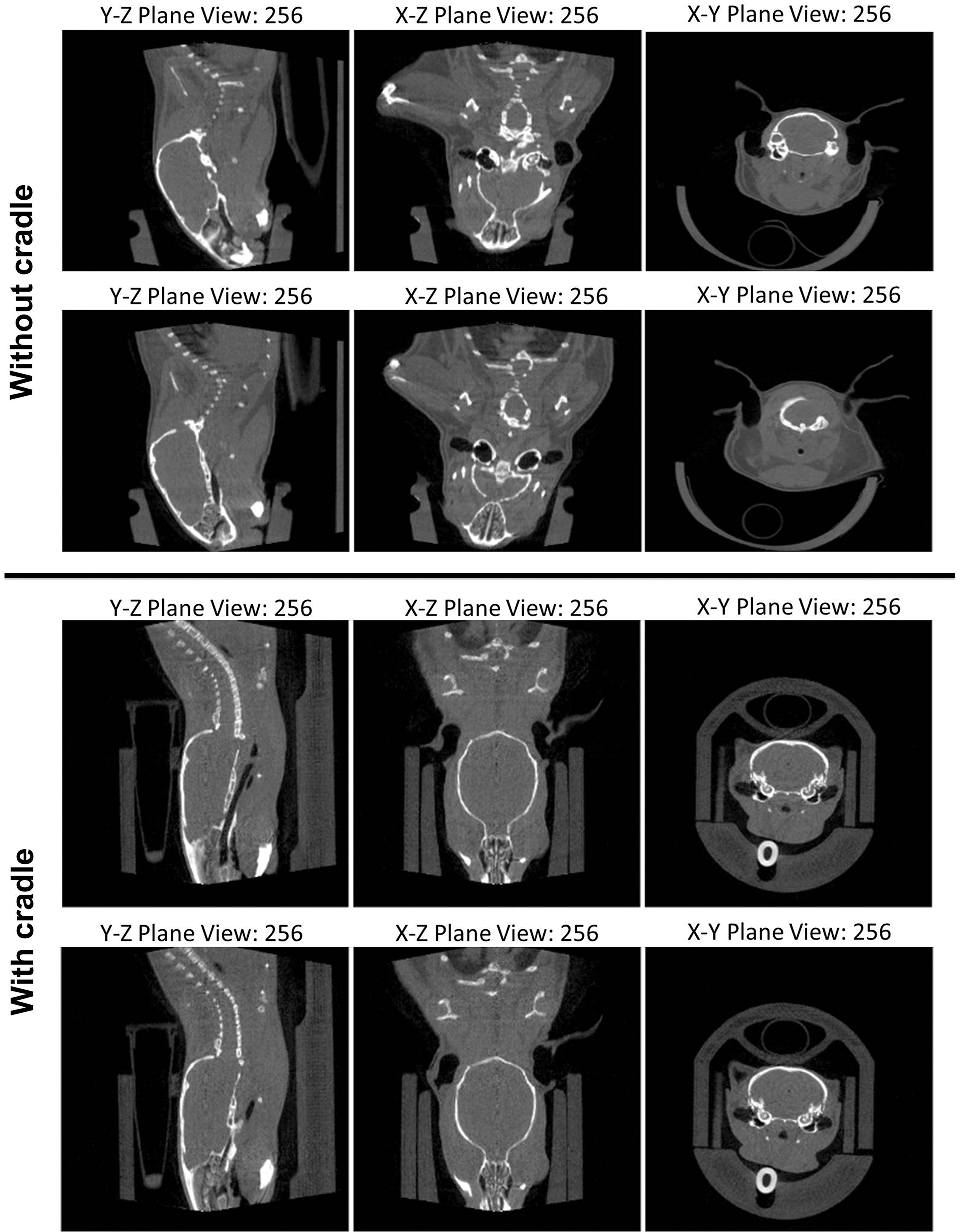
Tissue configuration variation in head and neck region is greatly reduced with the use of cradle during image acquisition. The top two rows show the sagittal, coronal, and transaxial views of X-ray CT images of the same mouse acquired on separate days without the cradle. The bottom two rows show the sagittal, coronal, and transaxial views of CT images of the same mouse acquired on separate days with the cradle.
The translation and rotation between images for each mouse model are presented as mean ± standard deviation.
Mouse placement in the cradle and subsequent imaging conducted by the second user.
Automatic segmentation of thyroid VOIs on t1 and t24 SPECT images
At 1 h post 123I injection, 123I accumulates not only in the thyroid gland but also in the salivary glands as both glands express the Na+/I− symporter, which mediates 123I influx against its concentration gradient. The mouse thyroid and salivary glands are in close proximity, and the boundaries of soft tissues are not discernable via the anatomical CT image. It is therefore difficult to separate 123I accumulation in the thyroid gland from adjacent salivary glands in t1 SPECT images. At 24 h post 123I injection, 123I has been organified into thyroglobulin and retained in the thyroid, whereas 123I in the salivary gland is not retained. Consequently, 123I accumulation in the thyroid gland is readily discernable in t24 SPECT images, and the thyroid VOI can be automatically segmented in a threshold-based manner. After t1 and t24 images are aligned, the thyroid VOI from the t24 image can be superimposed onto the t1 image to enable automatic segmentation of t1 thyroid VOI. The CT images of t1 and t24 prior to and after alignment are shown in Figure 3A versus 3D (sagittal view), 3B versus 3E (transaxial view), and 3C versus 3F (coronal). Automatic segmentation of the thyroid VOI in t24 image is shown in Figure 3G–I, wherein the perimeter of VOI is indicated with a red curve. The perimeter of t24 thyroid VOI is then superimposed onto the aligned t1 image, as shown in Figure 3J–L. It can be seen that the red curve encloses the region with highest gamma photon intensity from 123I, the anticipated thyroid VOI of the t1 image. This demonstrates the effectiveness of pose control using the cradle and the accuracy of image alignment. Taken together, the cradle along with the in-house CTViewer software allows the thyroid VOI to be automatically defined in t1 and t24 SPECT images without user subjectivity.
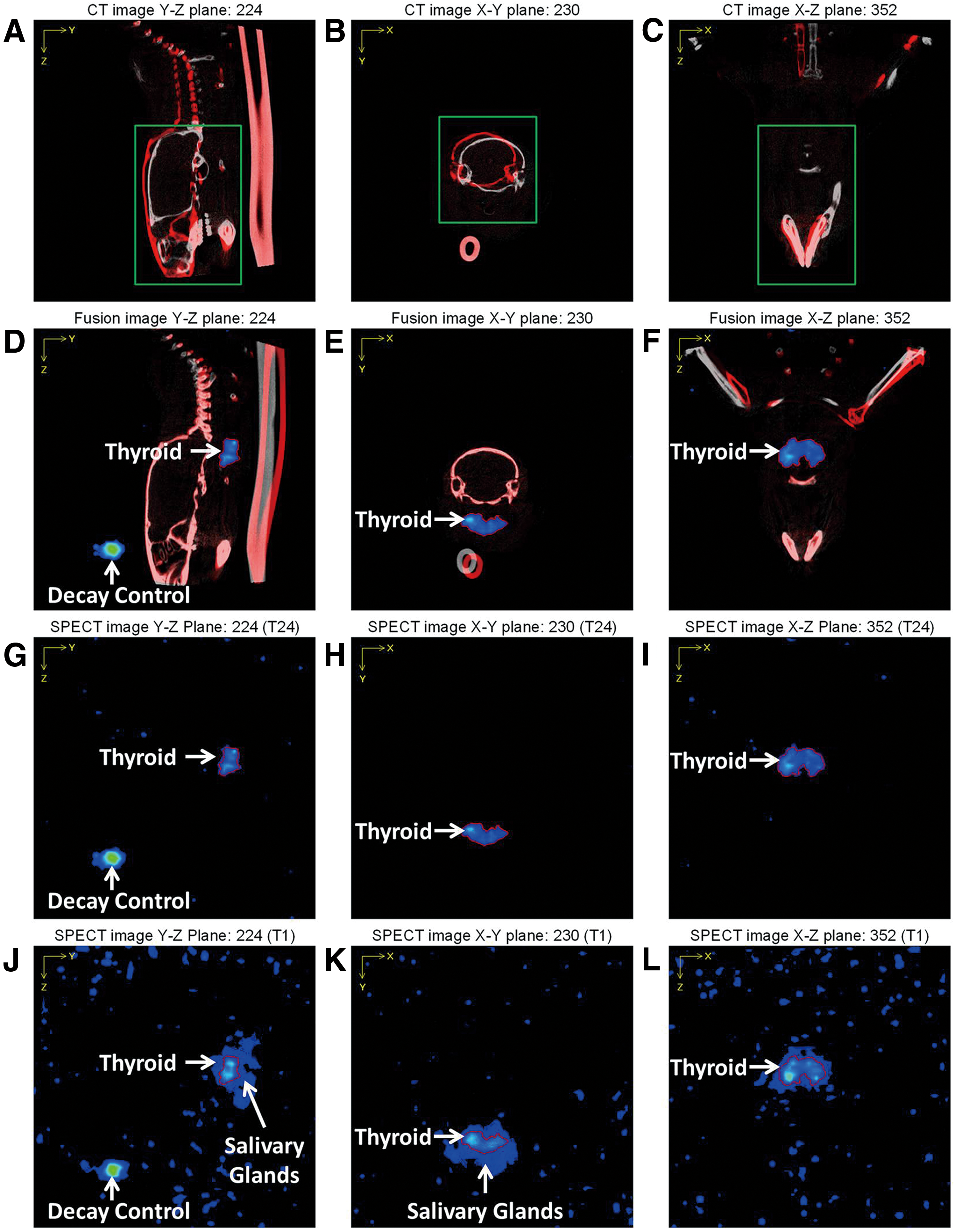
Automatic segmentation of thyroid volume of interest (VOI) on t1 image via superimposing thyroid VOI from t24 SPECT image. MicroSPECT/CT images are acquired at 1 h (t1) and 24 h (t24) post 123I injection. The sagittal (
Evaluating the extent of 123I uptake heterogeneity within the thyroid via 3D voxel-based 123I gamma photon intensity
MicroSPECT/CT imaging is used to locate tissues accumulating radioisotope tracer and quantify their radioisotope tracer uptake. Heterogeneity of radioisotope tracer uptake within target tissues of interest is rarely quantitatively evaluated. It is known that thyroid cancer patients with RAI-avid lesions of larger size are less responsive to 131I therapy. One could assume that 131I uptake in larger metastatic lesions would have a greater extent of heterogeneity in 131I uptake such that areas with insufficient 131I uptake would escape from 131I therapeutic effect. To investigate the effects of heterogeneity on 131I therapeutic efficacy, it must be possible to evaluate quantitatively the extent of 131I uptake heterogeneity within lesions of interest. The current CTViewer does not have the capability of 3D display. The 3D data sets of the thyroid VOI in the SPECT image were exported and loaded into MATLAB to display 3D voxel-based 123I gamma photon intensity. Accordingly, the spatial distribution of areas with different levels of gamma photon intensity can be visualized and analyzed.
In normal thyroid VOI of FVB/N mice (Fig. 4, left panel, and Supplementary Video S3), the center of both thyroid lobes had the highest level of 123I accumulation, indicating a higher metabolic activity and an increased iodinating capacity of centrally located small follicles. Follicles located peripherally are usually composed of flattened follicular epithelium that surrounds big colloids with low turnover rate and a reduced iodinating capacity.
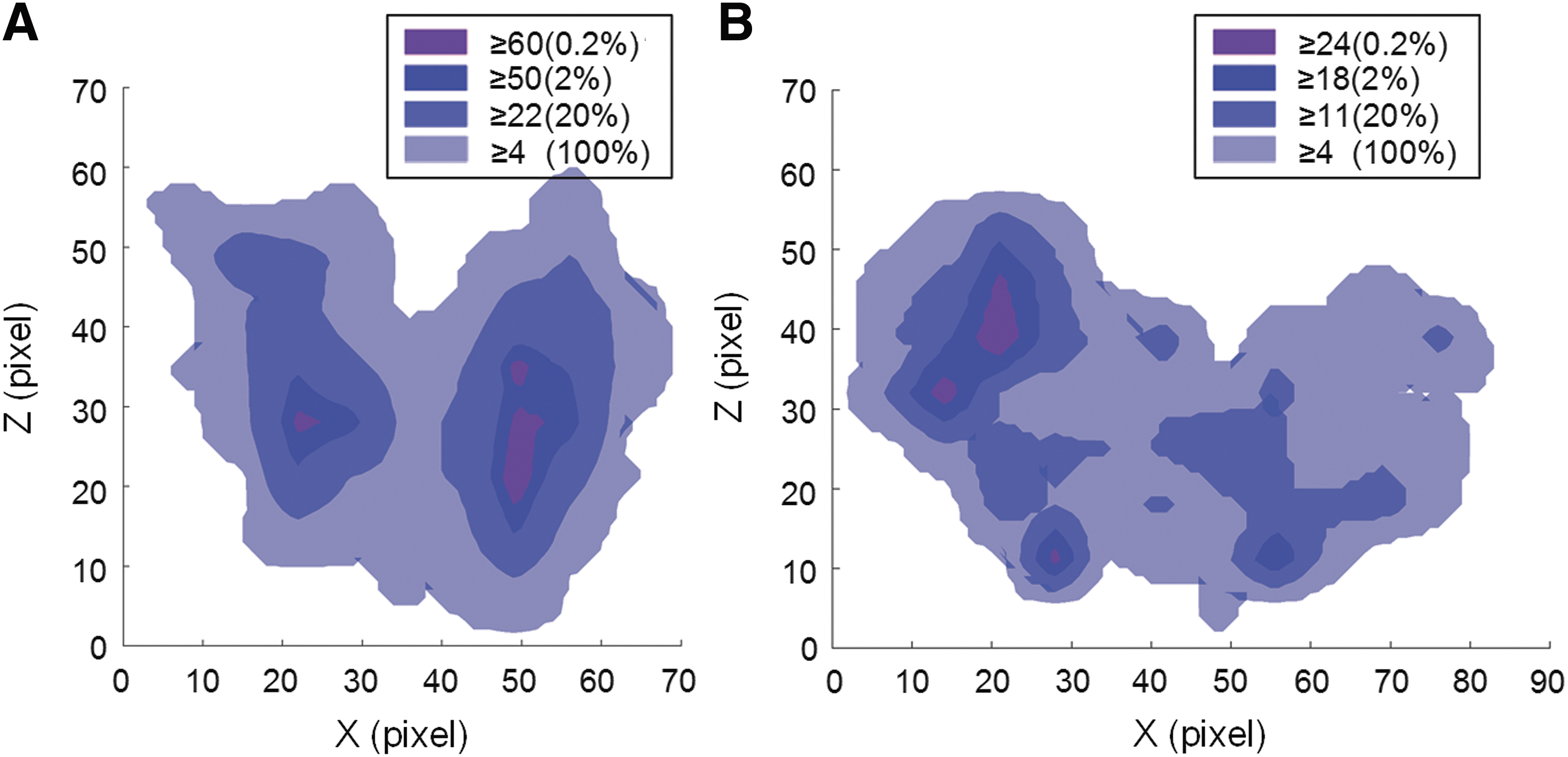
Coronal view of 3D voxel-based gamma photon intensity of a FVB/N mouse thyroid (
In thyroid tumor VOI of TgBRAFV600E mice (Fig. 4, right panel, and Supplementary Video S4), the spatial distribution of heterogeneity in 123I accumulation was asymmetric between thyroid lobes and had many areas with low 123I accumulation. As shown in Figure 4, gamma photon intensity in normal thyroid is much higher than BRAFV600E -driven thyroid tumors, with the top 2% of normal thyroid voxels having gamma photon intensity ≥50, while the top 2% of thyroid tumor voxels had a gamma photon intensity ≥18. Sixteen additional TgBRAFV600E mice were investigated, which were five months of age. Their 123I uptake was asymmetric between two thyroid lobes, and the extent of heterogeneity in 123I uptake within thyroid tumor varied greatly among these age-matched mice. Identifying the location and quantifying the volume of areas with high or low gamma photon intensity will help to explain and predict differential response to 131I therapy among 131I-avid lesions.
Discussion
This study utilized a mouse cradle during image acquisition to achieve consistent tissue configuration within the head and neck region. This allowed rigid body registration to be applied to align serial images taken at different times of the same subject mice with accuracy. Automatic segmentation of thyroid VOIs in t1 images is enabled by superimposing thyroid VOIs from the t24 images onto their corresponding aligned t1 images. Based on 3D voxel-based 123I gamma photon intensity, it is possible to evaluate the extent of heterogeneity of 123I uptake within thyroid VOIs in terms of spatial distance and volume of areas with high or low gamma photon intensity. These advances will greatly facilitate the effort to uncover novel strategies to improve 131I therapeutic efficacy.
Cradles have been routinely used to facilitate the accuracy of cranial irradiation in small animals (11 –14) or co-registration of multimodality and longitudinal images (15 –18). Among various cradles for the mouse head and neck region, the tooth bar anchoring upper incisors is essential. Ear bars (13,18) are effective but are difficult to use, leading to the use of alternatives such as a Styrofoam neck collar (11) or cheek bars in the cradle used in this study. With 3D printing technology, cradles can be easily customized and produced at low cost. For studies requiring consistency in tissue configuration, use of a cradle during image acquisition is necessary. Consistent tissue configuration allows rigid body registration among serial images of the same mice to correct any difference in position and orientation post-image acquisition easily. It is hoped that these studies will encourage cradles to be used widely among investigators.
VOI selection for targets with discrete signal in functional images is not problematic. VOI selection for multiple targets with close physical proximity in functional images requires co-registration of anatomic images that show discernable boundaries between targets. The boundaries of soft tissues are usually not discernable without contrast agent in CT images. While magnetic resonance imaging (MRI) is superior to CT imaging in visualizing the boundaries of soft tissues, MRI is time consuming, costly, and not widely available. Alternatively, the 3D MOBY digital mouse phantom (19) has been used as a common reference to facilitate VOI selection in functional images (20,21). However, this approach is less accurate, as the proportion of the target tissues to the rest of the mouse body may be different between subject mice and the mouse phantom. Most importantly, this approach cannot be applied to tumors with varying size and shape.
The fact that thyroid tissue has a discrete signal on the t24 image due to its unique ability to retain radioiodine allows automatic segmentation of the thyroid VOI in the t24 image to be conducted in a threshold-based manner. The thyroid VOI from the t24 image can then be superimposed onto its corresponding aligned t1 image to select the thyroid VOI in the t1 image automatically. In this study, consistent tissue configuration facilitated by the use of a cradle during image acquisition allowed the head and neck region to be treated as a rigid body, such that anatomic landmarks could be used for alignment of serial images by rigid body registration. While deformable registration is a powerful avenue to align images (20,22), the need to compensate difference in tissue configuration between images may lead to changes in voxel-based geometry of target tissues. Taken together, this approach can be applied to studies in which target tissue has differential radiotracer retention from surrounding tissues.
The overall response to 131I therapy is likely limited by the volume of areas with insufficient 131I accumulation (23). It is reasoned that insufficient 131I accumulation in micro-foci would have less impact than macro-foci on overall therapeutic outcome due to possible bystander effects of 131I. The 3D data set of voxel-based gamma photon intensity allows the volume of areas with low 131I accumulation and their spatial distance from areas with high 131I accumulation to be measured. This capability will facilitate the investigation of the impact of volume and spatial distance of areas with low 131I accumulation on the overall outcome of 131I therapy in patients with advanced thyroid cancer.
Conclusions
In summary, a customized mouse cradle facilitates consistent tissue configuration, such that rigid body registration can be applied to align serial images of the same mouse via in-house CTViewer software. The approach of superimposing thyroid VOIs from t24 images to select the thyroid VOI on corresponding aligned t1 images can be applied to studies in which target tissue has differential radiotracer retention from surrounding tissues. The ability to identify location with xyz coordinates and to measure the volume of areas with low 131I accumulation allows investigators to predict response to 131I therapy and to uncover novel strategies to improve 131I therapeutic efficacy.
Footnotes
Acknowledgments
We are grateful to Dr. James Fagin at Memorial Sloan Kettering for providing the TgBRAFV600E mice. We would like to thank Kevin Wolfe for sharing his expertise to help us determine the best material and resolution for 3D printing of the cradle and for printing and assembling the cradle. We would also like to thank Akram Hussein, Rodd Reinhart, and Andrew Brown for preparing the 123I doses for our SPECT/CT imaging studies. This work was supported in part by National Institutes of Health (NIH) grant P01CA124570 (project 3 leader: S.M.J; PI: M.D. Ringel), NIH grant P50CA168505 (project 2 leader; S.M.J.; PI: M.D. Ringel), National Science Foundation (NSF) grant CMMI-1067962 (PI: C.H.M.), and NSF grant CMMI-1200017 (PI: C.H.M.). B.H. is a recipient of NIH/NIDCR T32 DE014320.
Author Disclosure Statement
The authors have nothing to disclose.