
Abstract
Background:
Hypothyroidism is a common hormone deficiency condition. Regenerative medicine approaches, such as a bioengineered thyroid, have been proposed as potential therapeutic alternatives for patients with hypothyroidism. This study demonstrates a novel approach to generate thyroid grafts using decellularized rat thyroid matrix.
Methods:
Isolated rat thyroid glands were perfused with 1% sodium dodecyl sulfate to generate a decellularized thyroid scaffold. The rat thyroid scaffold was then recellularized with rat thyroid cell line to reconstruct the thyroid by perfusion seeding technique. As a pilot study, the decellularized rat thyroid scaffold was perfused with human-derived thyrocytes and parathyroid cells.
Results:
The decellularization process retained the intricate three-dimensional microarchitecture with a perfusable vascular network and native extracellular matrix components, allowing efficient reseeding of the thyroid matrix with the FRTL-5 rat thyroid cell line generating three-dimensional follicular structures in vitro. In addition, the recellularized thyroid showed successful cellular engraftment and thyroid-specific function, including synthesis of thyroglobulin and thyroid peroxidase. Moreover, the decellularized rat thyroid scaffold could further be recellularized with human-derived thyroid cells and parathyroid cells to reconstruct a humanized bioartificial endocrine organ, which maintained expression of critical genes such as thyroglobulin, thyroid peroxidase, and parathyroid hormone.
Conclusion:
These findings demonstrate the utility of a decellularized thyroid extracellular matrix scaffold system for the development of functional, bioengineered thyroid tissue, which could potentially be used to treat hypothyroidism.
Introduction
Hypothyroidism is one of the most common conditions of hormone deficiency and is caused by different etiological factors, such as autoimmune thyroiditis, thyroidectomy, or radioiodine therapy (1,2). Oral hormone administration is a relatively simple and inexpensive replacement therapeutic approach, but it commonly requires lifelong treatment (3). Nevertheless, it is difficult to maintain the complex homeostatic interactions of hormones, since fine regulation of thyroid status is almost completely lost (4). It has also been suggested that a small proportion of patients do not respond well to levothyroxine (mono)therapy (5).
Advances in tissue engineering have facilitated the development of replacement tissues or organs for the treatment of endocrine diseases (6,7). The generation of bioengineered thyroid tissue by appropriate combination of cells and biomaterial scaffolds could provide a potential alterative therapeutic modality for hypothyroidism.
Various synthetic and natural biomaterials have been used for the reconstruction of thyroid tissue, such as collagen for two-dimensional (2D) thyroid cell culture (8), three-dimensional (3D) collagen gel for thyroid culture (9 –12), and collagen hydrogel combined with thyrocytes for thyroid tissue 3D bioprinting (13). Most of these studies have been successful in achieving the formation of thyroid follicles in vitro or after implantation in vivo. Yet, the biomaterial components and resultant structure are far from reaching the natural intricacy of thyroid extracellular matrix (ECM) composition and complex 3D ultrastructure.
Recent advances in organ decellularization technology allow the generation of whole-organ scaffolds. The decellularized organ scaffold preserves the 3D organ architecture, organ-specific ECM, matrix-bound growth factors and cytokines (14), and, most importantly, the underlying matrix of the vascular network, which can be connected to the circulation for oxygen and nutrition delivery after transplantation. This natural innate ECM scaffold provides the necessary environment for cell welfare due to its indispensable information for cell growth and function (15,16). Additionally, numerous studies have shown that the decellularized organ ECM scaffold could signal stem cells for appropriate site-specific differentiation (17 –23). Successful decellularization and recellularization of organ ECM scaffolds has been reported in several organ systems, including the heart (24 –29), lung (30 –32), liver (33 –35), kidney (36 –38), and pancreas (39,40). It was hypothesized that the native thyroid ECM could provide such a scaffold that could mimic the complex in vivo microenvironment of the thyroid and that could be used for subsequent thyroid cell repopulation to reconstruct a thyroid.
The present study investigated the possibility of generating a decellularized thyroid scaffold by a perfusion decellularization technique and utilizing this scaffold for reconstruction of a functional tissue-engineered thyroid. With this aim in mind, the thyroid ECM scaffold was characterized for preservation of ECM composition and 3D structural integrity. Then, the decellularized thyroid scaffold was repopulated with FRTL-5 rat thyroid cells, as well as human thyrocytes. The function of the repopulated cells in the engineered thyroid was further characterized. Moreover, the thyroid scaffold slices were utilized for adipose-derived mesenchymal stromal cell (ADMSC) culture to determine its stem-cell compatibility. A work-flow diagram that describes the study is shown in Figure 1A.
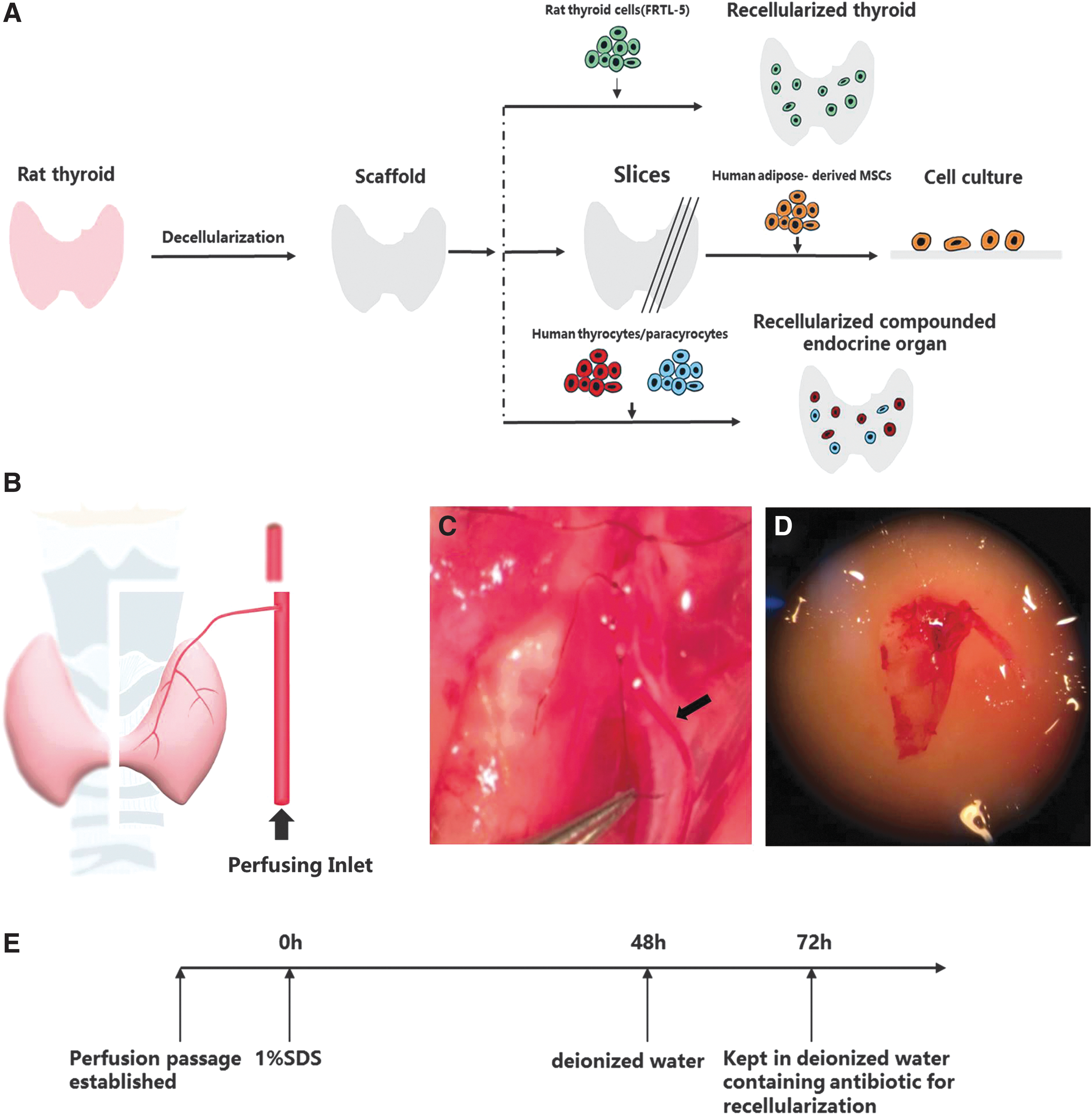
Work-flow diagram of the thyroid engineering and rat thyroid harvest. (
Methods
Rat thyroid harvest and cannulation
Male Lewis rats (250–300 g, aged 10 weeks, n = 5) were purchased from Vitalriver Company (Beijing, P.R. China). All the animals were housed in a specific pathogen free environment in the Laboratory Animal Center of Zhejiang Province, P.R. China. The experimental rats were individually caged at 21°C, exposed to a 12 h/12 h light/dark cycle, and fed with sterilized water and standard rat chow. All animals received humane care, with the study conducted in accordance to the Guide for the Care and Use of Laboratory Animals. The study was also approved by the Institutional Review Board, consistent with the Animal Protection Act of China.
The rats were euthanized using intraperitoneal injection of aqueous sodium chloralhydrate solution at a concentration of 0.7 mL/100 g body weight. Sterile conditions were observed when removing the thyroid. The cervical resection was performed according the following method. One thyroid lobe and partial trachea, which the thyroid lobe overlaid, were carefully dissected from adjacent structures (Fig. 1B–D). The homolateral common carotid artery (Fig. 1C, black arrow) and its branch, the superior thyroid artery (STA), were carefully preserved as the perfusion inlet.
Perfusion decellularization of rat thyroid
The isolated thyroid was connected to a perfusion system allowing perfusion at 1 mL/h, in which solutions flowed through the common carotid artery. An ionic detergent (i.e., 1% sodium dodecyl sulfate [SDS] in deionized water) was used as perfusate to rinse cells and cell debris out of the thyroid. The decellularized thyroid was then perfused for 24 hours with deionized water to clear the residual SDS, and it was kept in deionized water containing antibiotics for subsequent recellularization (Fig. 1E).
Histological assessment
The decellularized thyroid scaffold was fixed overnight in 10% neutral-buffered formalin. The tissue was then transferred in 60% ethanol and dehydrated in alcohol, immersed in chloroform, and embedded in paraffin wax. Sections (5 μm thick) were then obtained and stained with hematoxylin and eosin (black/pink, nuclei/tissue, respectively) and Masson's Trichrome staining (blue/black/red, collagen/nuclei/cytoplasm, respectively).
Immunostaining
Briefly, tissue samples were fixed with 4% formaldehyde (Thermo Fisher Scientific), cryoprotected with 30% sucrose, and cut into sections 7 mm thick. For immunostaining, the following primary antibodies were used: rabbit anti-collagen I (ab34710), rabbit anti-collagen IV (ab19808), rabbit anti-fibronectin (ab2413; Abcam; 1:100), rabbit anti-albumin (sc-50536; Santa Cruz Biotechnology; 1:100), anti-MHC class I antibody (ab22367), anti-MHC class II antibody (ab23990), anti-thyroglobulin antibody (ab156008), and anti-parathyroid hormone (ab154792; Abcam; 1:100). The secondary antibodies used were: goat anti-rabbit Alexafluor 647 and goat anti-mouse Alexafluor 488 (Invitrogen; 1:100). After incubation with primary antibody, an additional blocking step was included prior to the addition of the second antibody. The slides were then washed three times with 1% phosphate-buffered saline (PBS; 5–10 min) and were then mounted with ProLong® Gold Anti-fade Reagent with DAPI (Invitrogen) to demonstrate the nuclei. Images were recorded with Metamorph v7.5.6.0 (Molecular Device) on an Olympus IX81 inverted microscope.
Allura red dye and scanning electron microscopy
The decellularized thyroid was perfused with allura red dye via the common carotid artery to visualize the integrity of the vasculature. For scanning electron microscopy (SEM), the decellularized thyroid was fixed in 2.5% glutaraldehyde in 0.1 M PBS (pH 7.4) for 60 min. The samples were washed thoroughly in three changes of 0.1 M PBS for 15 min each. The samples were then fixed in 1% OsO4 in 0.1 M PBS for 60 min, followed by another three changes of PBS washing steps for 15 min each. Next, the samples were dehydrated in a gradient series of acetone for 15 min each. In addition, at the critical point, the samples were dried and coated with Au/Pd using a Cressington Coater 108A sputter coater. Electron microscope images were taken with a Jeol JSM- 6335F field emission SEM.
Seeding of decellularized thyroid scaffolds
Human adipose tissue was obtained in the course of local surgery with informed consent and ethical approval at the First Affiliated Hospital, School of Medicine, Zhejiang University, P.R. China. Human ADMSCs were prepared, as previously described (41). Decellularized thyroid glands were resected into slices with the intersecting surface just covering the hole of a 96-well cell culture plate. The scaffold slices (n = 3) were then placed in a 96-well cell culture plate with the cutting surface upwards and were seeded with hADMSCs in Dulbecco's modified Eagle's medium (DMEM; Gibco) containing 10% fetal bovine serum (FBS; Life Technologies) and penicillin–streptomycin (100 IU/mL) in a humidified environment at 37°C with 5% CO2. Proliferation of hADMSCs on acellular thyroid scaffolds was assessed with a Cell Counting Kit-8.
Cell culture
Rat thyroid FRTL-5 cells were cultured using Coon's modified Ham's F-12 medium supplemented with 5% bovine serum (Invitrogen) and a six-hormone mixture consisting of bovine thyrotropin (TSH; 1 mIU/mL), insulin (10 μg/mL), hydrocortisone (0.35 ng/mL), transferrin (5 μg/mL), glycyl-L-histidyl-L-lysine acetate (2 ng/mL), and somatostatin (10 ng/mL).
Human thyroid tissue and parathyroid tissue were collected with informed consent and ethical approval at the First Affiliated Hospital, School of Medicine, Zhejiang University, P.R. China. Thyroid cells were isolated from adjacent normal tissues patients undergoing thyroidectomy for differentiated thyroid carcinoma. Human thyroid cells were prepared, as previously described (42). Briefly, human thyroid tissues were minced into small fragments and treated with 2 mg/ml type II collagenase in PBS at 37°C for 45 min. Digested tissues were then mechanically dispersed to form a homogeneous suspension. The obtained cell suspension was cultured in DMEM/Ham's F-12 medium (1:1) supplemented with 10% FBS and 1% penicillin–streptomycin in a fully humidified atmosphere of 95% air–5% CO2 in 100 mm plastic dishes. Parathyroid cells were isolated from parathyroid tissues collected from patients undergoing surgery for secondary hyperparathyroidism (SHPT) (43). The patients met the criteria for therapeutic parathyroidectomy for SHPT based on the 2017 Chronic Kidney Disease-Mineral and Bone Disorder (CKD-MBD) Guidelines (44). Briefly, the parathyroid tissues were digested with 1.5 mg/mL type II collagenase and 0.1 mg/mL DNase. The cells were cultured in DMEM/F-12 (GlutaMAX; Gibco) supplemented with 10% FBS and 1% penicillin–streptomycin.
Recellularization and culture of seeded thyroid construct
FRTL-5 cells (106) were trypsinized and diluted in Coon's modified Ham's F-12 medium supplemented with 5% bovine serum (Invitrogen) and a six-hormone mixture consisting of bovine TSH (1 mIU/mL), insulin (10 μg/mL), hydrocortisone (0.35 ng/mL), transferrin (5 μg/mL), glycyl-L-histidyl-L-lysine acetate (2 ng/mL), and somatostatin (10 ng/mL). FRTL-5 cells (106) were suspended in 200 μL of the culture medium. The cell suspension solution was then perfused into the decellularized thyroid through the common carotid artery. The FRTL-5 seeded thyroid scaffold (n = 3) was cultured in medium described above for 12 h.
For the co-recellularization strategy, human primary thyrocytes and parathyroid cells isolated from thyroid and parathyroid tissues were mixed in DMEM/Ham's F-12 medium (1:1) supplemented with 10% FBS and 1% penicillin–streptomycin to form a cell suspension. About 200 μL cell suspension including thyrocytes (106) and parathyrocytes (106) was perfused through the common carotid artery. The reconstructed endocrine organ (n = 3) was cultured in medium for 12 h.
Statistical analysis
Quantitative data are expressed as mean ± standard deviation. Significant differences between groups were determined by the Wilcoxon rank-sum test for two-group comparisons or analysis of variance followed by post hoc analysis for multiple group comparisons. For statistical purposes, at least three independent cultures were considered. A p-value of <0.05 was considered statistically significant.
Results
Properties of decellularized thyroid matrix
Thyroid decellularization was achieved by thyroid artery perfusion with 1% SDS, an anionic detergent that lyses cells and solubilizes cytoplasmic components. After 48 hours of decellularization, a translucent acellular scaffold was obtained; the scaffold preserved the gross shape of the thyroid (Fig. 2A–C). Compared to the normal rat thyroid, no nuclei or cytoplasmic staining was found in the decellularized thyroid matrix (DTM; Fig. 2D and E). In addition, the perfusion decellularization preserved the structure of the thyroid ECM with an intact follicular basement membrane, the arterial elastic fiber network (from thyroid arteries and veins to microvessels forming multiple angio-follicular units). Moreover, Masson's trichrome staining demonstrated the removal of cellular material and preservation of collagen fibers (Fig. 2F and G).
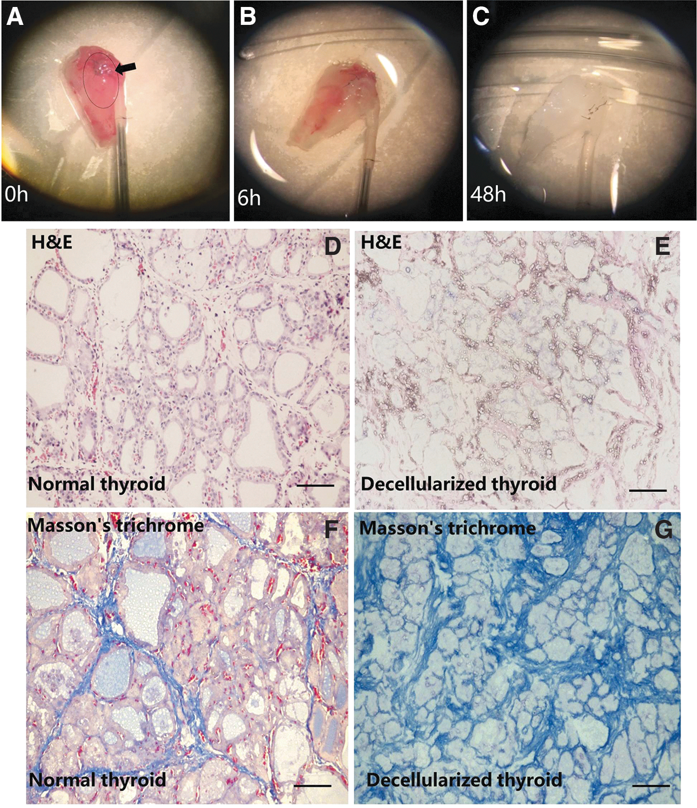
Perfusion decellularization of the rat thyroid lobe. (
Immunostaining for four ECM proteins—collagen type I, collagen type IV, fibronectin, and laminin-β1—demonstrated that the structure and component of the ECM was similar to native thyroid (Fig. 3A). In addition, a lack of DAPI staining confirmed the absence of cells in the DTM (Fig. 3A). Also, immunostaining for major histocompatibility complex I and complex II confirmed that the DTM was depleted of these cellular markers (Fig. 3B).
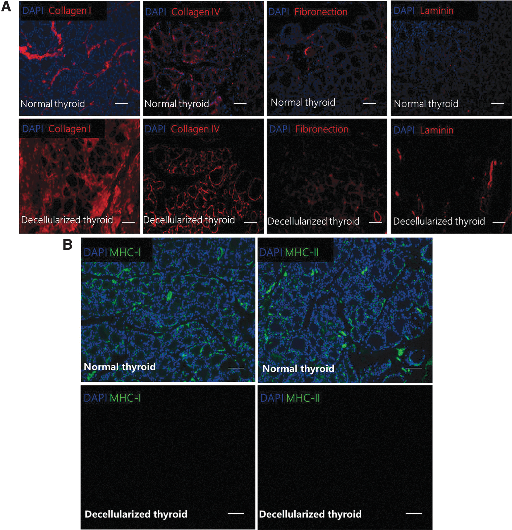
Characterization of acellular rat thyroid matrix. (
Furthermore, SEM images confirmed the presence of thyroid follicular scaffolds of spherical structure and numerous vascular matrix scaffolds, whereas the thyroid follicular cells and vascular endothelial cells were absent, retaining the thyroid cell-free hollow spaces (Fig. 4A). Nanofibrous structures of the ECM were retained within the parenchyme (Fig. 4B).
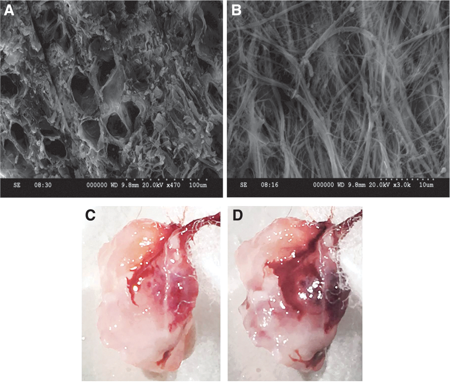
Decellularized thyroid matrix (DTM) retains intact follicular structure and vascular bed. (
To show further that the decellularization protocol preserved a functional vascular bed, the scaffold was perfused with Allura Red dye. The vascular tree was apparent in the translucent matrix (Fig. 4C and D). The perfusion of the dye was detected through the thyroid artery, gradually flowing from larger vessels (Fig. 4C) to smaller capillaries (Fig. 4D), suggesting that the microvasculature remained intact.
Cell seeding on decellularized thyroid ECM
To assess the cellular compatibility of the decellularized thyroid ECM scaffold, the slices of DTM were used as substrate for hADMSCs, and their growth was further examined on the scaffold (Fig. 5A). After seven days, hADMSCs were observed on the surface and inside the scaffold, and they displayed a rounded phenotype (Fig. 5B). The assessment of cell growth showed a significant increase in cell number of 39.5%, from 3185.68 ± 185.68 on day 3 to 4443.10 ± 448.59 on day 7 after seeding (Fig. 5C). This observation suggests that the substrate cues on the decellularized matrix are functional for hADMSCs attachment and replication.
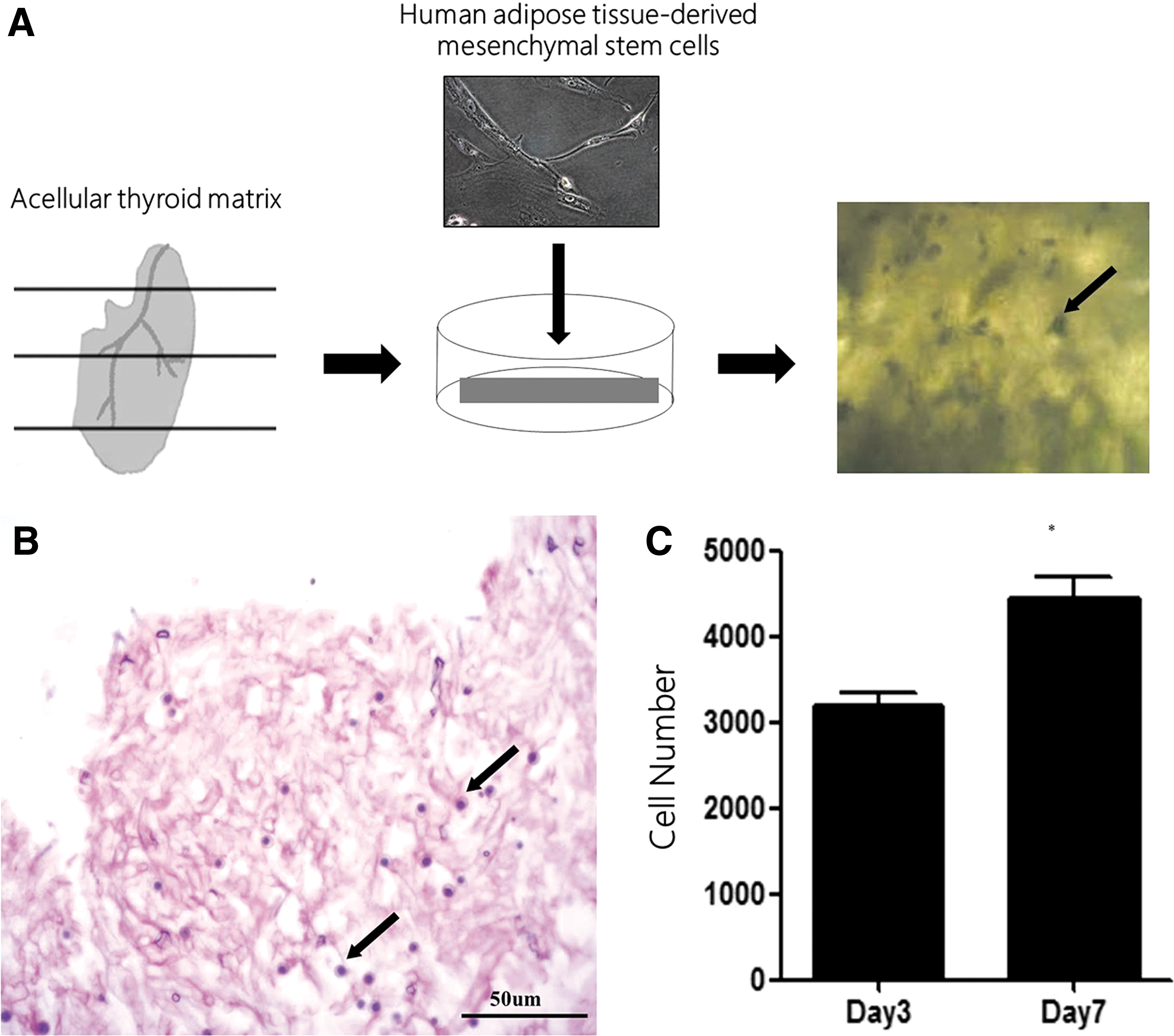
Cell growth in acellular rat thyroid. (
Recellularization of DTM
The presence of a functional vascular bed in the DTM offers the ability to control thyroid cells engraftment and support thyroid specific endocrine function in vitro. FRTL-5 cells (106) were trypsinized and diluted in 200 μL of medium. The cell suspension was then introduced into the decellularized thyroid via the common carotid artery for thyroid perfusion (Fig. 6A). The thyroid seeded with FRTL-5 cells was immersed and cultured in the same FRTL-5 culture medium described above for 12 hours. Histological analysis showed that seeding of the decellularized matrix with FRTL-5 generally resulted in adherence of the cells throughout the scaffold, and arrangement of the cells surrounding the follicular scaffolds resulting in spherical structures (Fig. 6B). In addition, immunohistochemical analysis indicated that thyroglobulin (Tg) and thyroid peroxidase (TPO) were both expressed in FRTL-5 cells, indicating maintained expression of critical genes (Fig. 6C and D).
Reseeding of the acellular thyroid matrix with FRTL-5 rat thyroid cells. (
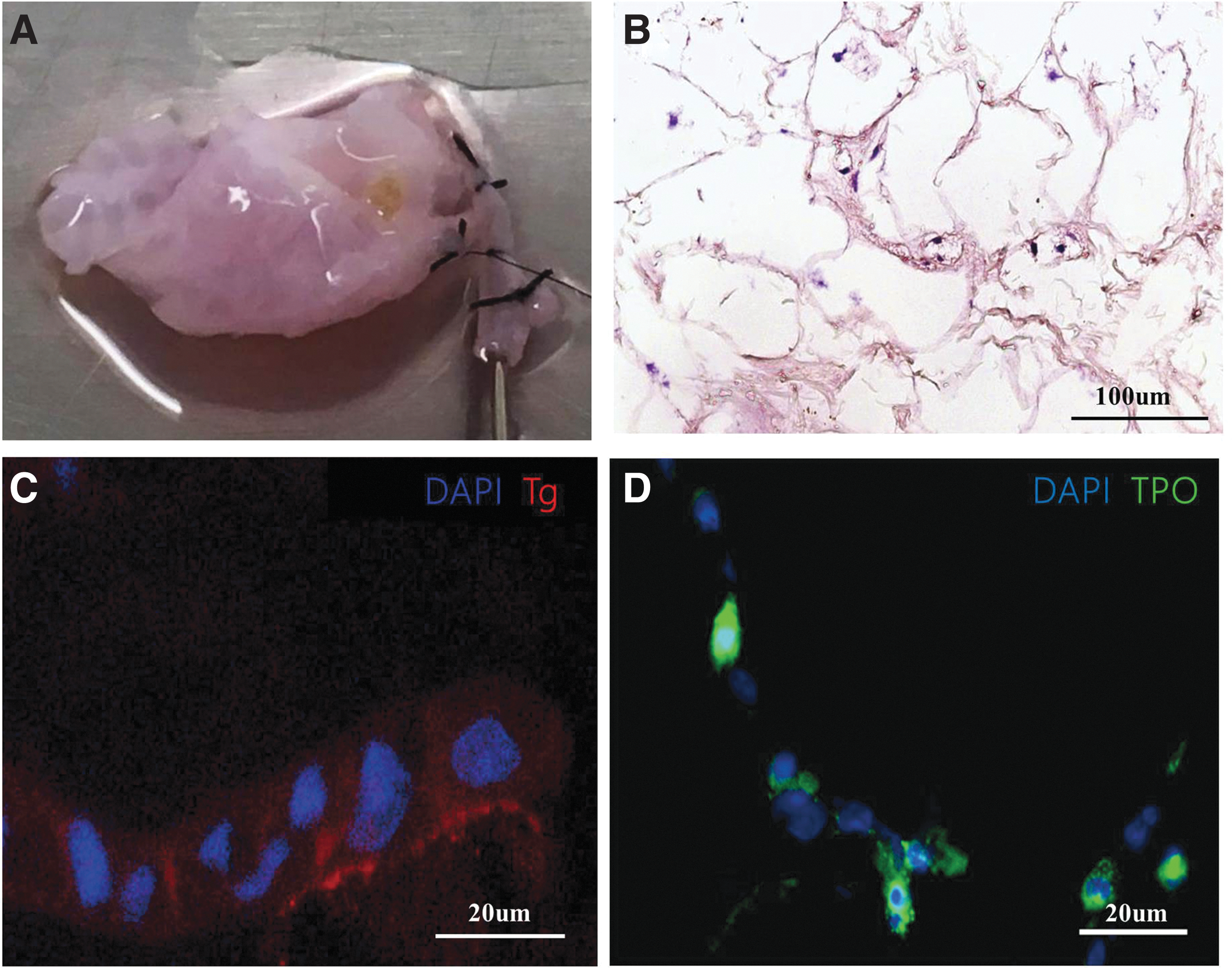
Further scalability of recellularization technique
As the parathyroids are close to the thyroid, they can sometimes become injured during the resection of the thyroid. Thus, as a preliminary test, this study attempted to construct a multifunctional endocrine organ by combining thyroid and parathyroid cells. Importantly, the decellularized rat thyroid scaffold is not immunogenic. The rat derived-decellularized thyroid scaffold (Fig. 7A and B) and human-derived cells were used to build a new organ. Primary thyrocytes were isolated from human thyroid tissues, and primary parathyrocytes were isolated from human parathyroid tissues (Fig. 7C). Then, human primary thyroid cells (106) were seeded with the DTM by incorporating parathyroid cells (106; Fig. 7D and E). Histological analysis showed that human thyroid cells and parathyroid cells were capable of distributing throughout the decellularized rat thyroid matrix and that they adhered to the scaffold (Fig. 7F and G). Immunohistochemical analysis showed expression of Tg (Fig. 7H) and TPO (Fig. 7I) by thyrocytes, and parathyroid hormone expression of parathyroid cells (Fig. 7J), again indicating maintained expression of critical genes.

Reseeding of the acellular rat thyroid matrix with human thyrocytes and parathyroid cells. (
Discussion
The results presented here illustrate first steps toward the development of a recellularized thyroid matrix as a promising alternate approach for thyroid tissue engineering and functional organ replacement. Here, the preservation of the thyroid's 3D architecture is demonstrated, including follicular scaffolds of spherical structure, a perfusable vasculature matrix, and a native ECM composition, which were used as a suitable template for thyroid tissue engineering.
The objectives for successful decellularization are complete removal of cellular material while preserving ECM composition (45). Previously reported decellularization protocols include mechanical agitation or freezing and thawing to remove cellular materials (46), which are suitable for tissue engineering. The approach of whole-thyroid perfusion decellularization builds on the techniques that have been previously reported for other organs (24,30,34,36,39), demonstrating its potential feasibility for rat thyroid decellularization and further reseeding of thyroid cells.
Hypothyroidism is one of the most common hormone deficiencies. Due to the limitations of lifelong oral hormone administration in the treatment of hypothyroidism, the use regenerative medicine approaches for thyroid hormone replacement are of interest (47). Several studies have tried various strategies to rebuild thyroid tissue (8,9,48). Bell et al. combined thyroid cells with a collagen gel so as to reconstruct thyroid-like tissue that was then implanted in vivo (8). Toda et al. used porcine thyroid cells to rebuild thyroid follicles using a 3D culture technology (9,48). Another study used a cell sheet engineering approach to construct thyroid tissue with similar structure to the normal thyroid with typical follicles and hormone secretory function (49).
The present study demonstrates that rat thyroids can be depleted of their cellular components and used to retain the sophisticated thyroid follicle spherical structure with perfusable vascular trees. Reseeding of the decellularized scaffold with thyrocytes may permit functional thyroid tissue to be generated. The perfusion decellularization and recellularization technique was applied for thyroid reengineering using the FRTL-5 rat thyroid cell line, and the approach was further extended by using a mixture of human thyrocytes and parathyroid cells to build a multifunctional endocrine organ. The 3D ECM microstructure developed from the thyroid could not completely imitate the parathyroid microenvironment. Yet, considering the adjacent anatomical location of the two organs and that injury to the parathyroid gland(s) is a common complication in thyroidectomies, this strategy is of potential clinical interest. The possible use of DTM for the regeneration of a specific endocrine organ combining both thyroid and parathyroid function is envisioned.
Recently, several studies have used stem cells for regeneration of the thyroid organoid. Lin et al. used mouse embryonic stem cells (mESCs) for differentiation into thyroid-like cells in vitro (50). Antonica et al. also succeeded in differentiating mESCs to thyroid follicular cells capable of synthesizing thyroid hormones (51). Recently, Kurmann et al. regenerated thyroid follicular cells differentiated from mouse and human pluripotent stem cells (PSCs), which could produce thyroid hormones and rescue mouse hypothyroidism (52). In their study, combined with a 3D culture scaffold, the stem cell–derived precursor cells showed a high expression of thyroid-restricted genes and the formation of follicular-like clusters of cells. The organ-derived decellularized native ECM scaffold, as previous studies have shown, has the potential ability to allow stem-cell differentiation into organ-specific mature cells. ADMSCs, which are being studied in preclinical trials for applications in tissue engineering and regenerative medicine, have self-renewal capacity and multilineage differentiation capacity toward adipocytes, chondrocytes, hematopoietic-supporting cells, neuronal cells, myocytes, and osteoblasts (53). Previous studies have shown that the decellularized organ scaffold could trigger organ-specific differentiation of ADMSC (22,23,54,55). The present study demonstrates the cell compatibility of the decellularized rat thyroid by the seeding of hADMSCs as a potential stem-cell source for the generation of thyrocytes. However, using the decellularized thyroid scaffold and hADMSCs for re-engineering of replaceable thyroid tissue still needs to be investigated.
Additionally, the decellularized thyroid scaffold holds the potential for providing a 3D environment for thyroid cell seeding to study the toxicity of chemicals, and for repopulation of thyroid cancer cells to investigate the effect of the 3D tumor microenvironment on thyroid tumor development.
Despite these advantages, further improvements of the decellularization and recellularization of thyroid matrix are necessary. Engineering of an entire thyroid will also require parafollicular cells and vascular endothelial cells. Ongoing studies are directed toward re-endothelialization and modification of the perfusate system, which could further improve the reconstruction of a functional thyroid graft. Moreover, the recellularized thyroid could be transplanted either in part such as thyroid slice or as an entire organ to cure hypothyroidism. However, to this end, the synthesis of thyroid hormones by the system and regulation by TSH need to be formally demonstrated.
Overall, this study suggests that decellularized thyroid ECM could serve as a platform for bioengineering a bioartificial thyroid. Further studies are required to investigate the differentiation of stem cells (ESC/MSC/induced PSCs) within the native DTM. In addition, in vivo studies of the performance of the reengineered thyroid organoid constructs are also required.
Conclusions
This study demonstrates successful decellularization of rat thyroid. The decellularized thyroid retained its native structure, intact vascular channels, and ECM components. The study further showed that the rat thyroid ECM scaffold could be repopulated with FRTL-5 rat thyroid cells and the human-derived compounded cells of thyrocytes and parathyroid cells. These results provide a foundation for the further development of endocrine organ engineering approaches to treat hormone deficiency disorders such as hypothyroidism and hypoparathyroidism.
Footnotes
Acknowledgments
The Project Supported by Zhejiang Provincial Natural Science Foundation of China (LQ18H180003, LQ18H050003).
Author Disclosure Statement
No competing financial interests exist.