
Abstract
Background:
Physiological changes in maternal thyroid function during pregnancy necessitate the use of pregnancy-specific reference ranges. Dynamic changes in thyrotropin (TSH) within the first trimester of pregnancy have been reported, but more evidence is needed to substantiate the findings. The objective of this study was to estimate pregnancy week-specific reference ranges for maternal TSH and free thyroxine (fT4) in early pregnancy.
Methods:
The study consecutively recruited serum residues from blood samples collected as part of the prenatal screening in the North Denmark Region, 2011–2015. TSH, fT4, thyroid peroxidase antibodies (TPOAb), and thyroglobulin antibodies (TgAb) were measured using an ADVIA Centaur XPT immunoassay. The reference cohort included 10,337 pregnant women who had no thyroid disease or other autoimmune diseases and were TPOAb- and TgAb negative. The main outcome measures were lower and upper reference limits (2.5th and 97.5th percentiles) for TSH and fT4 stratified by week of pregnancy.
Results:
Blood samples were drawn in pregnancy weeks 4–20 (median week 10), and 92% of the pregnancies ended with a live birth. TSH varied considerably in the first trimester of pregnancy, and the levels were highest in early pregnancy (weeks 4–6: 0.6–3.7 mIU/L) followed by a gradual decline to lower levels in weeks 9–11 (0.1–2.8 mIU/L) and 12–14 (0.03–2.8 mIU/L). Maternal fT4 showed less variation (weeks 4–6: 12–20 pmol/L; weeks 9–11: 13–21 pmol/L; weeks 12–14: 13–20 pmol/L).
Conclusions:
The results corroborate dynamic week-specific changes in maternal TSH in early pregnancy. The use of uniform lower and upper reference limits for TSH in early pregnancy may be too simple.
Introduction
Thyroid function in pregnant women is of clinical relevance (1). Thyroid hormones in the fetus are solely of maternal origin in early pregnancy, and the crucial role of thyroid hormones in fetal development emphasizes the importance of adequate maternal thyroid hormone levels (2). The physiological changes in maternal thyroid function in early pregnancy are mediated via estrogen, human chorionic gonadotropin (hCG), and alterations in the metabolism of thyroid hormones, and these changes necessitate the use of pregnancy-specific reference ranges for the evaluation of maternal thyroid function (3). Much emphasis has been on the use of trimester-specific reference ranges (1), but recent data indicate that the physiological changes in maternal thyrotropin (TSH) in early pregnancy are marked, even within the first trimester of pregnancy, and that the use of a uniform reference range may be too simple (4,5).
Thyroid dysfunction in women of reproductive age is predominantly of autoimmune origin (6,7). Thyroid peroxidase antibodies (TPOAb) and thyroglobulin antibodies (TgAb) are associated with the presence of hypothyroidism, and TSH is higher in individuals who are positive for thyroid autoantibodies (8). Thus, it is recommended to exclude individuals with TPOAb from a reference cohort (1). Previously, the week-specific changes in maternal TSH and free thyroxine (fT4) were described in a large cohort of pregnant women from the Danish National Birth Cohort established in 1997–2003 (5). Pregnant women with known thyroid disease were excluded, but the measurement of thyroid autoantibodies was not possible in this cohort. Furthermore, mandatory iodine fortification of salt was introduced in Denmark in 2000 (9), and data on the level of TSH in Danish pregnant women after iodine fortification are warranted.
The present study aimed to evaluate the week-specific reference ranges for TSH and fT4 in a recent large cohort of pregnant women from the North Denmark Region established in 2011–2015. Blood samples were consecutively collected as part of the prenatal screening for chromosomal anomalies in early pregnancy and used for the measurement of maternal thyroid function (TSH and fT4) and thyroid autoantibodies (TPOAb and TgAb).
Methods
Study population
Nationwide prenatal screening for fetal chromosomal anomalies was implemented in Denmark in 2004 (10). It includes the measurement of biochemical markers in a blood sample from the pregnant woman drawn at the first pregnancy visit in general practice. All serum samples from pregnant women in the North Denmark Region are analyzed at the Department of Clinical Biochemistry, Aalborg University Hospital (Aalborg, Denmark). Serum residues from all samples received in the laboratory from June 28, 2011, to January 5, 2015, were collected for the present study and stored at −80°C until the measurement of maternal thyroid function and thyroid autoantibodies. Serum residues from January to March 2012 and January to March 2013 were not available due to logistics in the laboratory, and a total of 18,186 samples were included in the study (Fig. 1). The study was approved by the North Denmark Region Committee on Health Research Ethics (N-20150015) and the Danish Data Protection Agency (
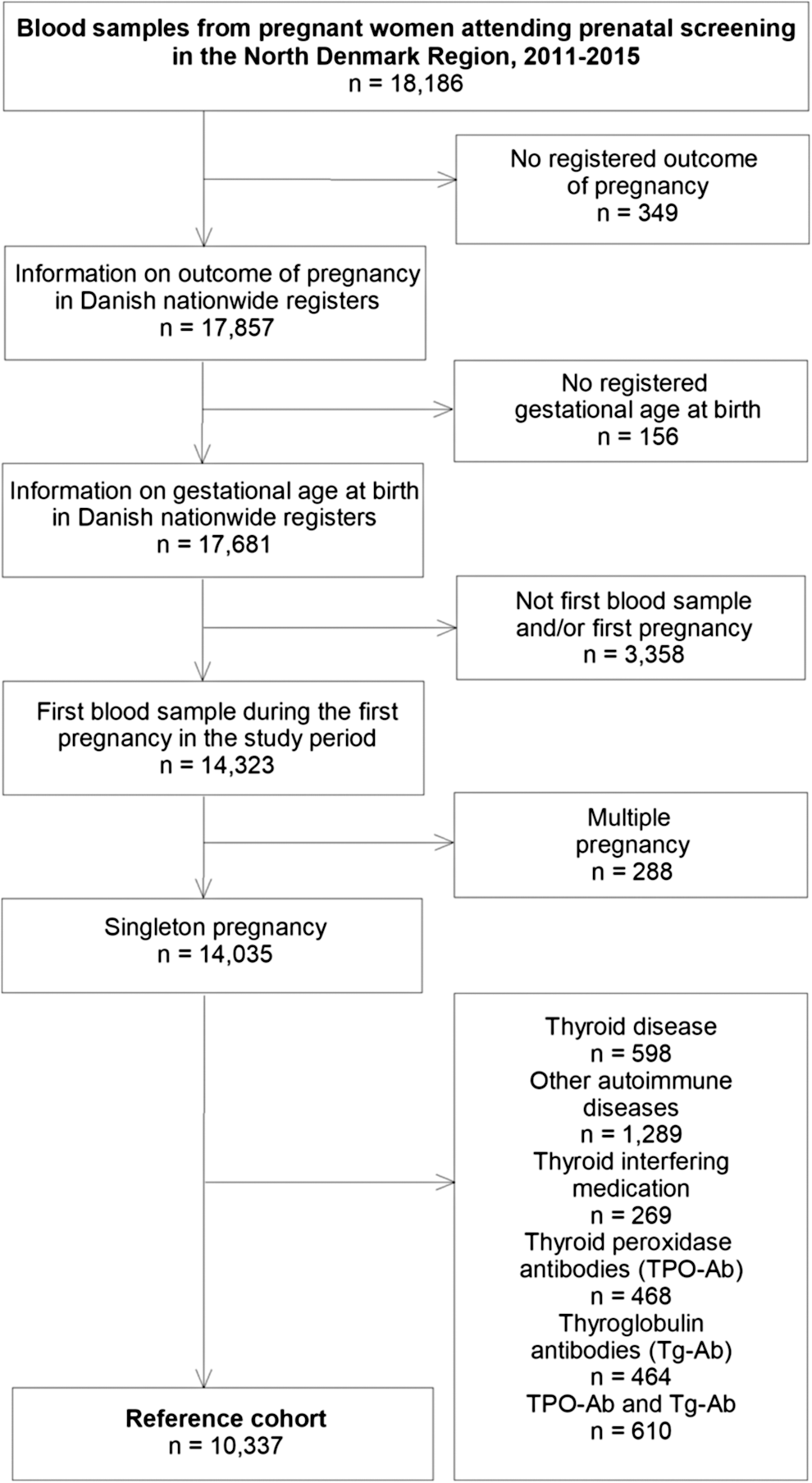
Flow chart illustrating the selection of the reference cohort.
Laboratory procedures
Serum TSH, fT4, TPOAb, and TgAb were measured from September 2015 to May 2016 in the Department of Clinical Biochemistry, North Denmark Regional Hospital (Hjørring, Denmark) using an ADVIA Centaur XPT automatic immunoassay (Siemens Healthineers, Erlangen, Germany). For TSH (TSH3-Ultra), the functional sensitivity provided by the manufacturer was 0.008 mIU/L, and the analytical measurement range was 0.008–150 mIU/L. Intermediate precision was 3.6–5.2% for six patient serum samples in the range 1.0–132.8 mIU/L. The reference range for nonpregnant adults was 0.55–4.78 mIU/L. For fT4, the analytical measurement range was 1.3–155 pmol/L, and intermediate precision was 3.4–4.6% for three concentrations in the range 9.3–38.8 pmol/L. The reference range for nonpregnant adults was 11.5–22.7 pmol/L. For TPOAb, the analytical measurement range was 28–1300 U/mL, and intermediate precision was 7.6% and 3.1% for concentrations of 71 and 459 U/mL, respectively. For TgAb, the analytical measurement range was 15–500 U/mL, and intermediate precision was 3.9–6.6% for three concentrations in the range 71–344 U/mL. A cutoff value of 60 U/mL was given by the manufacturer for both TPOAb and TgAb, and women with measurements above this cutoff were considered thyroid autoantibody positive in the present study. The cutoff for TPOAb in pregnant women has previously been evaluated for the ADVIA Centaur assay and was at a comparable level (54 U/mL) (11). A sub-analysis was included in which the cutoff was halved to 30 U/mL to evaluate a possible impact of subtle thyroid autoimmunity.
Data linkage
The date of blood sampling and results of thyroid function parameters and thyroid autoantibodies were linked to information in Danish nationwide registers via Statistics Denmark. All citizens in Denmark are provided a unique personal identification number, which is used in all registers and enables data linkage (12). Data were available in Statistics Denmark in encrypted form so that no individuals could be identified by the researcher. First, pregnancy outcome and the week of blood sampling during pregnancy were established via linkage to the Medical Birth Register (MBR) (13) and the Danish National Hospital Register (DNHR) (14). The MBR holds information on all live births and stillbirths in Denmark, including information on whether it was a singleton or multiple birth, and on maternal age, parity, origin (country of birth), smoking in pregnancy, and pre-pregnancy body mass index (BMI). The DNHR holds information on all in- and outpatient visits to Danish hospitals with a diagnosis of pregnancy loss, including induced abortion, spontaneous abortion, and molar and ectopic pregnancy coded according to the 10th International Classification of Disease (ICD-10). In both registers, information on date and gestational age at the time of pregnancy termination is available, and this information was linked to the date of blood sampling in early pregnancy and used to establish the pregnancy week of blood sampling. Gestational age is established by ultrasound in Denmark (15) and registered in full weeks plus days counted from the first day of the last menstrual period (e.g., 8 + 2, which corresponds to the 9th week of pregnancy). Information on maternal diseases before, during, and after the pregnancy was obtained from (i) hospital diagnosis of disease registered from January 1, 1994, to December 31, 2016, in the DNHR and coded according to the ICD-10, and (2) redeemed prescriptions of drugs registered from January 1, 1995, to December 31, 2016, in the Danish National Prescription Register (16) and coded according to the Anatomical Therapeutic Chemical Classification System.
Reference cohort
Among the 18,186 blood samples collected in early pregnancy and used for measurement of maternal thyroid function and thyroid autoantibodies, altogether 17,681 were linked in Statistics Denmark with information on outcome of the pregnancy and gestational age (Fig. 1). Some women had more than one blood sample drawn during a pregnancy or consecutive pregnancies in the study period, and in those cases the first blood sample during the first pregnancy in the study period was selected for each woman (Fig. 1). Multiple pregnancies were excluded from the reference cohort, as were women with a registration of thyroid or other autoimmune diseases (diabetes, rheumatoid arthritis, and inflammatory bowel disease) before, during, or after the pregnancy, women with a registration of thyroid-interfering medication (antiepileptic drugs, dopamine agonists, lithium, and prednisolone) in pregnancy defined by redeemed prescriptions in the one-year period prior to pregnancy termination, and women who were positive for TPOAb and/or TgAb, leaving 10,337 pregnant women in the reference cohort (Fig. 1).
Statistical analyses
Pregnancy week-specific results of TSH and fT4 analyses are reported as median, 2.5th percentile, and 97.5th percentile with 95% confidence interval (CI). The early (weeks 4–6) and the late (weeks 15–20) pregnancy weeks were collapsed to obtain a sufficient number of individuals in the groups. Within each pregnancy week group, Box–Cox transformation was used to obtain normal distribution prior to the exclusion of outliers using Tukey's fences (17). Analyses were performed both in the full reference cohort (n = 10,337) and among women who ended the pregnancy with the birth of a live-born child (n = 9515). Predictors of maternal TSH and fT4 were evaluated in pregnancy weeks 9–11, which included the largest number of individuals. This evaluation was performed among women who ended the pregnancy with the birth of a live-born child and had available information on maternal characteristics (n = 7438). All predictors (maternal age, parity, origin, smoking, and BMI) were assessed as independent categorical variables in multivariate linear regression with Box–Cox transformed TSH or fT4 as the dependent variable, and significant predictors were selected for univariate stratified analyses. Statistical analyses were performed using STATA v15 (StataCorp, College Station, TX).
Results
Reference cohort
Altogether, 10,337 pregnant women were included in the reference cohort (Fig. 1). The median maternal age was 29 years (range 16–51 years), and 88% of the women were born in Denmark. The median week for the blood sample being drawn was week 10 (9 + 0 to 9 + 6) ranging from the 4th week (3 + 0 to 3 + 6) to the 20th week (19 + 0 to 19 + 6). The birth of a live-born child was the most frequent outcome of pregnancy (n = 9515), whereas induced abortion (n = 140), spontaneous abortion (n = 639), molar and ectopic pregnancy (n = 12), and stillbirth (n = 31) accounted for a smaller proportion of the cohort.
Reference ranges
Week-specific median and upper and lower reference limits with CI for TSH and fT4 showed a similar trend when established within the full cohort of 10,337 pregnant women and among the 9515 women who ended the pregnancy with the birth of a live-born child (Fig. 2 and Table 1). TSH showed considerable variation in the median and upper and lower reference limits during early pregnancy, being high in the early pregnancy weeks followed by a gradual decline to low levels in pregnancy weeks 9–11 and 12–14, which was most pronounced for the lower reference limit. Maternal fT4 showed an opposite pattern, but less pronounced variation with the highest levels in pregnancy weeks 9–11. A uniform reference range for TSH and fT4 in the first trimester of pregnancy (weeks 4–12) was 0.14–2.9 mIU/L for TSH and 13.0–20.5 pmol/L for fT4, with similar figures within the full cohort and within the cohort of women with live births. However, the week-specific changes during the first trimester (Fig. 2 and Table 1) indicated that shorter distinct intervals could be described (Table 2), particularly for TSH, with an initial high TSH period followed by subsequent lower TSH periods in weeks 9–11 and 12–14. When the cutoff for TPOAb and TgAb was halved to 30 U/mL, the study population was restricted to 6216 pregnant women. Reference limits for maternal thyroid function tests were similar, with the same trend during the first trimester of pregnancy (weeks 4–8: TSH 0.29–3.2 mIU/L, fT4 12.4–19.3 pmol/l; weeks 9–11: TSH 0.13–2.8 mIU/L, fT4 12.8–20.2 pmol/L; weeks 12–14: TSH 0.04–2.8 mIU/L, fT4 12.7–19.5 pmol/L). Considering the exclusion of TPOAb- and/or TgAb-positive women, the TSH reference limits prior to exclusion were 0.43–3.8 mIU/L for weeks 4–8, 0.17–3.2 mIU/L for weeks 9–11, and 0.08–3.1 mIU/L for weeks 12–14. After exclusion of TPOAb-positive women only, the TSH reference ranges were 0.40–3.4 mIU/L for weeks 4–8, 0.14–2.9 mIU/L for weeks 9–11, and 0.03–2.8 mIU/L for weeks 12–14. They were fairly the same after exclusion of TgAb-positive women only (weeks 4–8: 0.40–3.5 mIU/L; weeks 9–11: 0.15–2.9 mIU/L; weeks 12–14: 0.03–2.7 mIU/L). Reference limits for fT4 were similar to those presented in Table 2, irrespective of whether TPOAb- and/or TgAb-positive women were excluded (data not shown).
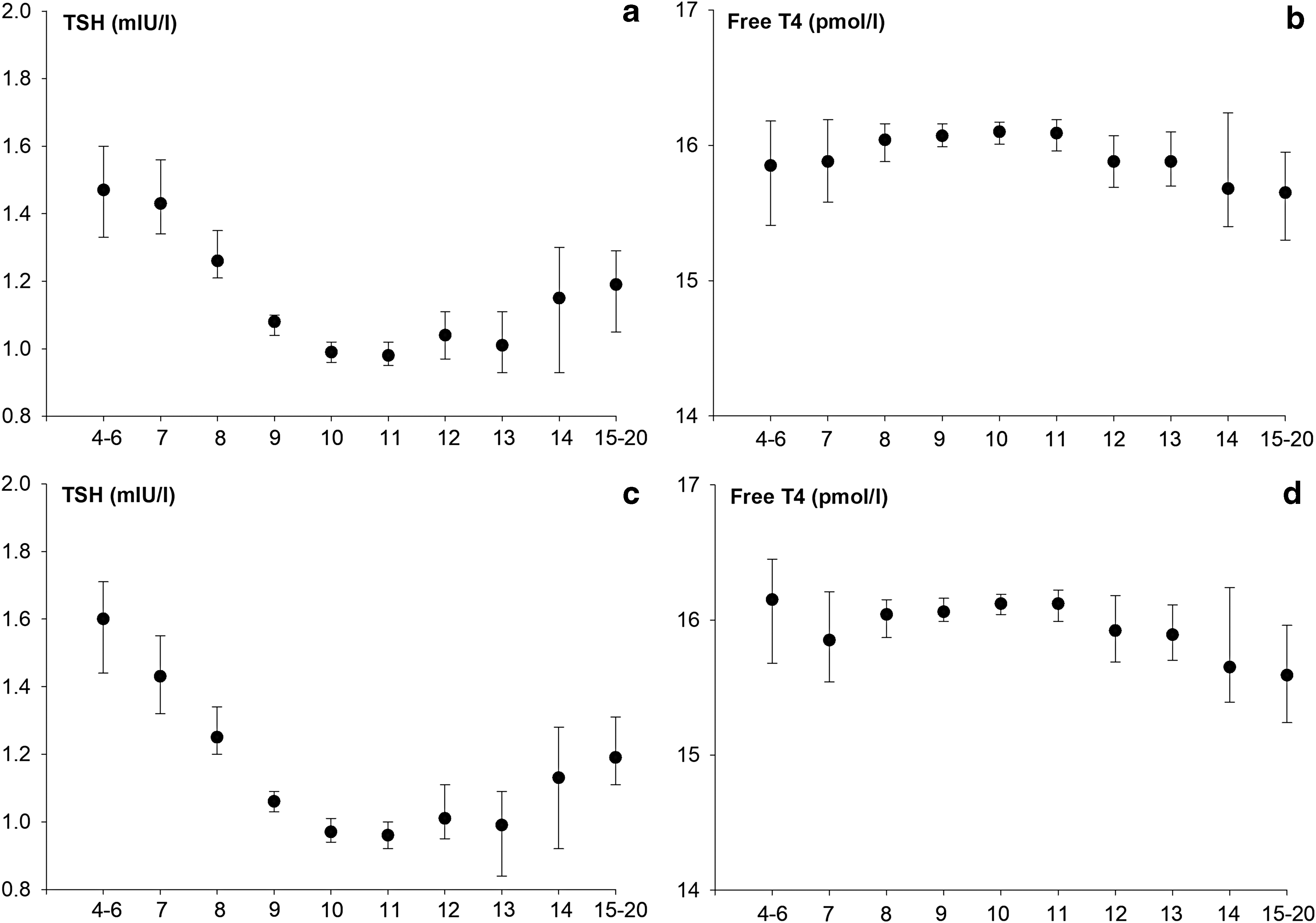
Pregnancy week-specific median with 95% confidence intervals for maternal thyrotropin and free thyroxine in all 10,337 pregnant women from the North Denmark region (
Pregnancy Week-Specific Reference Limits with Confidence Intervals for Maternal TSH and fT4 in all Pregnant Women from the North Denmark Region and in Pregnant Women who Ended the Pregnancy with a Live Birth
Number of individuals in each group is reported after the exclusion of outliers using Tukey's fences.
Lower reference limit was held at minimum of the sample in weeks 4–6, 14, and 15–20.
Upper reference limit was held at maximum of the sample in weeks 4–6, 14, and 15–20.
TSH, thyrotropin; fT4, free thyroxine; 2.5p, 2.5th percentile; 97.5p, 97.5th percentile; CI, 95% confidence interval.
Reference Limits of Maternal TSH and fT4 with Confidence Intervals in Different Periods of Early Pregnancy
Number of individuals in each group is reported after the exclusion of outliers using Tukey's fences.
Maternal characteristics
Different maternal characteristics were assessed for women who ended the pregnancy with the birth of a live-born child (Table 3). In multivariate analyses, multiparity and smoking in pregnancy were significant predictors of lower maternal TSH, whereas higher maternal BMI was associated with higher TSH. Maternal age and origin were not significant predictors of TSH. For maternal fT4, higher maternal age, higher BMI, and smoking in pregnancy were significant predictors of lower fT4, whereas multiparity and origin (not born in Denmark) were associated with higher maternal fT4. However, when stratified reference limits for TSH and fT4 in weeks 9–11 of pregnancy were calculated, the impact of individual predictors was modest, and confidence limits were overlapping (Table 4). A disparity was most pronounced for maternal BMI, which showed higher TSH and lower fT4 limits in obese women with a pre-pregnancy BMI ≥30 kg/m2. Reference limits among overweight women with a BMI in the range 25–29.9 kg/m2 (TSH: 0.13–2.73 mIU/L; fT4: 13.1–20.6 pmol/L) were comparable to the reference limits observed among women with a BMI <25 kg/m2 (TSH: 0.11–2.75 mIU/L; fT4: 13.2–20.6 pmol/L).
Characteristics of the 9515 Pregnant Women who Gave Birth to a Live-Born Singleton Child
Missing value n = 41 not included.
Missing value n = 83 not included.
Missing value n = 47 not included.
BMI, body mass index.
Reference Limits with Confidence Intervals for Maternal TSH and fT4 in Weeks 9–11 of Pregnancy Stratified by Maternal Characteristics
Number of individuals in each group is reported after the exclusion of outliers using Tukey's fences.
Discussion
Main findings
The establishment of pregnancy week-specific reference ranges for maternal thyroid function parameters in a large cohort of thyroid autoantibody–negative Danish pregnant women corroborates the dynamics of TSH during the first trimester of pregnancy. The findings imply that the clinical assessment of smaller time intervals within the first trimester of pregnancy may be preferable to the use of a uniform reference range for evaluation of maternal thyroid function. Reference limits for maternal fT4 showed opposite but less marked dynamics during the first trimester, and a uniform reference range may apply. Maternal characteristics such as age, parity, origin, smoking, and BMI were associated with maternal thyroid function parameters, but only small differences were observed in stratified reference limits for TSH and fT4.
Reference ranges
Valid reference ranges are an important part of diagnosis and management in clinical patient care and as part of exposure and outcome definitions in clinical research. Comprehensive guidance exists on how to establish reference ranges (17), but still many aspects remain debatable on the selection of the reference cohort, and it may further pose a challenge to obtain a sufficient number of samples. Whereas some laboratory tests are hardly affected by pregnancy, maternal thyroid function is considerably influenced by physiological changes in pregnancy, and nonpregnant reference ranges should not be used (3). Another challenge related to thyroid function parameters determined by automatic immunoassays is the potential disparity in results obtained with different methods. This is particularly a concern for the measurement of free thyroid hormones, because the determination of free thyroid hormone concentration by an automatic immunoassay is an indirect approach with no complete separation of free and protein-bound hormones (18 –20). Thus, clinical guidance recommends the use of method- and pregnancy-specific reference ranges (1).
The present study and previous investigations have questioned the use of a uniform reference range in the first trimester of pregnancy (4,5,21 –25). It has now been observed in two separate large cohorts of Danish pregnant women that TSH reference limits are highly dynamic in the first trimester of pregnancy, with an initial high TSH period followed by a gradual decline in weeks 7–8 to lower levels from week 9 onward. The present study suggests that the lower TSH limit continues to be low at the beginning of the second trimester, and is lower in weeks 12–14 compared to weeks 9–11, whereas no difference was observed in the upper limit between these periods of pregnancy. These findings are in keeping with the clinical experience that TSH may remain low at the beginning of the second trimester. It can be speculated whether this relates to a prolonged effect of hCG and if women with lower TSH levels are more sensitive to the effect of hCG. This also conforms to the more pronounced dynamics in the lower TSH reference limit within the first trimester observed in the present study and in a previous report (5). Uncertainty may prevail on the exact week of pregnancy in early pregnancy, and the use of pregnancy week-specific reference ranges may not be superior in clinical practice. However, the data suggest that clinical awareness at different time points in early pregnancy may be warranted for the classification of maternal thyroid function, with a higher TSH in the time period up to week 8 of pregnancy followed by a lower TSH during weeks 9–11 and by weeks 12–14.
The mechanisms underlying the dynamic changes in TSH likely relate to the balance between the hCG effect in early pregnancy, which tends to suppress TSH, and the activity of the type 3 deiodinase (DIO3) in the placenta, which tends to increase TSH (3). DIO3 is expressed from the early weeks of pregnancy (26 –29), whereas hCG peaks in weeks 9–10 of pregnancy (30). Thus, the initial high TSH period may reflect that the DIO3 effect is predominant in the early weeks of pregnancy, and the later low TSH period may reflect a dominant hCG effect.
Strengths of the present study are that thyroid autoantibodies were measured in a large cohort of pregnant women, and women who were positive for TPOAb and/or TgAb were excluded from the reference cohort. Such an approach was not possible in a previous investigation of Danish pregnant women (5), but it is noteworthy that similar dynamics of TSH levels in the first trimester were observed in the two Danish investigations. A cutoff value of 60 U/mL provided by the manufacturer was used to identify women with thyroid autoantibodies, and 13% of the women were TPOAb- and/or TgAb-positive. For comparison, the frequency was 16% when evaluated in a cohort of Danish pregnant women in East Denmark in 2007–2008 using the same cutoff level with a different immunoassay (31). The selection of reference individuals for the establishment of reference ranges is debatable, and a main focus regarding thyroid function analyses is on how to handle individuals with TPOAb and/or TgAb. In the present study, women with known or later diagnosed thyroid disease were excluded, as were outliers of TSH and fT4. Slightly lower TSH reference limits were found following the subsequent exclusion of women with thyroid autoantibodies. Notably, the reference limits were consistent, irrespective of the cutoff used or whether TPOAb- and/or TgAb-positive women were excluded. Adding to this, a recent study of pregnant women in a Swedish cohort showed that the additional exclusion of TgAb-positive women on top of TPOAb hardly changed the reference limits (32). More evidence is needed as part of the discussion on how to manage pregnant women who are positive for thyroid autoantibodies and who have no known thyroid disease.
The clinical guidance from the America Thyroid Association on the management of thyroid disease in pregnant women recommends reducing the upper reference limit by approximately 0.5 mIU/L in the first trimester of pregnancy if local method-specific reference ranges are not available (1). It was observed that the upper limit of TSH was below this limit throughout pregnancy in this study and in a previous investigation conducted around the time when iodine fortification was implemented in Denmark (5). The uniform upper limit of TSH in the first trimester was 3.4 mIU/L in the study of pregnant women in East Denmark in 2008–2009 (33) and was therefore slightly higher than the limit of 2.9 mIU/L observed in the present study conducted in West Demark. Methodological differences in thyroid function assays may play a role, but the discrepancy is also compatible with observations in the general Danish population of regional differences in TSH (34). Considering the clinical proposal of a uniform TSH reference limit in the first trimester of pregnancy (1), the present results indicate that reference limits vary considerably within the first trimester of pregnancy and suggest that it may be feasible to establish TSH reference limits for shorter time periods during pregnancy.
Predictors
Hyperthyroidism and hypothyroidism in women of reproductive age is mainly of autoimmune origin (6,7). Autoimmune thyroid disease is considered multifactorial, and genetic as well as environmental risk factors have been described (35). For the establishment of reference ranges, it is often relevant to consider if patient characteristics influence the reference limits and whether stratified reference ranges are needed in clinical practice. This study was able to evaluate the impact of different maternal characteristics on the reference limits for TSH and fT4 in early pregnancy. In line with a previous investigation of Danish pregnant women (5), it was observed that many factors were associated with TSH and fT4, but the impact of the individual factors in stratified reference limits was limited. These findings question the clinical usefulness of stratified reference ranges and may favor the use of the pregnancy week-specific reference ranges combined with clinical awareness of maternal characteristics that may affect thyroid function results, particularly in the evaluation of patients with borderline results. Most pronounced was the effect of maternal BMI with higher TSH and lower fT4 limits in obese women, which is compatible with other reports (36 –38). The role of other maternal characteristics has been less consistently reported in the literature (39), and the findings are less clear. A trend toward lower TSH levels was observed in multiparous women, which is in line with some studies, whereas others found no effect (39). In this study, ethnicity was not a predictor of maternal TSH. This is in contrast with other reports (40,41). These studies had the possibility of stratifying maternal ethnic background by subtype, whereas the register-based design of the present study only provided the possibility of investigating reference ranges according to information on whether the pregnant woman was born in or outside of Denmark. Considering maternal smoking, the effect was smaller, but the results suggested lower TSH and fT4 limits in smokers. Studies of the population in general have shown that smoking tends to be associated with lower TSH levels and an increase in peripheral thyroid hormone concentrations, possibly caused by increased activity in the sympathetic nervous system (42). A previous Danish investigation (5) and other reports (43) similarly showed lower fT4 levels associated with smoking in pregnancy, whereas other studies found no difference (32). Further studies are needed to corroborate and extend these findings, and to evaluate the clinical impact on maternal and fetal outcomes.
Methodological comments
The present large cohort of pregnant women allowed for the stratification of reference ranges by the week of pregnancy with an appropriate number of individuals in each of the stratified groups. However, the number of samples in the early and late pregnancy weeks was limited, and groups were collapsed to obtain sufficient sample size. Women with autoimmune disease were excluded from the reference cohort via linkage to information in Danish nationwide registers, and a recommended method of outlier detection was used (17).
Serum samples for the present study were consecutively collected as part of prenatal screening in Denmark, and the rate of participation is high (10). This method of inclusion is expected to minimize the risk of selection bias, and the number of samples obtained was representative of the expected number of pregnant women in the North Denmark region around the time of inclusion.
Determination of pregnancy outcome was performed via linkage to Danish nationwide registers, and the validity is considered high (44). This provided the possibility of evaluating reference ranges in the full cohort and in pregnancies with live births. Notably, results are consistent in both cohorts, which may apply to the design of future studies and considerations on the selection of reference cohorts. Information was available on pregnancy loss and births, including spontaneous and induced abortions as well as live births and stillbirths at hospital and at home. However, pregnancies terminating with spontaneous abortion without clinical recognition and hospital contact would not have been identified with an outcome of pregnancy.
Information on a number of maternal characteristics was available for women who terminated the pregnancy with the birth of a live-born child. Pre-pregnancy BMI and smoking status were self-reported by the pregnant women, but any misclassification is expected to be non-differential regarding thyroid function.
All samples were analyzed consecutively in the same laboratory after the last sample had been collected, and storage time until the present analyses ranged from two to six years. The storage time is not expected to influence the results, since thyroid function parameters and thyroid autoantibodies in pregnant women have been shown to be stable for >20 years of storage at −25°C (45).
Conclusion
Reference limits for maternal TSH are dynamic during the first trimester of pregnancy, with an initial high TSH period followed by a gradual decline to a low TSH period. A clinical focus on shorter time intervals within the first trimester of pregnancy may be preferable. Future investigations should evaluate the dynamics of maternal thyroid function parameters in other populations using different thyroid function assays. It remains to be addressed whether clinical awareness of the dynamic changes in maternal TSH in early pregnancy and of maternal characteristics will improve patient care and clinical research.
Footnotes
Acknowledgments
We deeply acknowledge Professor Peter Laurberg, Department of Endocrinology, Aalborg University Hospital (Aalborg, Denmark), who tragically died on June 20, 2016. Professor Peter Laurberg conceptualized the study and was responsible for the biochemical measurements. His contributions were invaluable and are greatly missed.
Author Disclosure Statement
No competing financial interests exist.