
Abstract
Background:
The lack of facile methods for the specific characterization of malignant thyroid nodules makes the diagnosis of thyroid cancer (TC) challenging. Due to its restricted expression in such nodules, the cell-associated lectin galectin-3 (Gal3) has emerged as a marker for TC with growing interest for in vivo imaging as well as targeted radionuclide therapy. To accelerate translation into clinical application, we have developed a cognate chimeric human antigen-binding fragment (Fab) derived from the rat anti-Gal3 monoclonal antibody M3/38.
Methods:
The variable immunoglobulin (Ig) light and heavy chain sequences were cloned from the hybridoma cell line, and the corresponding Fab carrying human IgG1/κ constant genes was functionally produced in the periplasm of Escherichia coli and purified to homogeneity. To moderately prolong its plasma half-life and, thus, increase tumor uptake, the recombinant Fab was fused with a long disordered amino acid chain comprising in total 200 Pro, Ala, and Ser residues (PASylation). This novel tracer was subjected to in vitro characterization and in vivo validation by using two thyroid cancer orthotopic murine models. To this end, the αGal3-Fab-PAS200 was conjugated with deferoxamine (Dfo), labeled with 89Zr under mild conditions and tested for binding on TC cell lines. Athymic nude mice were inoculated either with FRO82-1 or with CAL62 tumor cells into the left thyroid lobe. After intravenous injection with ∼3.0 MBq of 89Zr-Dfo-PAS200-Fab, these mice were subjected to positron emission tomography (PET)/computed tomography imaging followed by quantification of tumor accumulation and immunohistochemical analysis.
Results:
The αGal3-Fab-PAS200 revealed high affinity toward the recombinant Gal3 antigen, with a dissociation constant ≤1 nM as measured via enzyme-linked immunosorbent assay, surface plasmon resonance spectroscopy, and radioactive cell binding assay. The in vivo Gal3-targeting by the 89Zr(IV)-labeled protein tracer, as investigated by immuno-PET, demonstrated highly selective and fast accumulation in orthotopically implanted tumors, with strong contrast images achieved 24 hours postinjection, and no uptake in the tumor-free thyroid lobe, as also confirmed by biodistribution studies.
Conclusions:
The chimeric αGal3 89Zr-Dfo-PAS200-Fab tracer exhibits selective accumulation in the tumor-bearing thyroid lobe of xenograft mice. Thus, this novel radioactive probe offers potential to change TC management, in addition to current diagnostic procedures, and to reduce unnecessary thyroidectomies.
Introduction
Thyroid cancer (TC) comprises a spectrum of malignancies that range from indolent papillary thyroid microcarcinoma to anaplastic thyroid carcinoma (ATC), an almost universally fatal disease. The incidence of TC has significantly increased during the past 15 years, with >52,000 new cases in the United States every year, mostly due to the detection of small and clinically insignificant papillary thyroid carcinoma (PTC) by screening ultrasound (US) scans (1,2). However, differentiating between malignant and benign thyroid nodules by US remains difficult. The detection of thyroid nodules on US that do not have benign features, therefore, frequently prompts further investigation, including fine-needle aspiration (FNA) biopsy analysis. Despite the crucial role of FNA in the work-up of thyroid nodules, there is a high variation in terms of sensitivity, specificity, as well as positive and negative predictive value among centers and events when adjunct molecular testing is done. Moreover, only about 25% of nodules categorized as Bethesda IV stage (follicular neoplasia) harbor malignancy (3). Consequently, many unnecessary thyroidectomies are performed for nodules that eventually turn out to be benign (4). Therefore, a specific imaging method that is able to identify nodules that should undergo FNA biopsy followed by cytological analysis and/or molecular testing would be of great clinical utility.
The majority of patients with malignant thyroid nodules are affected by well differentiated thyroid carcinoma (DTC) and have a good response to total thyroidectomy and, if applicable, radioactive iodine as well as thyroid hormone for thyrotropin (TSH) suppression (5). The major clinical problem arises for up to about 5–10% patients diagnosed with poorly DTC (PDTC) or ATC, both of which represent the most frequent cause of TC-related death (6). This is mainly due to the lack of human sodium iodide symporter (NIS) expression as a consequence of oncogenic activation, which renders radioiodine ablation ineffective and monitoring of disease progression difficult (7,8). Radioiodine imaging is not sensitive for detecting PDTC and ATC and, therefore, not suitable for monitoring (using123I, 124I or 131I) or treatment (131I). 18F-FDG positron emission tomography (PET) is a useful imaging modality for these patients, but it is usually applied in combination with recombinant human TSH (rhTSH) to increase sensitivity. Patients with ATC face a fatal prognosis despite undergoing currently available treatment options. Therefore, the identification of these patients at an early stage is critical for optimal management.
Galectin-3 (Gal3), a member of the β-galactoside binding protein family, has been recognized as a clinically useful marker of TC on histology, as this lectin is overexpressed by the overwhelming majority of TC but not by normal thyroid cells (9,10). In fact, in a large international multicenter retrospective study of >1000 well-characterized thyroid lesions, the sensitivity, specificity, positive predictive value, and negative predictive value for Gal3 immunostaining were 88%, 98%, 91%, and 97%, respectively (9). Further, successful in vivo visualization of thyroid tumors by targeting Gal3 has recently been demonstrated by our group using a radiolabeled monoclonal antibody (mAb) in xenograft mice (11). In particular, we identified Gal3 as a suitable marker for thyroid imaging in animal models bearing orthotopic TC that recapitulate human tumors (12). In experiments using a 89Zr-labeled proteolytic F(ab′)2 antibody fragment, high tracer accumulation was achieved in PET compared with radioiodine imaging, which is usually used in the clinic for the staging of TC.
Although our prior studies have indicated the potential of targeting Gal3 for imaging of TC, the imaging agents we used were not ideal for translation into the clinic. In humans, full-size antibodies typically require a period of seven days after uptake for cancer imaging. This makes immuno-PET with full-size antibodies logistically difficult. More importantly, imaging with 89Zr-labeled full-size antibodies is associated with radiation doses that are several-fold higher than for small molecules. Conversely, the tumor uptake of antigen-binding fragments (Fabs) may be limited by the very rapid clearance of these molecules (13,14). Therefore, the goal of this study was to develop a recombinant Fab with optimized pharmacokinetics for tumor imaging. PASylation is a biological alternative to polyethylene glycol conjugation (PEGylation) that involves the genetic fusion of a biopharmaceutically active protein with a conformationally disordered polypeptide chain comprising the small natural amino acids Pro, Ala, and Ser (PAS). The PAS chain has precise composition and can be varied in length, which offers the opportunity to fine tune the circulation half-life of a recombinant protein (15), leading to benefits for in vivo imaging as shown for other established targets (13,14). Here, we describe the generation of a PAS-conjugated (PASylated) chimeric Fab tracer directed against Gal3. The clinical potential of this novel agent for imaging, and also therapy planning, of TC is demonstrated via high and specific uptake by orthotopic tumor xenografts in mice at early time points postinjection (p.i.).
Materials and Methods
Cell culture
Two DTC cell lines were chosen for this study: the poorly differentiated FRO82-1 cells (RRID: CVCL_6287) kindly provided in 2016 by Dr. Silvia Soddu (Regina Elena National Cancer Institute, Rome, Italy) and the anaplastic cells CAL62 (RRID: CVCL_1112) from the German Collection of Microorganisms and Cell Cultures (DSMZ, Leibniz, Germany). FRO82-1 and CAL62 cells were maintained as monolayer cultures in Dulbecco's modified Eagle medium and RPMI-1640 (both from Invitrogen, Carlsbad, CA), respectively, at 37°C under a 5% carbon dioxide atmosphere for <3 weeks. Hybridoma cells TIB-166 from the American Type Culture Collection (Rockville, MD) secreting the αGal3 mAb clone M3/38 (16) were cultured in RPMI-1640 medium supplemented with 1% (w/v) L-glutamine, 1% (w/v) penicillin/streptomycin, and 10% (v/v) low IgG fetal bovine serum (Bio&Sell, Feucht, Germany). All cell lines were tested for integrity and phenotype via simultaneous amplification of multiple short tandem repeat loci by the suppliers. Cells were cultured ≤5 months after testing. All in vitro experiments using these cell lines were repeated at least three times unless otherwise stated in the text.
Cloning of V-genes from hybridoma cells and preparation of a PASylated chimeric Fab
Total RNA was extracted from the TIB-166 hybridoma cells by using the RNeasy Mini Kit (Qiagen, Hilden, Germany). Complementary DNA (cDNA) synthesis was accomplished with the First Strand cDNA Synthesis Kit (Fermentas/Thermo Fisher Scientific, St. Leon-Rot, Germany) by using an oligo(dT)18 primer. The pair of immunoglobulin (Ig) V-regions was amplified via polymerase chain reaction (PCR) by using Q5 High-Fidelity DNA Polymerase (New England Biolabs, Ipswich, MA) and the primer pairs (17,18); Rat-VL-13B (5′-GATCGCCGGCGATGTTGTGATGACCCAG-3′) with RMK (5′-GACCTCCACGGAGTCAGC-3′) for the light chain and Rat-VH-F6 (5′-GATCGGCGCGCCAGGCCCAGCTGCAGTCTGG-3′) with RMG (5′-AGGTCGCCACACGTGTGG-3′) for the heavy chain (all oligonucleotides from Eurofins Genomics, Ebersberg, Germany) and then subjected to agarose gel electrophoresis.
To introduce suitable flanking restriction sites for subcloning on the bacterial expression vector pASK88 (19), which already encodes the human CH1 and Cκ regions as well as N-terminal bacterial signal peptides to direct secretion into the periplasm of Escherichia coli, the V-regions were re-amplified from gel-purified bands with appropriate primers according to a published protocol (18). Based on the results from DNA sequencing (Eurofins) of the gel-purified Ig V-regions from above, the following specific primers were designed: 5′-GATGTT
Antigen detection with the αGal3-Fab-PAS200 on tissue samples
Paraffin-embedded tissue sections from human malignant and benign thyroid tumors were tested to confirm the immunoreactivity of the new αGal3-Fab. In particular, tissue sections from papillary TC (n = 5), follicular TC (n = 5), nodular hyperplasia (n = 5), follicular adenoma (n = 5), and thyroiditis (n = 5) were prepared as previously described (11) and incubated with the αGal3-Fab-PAS200 (30 μg/mL) in 20 mM HEPES/NaOH, 150 mM NaCl, pH 7.5 for 1 hour at room temperature. The sections were then incubated with a polyclonal rabbit anti-human κ light chain antibody (Dako/Agilent) for 30 minutes. After washing with phosphate-buffered saline (PBS), the signal was revealed via incubation with an horseradish peroxidase (HRP)-conjugated polyclonal mouse anti-rabbit secondary antibody for 30 minutes, followed by development with 3,3’-diaminobenzidine (Dako EnVision FLEX System; Agilent) as a chromogenic substrate. Images were recorded by using an Aperio CS2 ScanScope image capture device (Leica Microsystems, Wetzlar, Germany) and analyzed with the Aperio slide Viewing Software (Aperio Technologies, Vista, CA). The study was carried out according to the ethical guidelines of the Helsinki Declaration, under approval of the Scientific Board of Sant'Andrea Hospital (Prot. CE No. 8391/2013).
Labeling of proteins and characterization of 89Zr-Dfo-αGal3-Fab-PAS200
The purified αGal3-Fab-PAS200 was conjugated with Sulfo-Cyanine 5.5 N-hydroxysuccinimide (NHS) ester (Lumiprobe, Hannover, Germany) in dimethyl-sulfoxide (DMSO), to be used as a probe for orthotopic tumor growth monitoring, or with p-SCN-Bn-Deferoxamine (Dfo; Macrocyclics, Plano, TX) in DMSO, for preclinical PET imaging experiments (Supplementary Data). For immuno-PET imaging, 130 μg Dfo-αGal3-Fab-PAS200 was labeled with 1 mCi 89Zr(IV)-oxalate (BV Cyclotron, PerkinElmer, Amsterdam, Netherlands) as previously described (13). Integrity of the radiolabeled protein tracer was verified by sodium dodecyl sulphate-polyacrylamide gel electrophoresis (SDS-PAGE). Twenty microliters of purified 89Zr-Dfo-αGal3-Fab-PAS200 dissolved at 1 mg/mL in PBS, or intact (unlabeled) mAb M3/38 (Mabtech, Nacka Strand, Sweden) as a control, was mixed 4:1 with Roti-Load reducing or non-reducing sample buffer (Carl Roth, Karlsruhe, Germany); then heated at 90°C; and finally applied to the wells of a 10% precast polyacrylamide MP TGX Gel (Bio-Rad Laboratories, Munich, Germany). Signals in the gel were quantified by 15 minutes incubation on a phosphorimaging plate (Fujifilm, Tokyo, Japan), followed by readout with an HDCR35 Bio imaging plate scanner (Dürr NDT, Bietigheim-Bissingen, Germany). The immunoreactive fraction of the tracer was assessed on FRO82-1 cells, which served as a representative cell line, using a published procedure (23). The dissociation constant (KD) of the 89Zr-Dfo-αGal3-Fab-PAS200 was calculated from a saturation binding assay by using FRO82-1 cells as previously described (11). All experiments were performed in triplicate.
In vitro stability test of 89Zr-Dfo-αGal3-Fab-PAS200
Stability of the protein tracer was evaluated under three conditions by incubation at 37°C (i) in storage buffer (0.25 M Na-acetate, 0.5 mg/mL gentisic acid, pH 5.5), (ii) in human serum isolated from a healthy volunteer, and (iii) in 50 mM diethylenetriaminepentaacetic acid (DTPA) (Sigma-Aldrich, Taufkirchen, Germany) adjusted to pH 4.0 with NaOH via size-exclusion radio-high performance liquid chromatography (HPLC) (SE-radio-HPLC) and radio-thin-layer chromatography (radio-TLC) at different time points (1, 12, and 24 hours). For each time point, 2 μL of the solution was spotted onto a silica gel 60 F254 aluminum TLC strip (Merck Millipore, Darmstadt, Germany) and eluted with 50 mM DTPA/NaOH pH 5.0 (Rf tracer = 0.1, Rf free 89Zr = 0.8); then, the activity was quantified on a radio-TLC scanner (Eckert & Ziegler, Berlin, Germany). At each time point, the stability of the protein tracer incubated in storage buffer was additionally evaluated by SE-radio-HPLC.
Orthotopic tumor xenograft models
Two TC orthotopic models were established in six-week healthy female athymic Nude-Foxn1nu/nu mice (Charles River Laboratories, Sulzfeld, Germany) by using FRO82-1 and CAL62 cell lines as previously described (12). All experimental protocols were approved by the local authorities (Regierung von Oberbayern, Germany; License: 55.2-1-54-2532-216-15). Tumor growth was monitored weekly by US scan and fluorescent imaging (Supplementary Data).
PET/computed tomography tumor imaging
Xenograft tumors exceeding a volume of 65 mm3 were considered appropriate to perform PET/computed tomography (CT) imaging. A group of mice bearing FRO82-1 orthotopic tumors (N = 3) were injected via catheter in the tail vein with 3 MBq (∼9 μg) of 89Zr-Dfo-αGal3-Fab-PAS200 in storage buffer; the time course of tracer uptake into the tumor and into normal organs was studied at 6, 12, and 24 hours p.i. to determine the best time point for imaging, by using a dedicated small animal scanner (Inveon; Siemens, Knoxville, TN). For each cancer cell line, two groups (N = 5) of tumor-bearing mice were studied. One group was injected with 3 MBq (∼9 μg) of 89Zr-Dfo-αGal3-Fab-PAS200 and the other one, for blocking tests, additionally with a 1000-fold amount (∼9 mg) of unlabeled αGal3-Fab-PAS200, followed by PET/CT scan 24 hours p.i. Another group of five healthy mice without orthotopic tumors were injected with 3 MBq of tracer and scanned 24 hours p.i. as a control. Image reconstruction was performed as previously described (12).
Tracer accumulation studies
The in vivo tumor uptake of 89Zr-Dfo-αGal3-Fab-PAS200 was evaluated by analysis of the PET images via Inveon Research Workplace software (Siemens). An approximate region of interest (ROI) was drawn on the left thyroid lobe of each mouse, encompassing the tumor signal with a threshold of 50%. The percentage of injected dose per gram (%ID/g) value was calculated as a ratio of the mean radioactivity in each ROI per tumor weight and total injected radioactivity. Biodistribution studies were performed as previously described (12).
In vivo stability of 89Zr-Dfo-αGal3-Fab-PAS200
To evaluate the in vivo stability of the protein tracer, the radioactivity was extracted from tumors, blood, and selected tissues (liver, kidneys, and muscles) 24 hours p.i. Tumors and organs were first frozen in liquid nitrogen; then, they were homogenized in an Ultra Turrax homogenizor (IKA, Staufen im Breisgau, Germany) using glass balls. To separate plasma from blood cells, the blood sample was centrifuged at 3200 rpm for 15 minutes (Hereus Fresco 21 centrifuge, Thermo Scientific, Schwerte, Germany). Pellet and supernatant were measured separately in a 2480Wizard2 gamma-counter (PerkinElmer, Waltham, MA). The extracts were analyzed via radio-TLC and SDS-PAGE followed by autoradiography.
Statistics
All data are presented as mean ± standard deviation. Statistical analyses were performed by using Prism 4.0 software (GraphPad, San Diego, CA), and Student's t-test was used for unpaired data. The two-sided significance level was calculated, and values p < 0.05 were considered statistically significant.
Results
Cloning, bacterial production, and characterization of the αGal3-Fab
The Ig variable genes from the rat mAb clone M3/38 (16) were amplified from a cDNA preparation of total hybridoma RNA. To this end, primer pairs were used that hybridize in the region encoding the leader sequence and mature N-terminus of the Ig variable domain (17) and within the region encoding the first constant domain (rat CH1γ1 and Cκ, respectively) (18), resulting in PCR products with a size of 400–500 bp, as expected. After sequencing of the PCR products, the DNA fragments were equipped with compatible restriction sites and cloned on a bacterial expression vector (pASK88) for the functional secretion of the first-generation chimeric Fab* carrying human constant domains (19,24). Immobilized metal ion affinity chromatography from the bacterial periplasmic extract via the His6-tag attached to the heavy chain, followed by size-exclusion chromatography (SEC), led to ∼0.5 mg soluble protein per 2 L E. coli culture. The purified Fab* was tested for binding to recombinant human galectin-3 (hGal3) by enzyme-linked immunosorbent assay (ELISA) and surface plasmon resonance (SPR) spectroscopy, revealing KD values of 9.5 ± 0.5 and 0.81 ± 0.01 nM, respectively. In contrast, the proteolytically prepared Fab′ (Supplementary Data) of the hybridoma mAb showed higher affinities, with corresponding KD values of 2.7 ± 0.6 nM (ELISA) and 0.27 ± 0.01 nM (SPR) (Fig. 1). Functional activity and target specificity of the recombinant αGal3-Fab* was further demonstrated by positive staining for hGal3 visualized on tissue sections of human papillary TC and follicular TC (Fig. 2A–C), whereas all benign thyroid tissues, including normal thyroid tissue, were negative (Fig. 2D–E).

Biochemical characterization of the recombinant αGal3-Fab. (
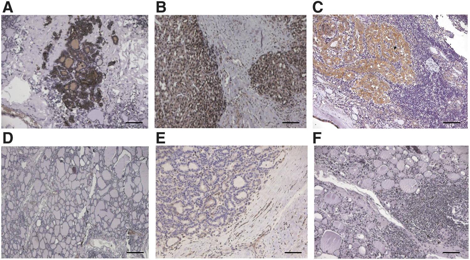
Reactivity of αGal3-Fab-PAS200 assessed on tissue sections of benign and malignant human thyroid conditions (20 × magnification, scale bar 200 μm). Four micrometers of formalin-/paraffin-embedded tissue sections taken from papillary TC, follicular TC, nodular hyperplasia, follicular adenoma, and thyroiditis were incubated with αGal3-Fab-PAS200 (30 μg/mL) followed by a rabbit anti-human kappa light chain antibody and the EnVision FLEX/HRP secondary antibody. Strongly positive staining of hGal3 is restricted to the papillary and follicular TC cells (
Optimization of αGal3-Fab antigen affinity
The slightly diminished antigen affinity of the initial recombinant Fab* may have been a consequence of the cloning procedure whereby the N-terminal 4–5 amino acids of each mature V-region were determined by the expression vector (24). Moreover, the original N-terminal sequences of the mAb M3/38 were biased due to the location of the hybridization sites of the forward primers (17) that were used for the V-gene amplification from the hybridoma cDNA. Hence, an attempt was made to identify the corresponding rat germline L/V-gene segments in the IMGT/Laboratoire d'ImmunoGénétique Moléculaire (LIGM) database (25). Therefore, the nucleotide sequences encoding residues L8 to L88 and H8 to H94, respectively—which cover the unbiased amplified light and heavy chain V-gene sequences from frameworks 1–3 of the M3/38 mAb—were compared in a BLAST search against the Ig nucleotide sequence database for Rattus norvegicus. The most similar V-segments showed 96.7% nucleotide sequence identity with a rat germline IGHV segment (IMGT/LIGM ID: BC092592) and 93.1% nucleotide sequence identity with a rat germline IGKV segment (IMGT/LIGM ID: L22655). In both cases, the encoded N-terminal amino acid sequence (Fig. 3) differed considerably from the corresponding stretch encoded on the expression vector, including an additional inserted residue in case of the rat heavy chain V-segment. Consequently, these changes in the N-terminal amino acid sequences were introduced into the cloned coding regions of both light and heavy chains, and the second-generation αGal3-Fab was produced, purified, and characterized.
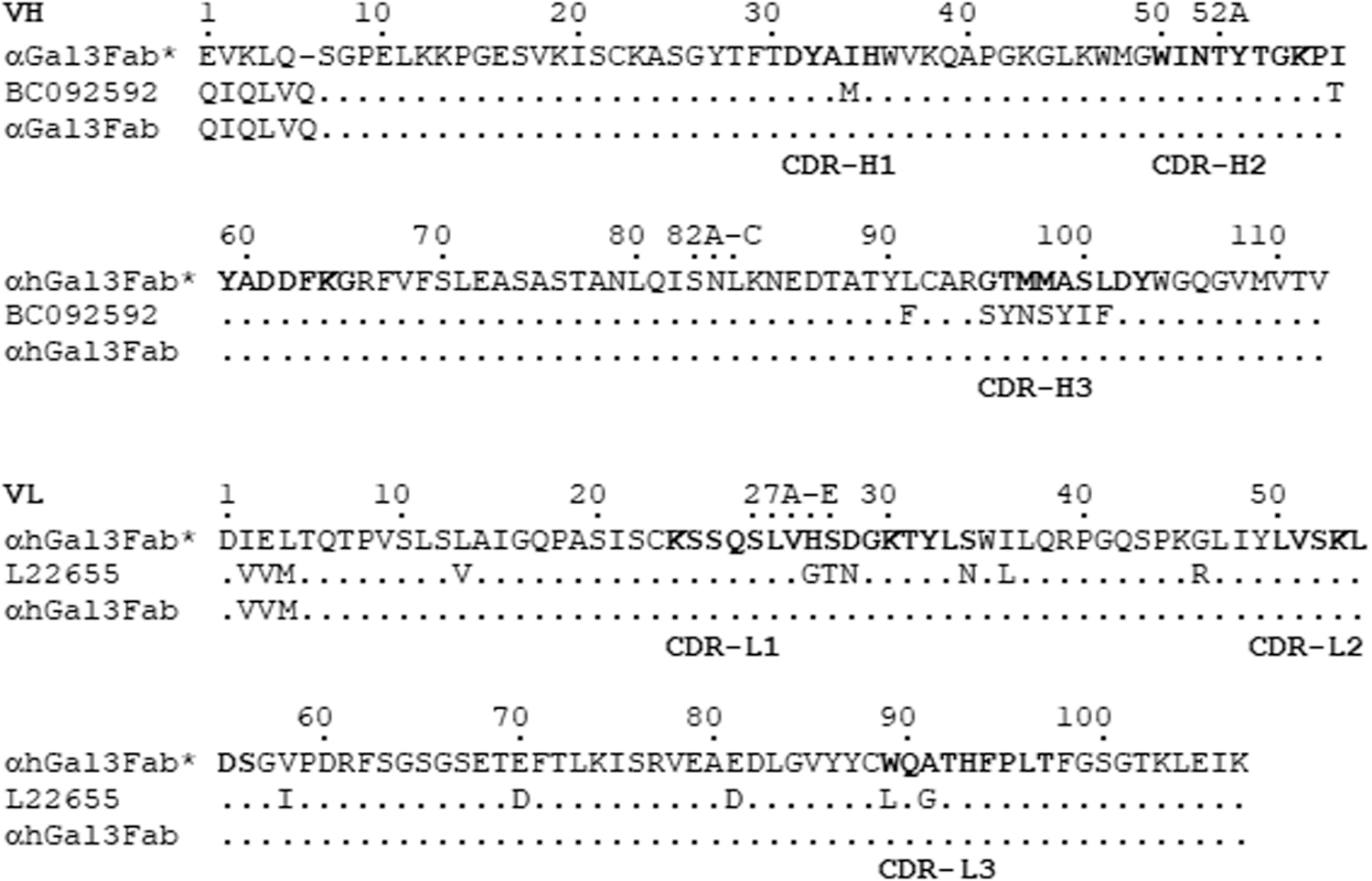
Amino acid sequences of the VH and VL regions cloned from the hybridoma M3/38, starting with the first residues of each mature rat antibody chain and ending with the last residue of framework region 4. Residues are numbered according to Kabat (21); note that residue H6 was missing in the initially cloned V-genes due to the use of the PCR primer RatVHF06 (17), which hybridizes to the Rattus norvegicus V-gene allele IGHV1-48*01 P (25). The upper line shows the initially cloned αGal3-Fab* wherein the 5/4 N-terminally encoded residues were originating from the expression vector pASK88 (19,24). The second line shows the most closely related sequences found in an IMGT/LIGM search (20) for each heavy and light chain. The lower line depicts the N-terminally mutated αGal3-Fab, which showed unchanged affinity compared with the original mAb. IMGT, immunogenetics; LIGM, Laboratoire d'ImmunoGénétique Moléculaire; mAb, monoclonal antibody; PCR, polymerase chain reaction.
The αGal3-Fab had KD values of 1.7 ± 0.5 nM (ELISA) and 0.25 ± 0.01 nM (SPR) (Fig. 1A, B). These numbers matched those of the proteolytic Fab′, thus confirming that the fully functional Fab of the M3/38 mAb had been successfully cloned and recovered as a recombinant protein (Fig. 3).
Plasma half-life tuning of the recombinant αGal3-Fab
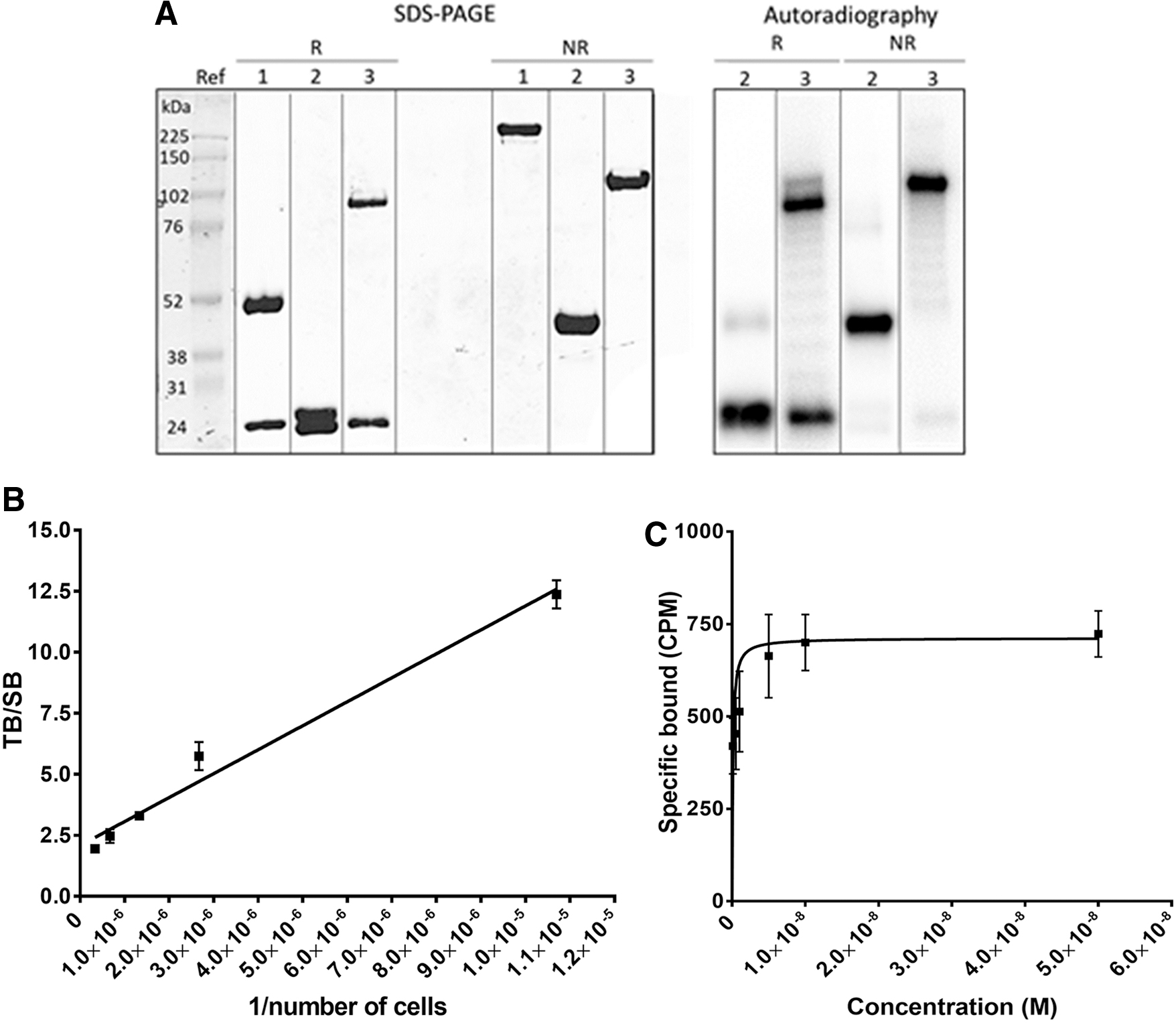
Motivated by a previous PET imaging study on the use of recombinant Fabs directed against the HER2 and CD20 tumor antigens demonstrating that a moderate increase in plasma half-life of the otherwise quickly eliminated Fab leads to improved tumor-to-blood ratio (13,14); a similar approach was followed for the αGal Fab cloned here. To prolong the plasma half-life of the chimeric M3/38 Fab, a genetically encoded structurally disordered sequence of the small L-amino acids PAS, which enlarges the hydrodynamic molecular volume beyond the glomerular pore size (22) similar to PEGylation, was appended in the form of a fusion protein (15). Thus, a 200-residue PAS sequence was inserted into the expression vector either at the C-terminus of the heavy chain or of the light chain and both variants were expressed and purified in the same manner as the original Fab. The version carrying the PAS sequence at the light chain led to a soluble purified protein yield of 0.5 mg per 2 L bacterial culture, similar to the unfused αGal3-Fab, whereas the heavy chain fusion showed much lower yield. Affinity measurements with the recombinant hGal antigen resulted in KD values of 1.3 ± 0.1 nM (ELISA) and 0.27 ± 0.01 nM (SPR) for the light chain PAS fusion, αGal3-Fab-PAS200 (Fig. 1A, B), indicating full retention of antigen-binding activity. Similar to the unfused αGal3-Fab, the purified αGal3-Fab-PAS200 showed quantitative disulfide bridge formation between light and heavy chains, demonstrated by SDS-PAGE (Fig. 1C), as well as singular peaks in the SEC and electrospray ionization mass spectrometry (ESI-MS) analyses (Supplementary Fig. S1A, B), thus confirming a high degree of biochemical definition. Subsequently, the αGal3-Fab-PAS200 was conjugated with Cy5.5 and with Dfo and the coupling efficiency was analyzed by using ESI-MS, yielding a degree of labeling (DOL) of 0.90 ± 0.05 for Cy5.5 and of 1.40 ± 0.09 for Dfo (Supplementary Fig. S2). Absorption measurements were performed to verify the DOL in the case of the fluorescence-labeled protein (Supplementary Data), revealing a similar value of 0.80 ± 0.02 (Supplementary Fig. S3).
αGal3-Fab radiotracer preparation and characterization
The αGal3-Fab-PAS200 conjugate with Dfo was labeled with 89Zr(IV), yielding a specific activity of 19.8 ± 1.2 GBq/μmol and a radiochemical purity >98%, as measured by radio-TLC and SE-radio-HPLC (Supplementary Fig. S4A, B). Biochemical integrity of the protein tracer was confirmed by SDS-PAGE, where the Coomassie-stained band coincided with the signal seen in the autoradiography (Fig. 4A). Binding tests performed with 89Zr-Dfo-αGal3-Fab-PAS200 on FRO82-1 TC cells revealed a KD value of 0.28 ± 0.09 nM and an immunoreactive fraction (23) equal to 48 ± 6% (Fig. 4B, C). Radio-TLC performed at different time points after labeling indicated that the stability of this protein tracer was >95% up to 24 hours in storage buffer and in human serum, whereas trans-chelation phenomena occurred in the presence of DTPA (Supplementary Fig. S4C). SE-radio-HPLC analysis of the samples kept in storage buffer revealed a slow release of 89Zr up to 5% of the total activity within 24 hours (Supplementary Fig. S4D).
Characterization of the 89Zr-Dfo-αGal3-Fab-PAS200 protein tracer. (
PET/CT imaging of orthotopic tumors using 89Zr-Dfo-αGal3-Fab-PAS200
The growth of orthotopic tumors from the TC cell lines FRO82-1 and CAL62 within the left thyroid lobe of athymic Nude-Foxn1nu/nu mice was visualized by US scanning as hypoechoic areas (Fig. 5A). Fluorescence molecular tomography (FMT) imaging of the neck region performed 24 hours p.i. of 100 μg Cy5.5-αGal3-Fab-PAS200 showed intense uptake of the fluorescent tracer by the tissue mass visualized in the US scan (Fig. 5B). PET/CT images of 89Zr-Dfo-αGal3-Fab-PAS200 demonstrated the best tumor-to-background ratio for the PASylated protein if recorded 24 hours p.i. (Supplementary Fig. S5). Both FRO82-1 and CAL62 orthotopic thyroid tumors were clearly visualized in this experiment, showing specific accumulation of the Fab tracer in the left thyroid lobe, with a high tumor-to-background contrast (Fig. 6A). Mice that were concomitantly treated with a 1000-fold amount of unlabeled αGal3-Fab-PAS200 in blocking experiments exhibited a strong decrease of the signal for the protein tracer in the left thyroid lobe, confirming the specificity and high targeting performance of 89Zr-Dfo-αGal3-Fab-PAS200 (Fig. 6A). There was accumulation of radioactivity seen in kidneys and liver consistent with the known function of these organs in tracer metabolism and excretion, respectively (14). PET images of control mice did not reveal any signal in the neck area (Fig. 6A, right). Xenograft uptake of 89Zr-Dfo-αGal3-Fab-PAS200 on the PET images correlated well with the shape and location of the orthotopic tumors on necropsy (Fig. 6B).
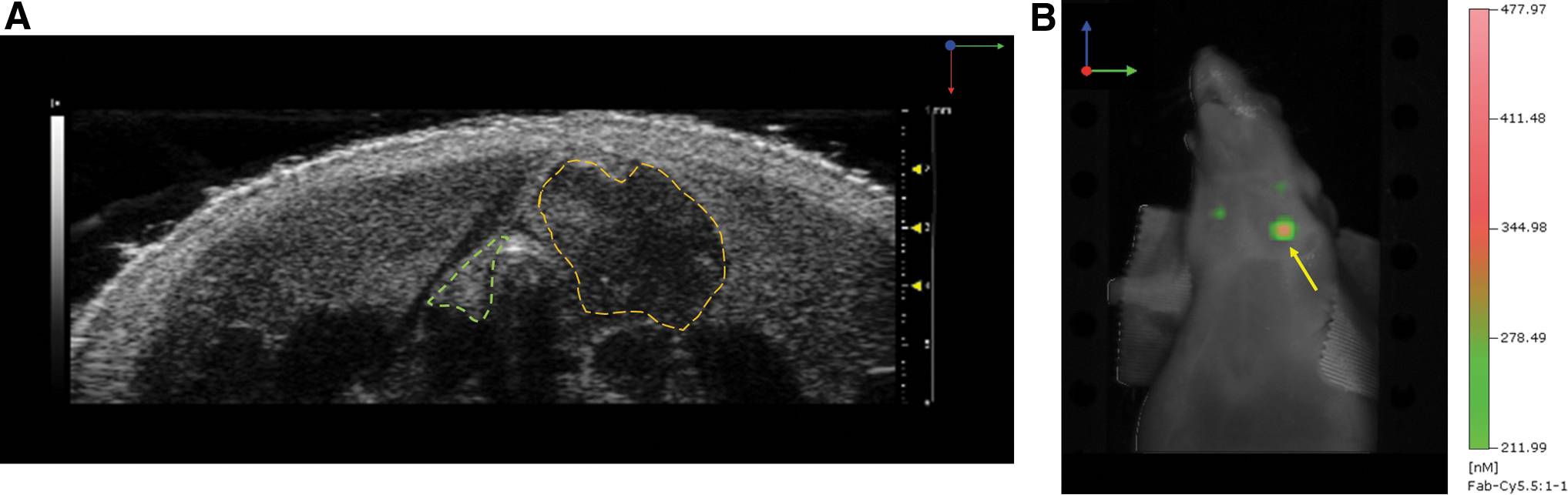
Monitoring of orthotopic tumor growth in a mouse xenograft model. (
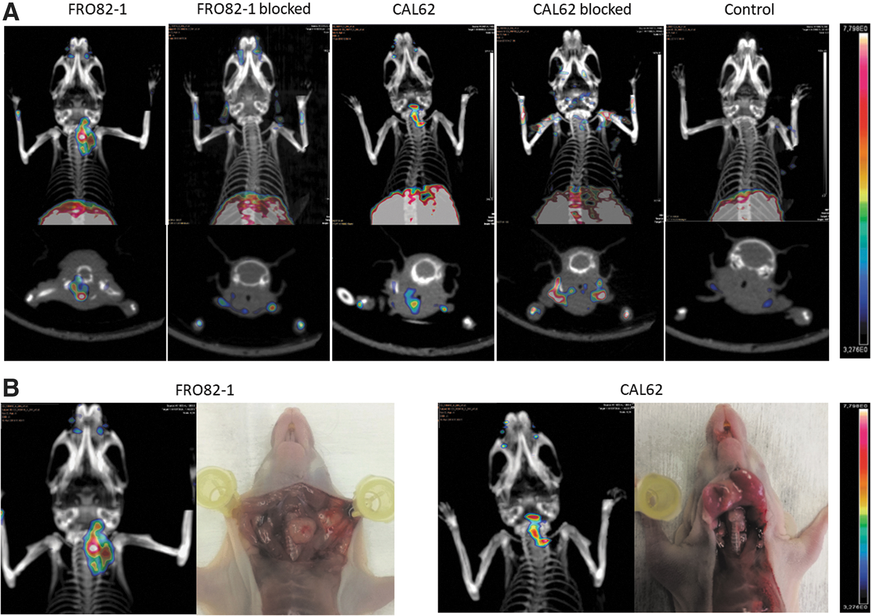
PET/CT imaging of mice bearing orthotopic FRO82-1 and CAL62 thyroid cancer xenografts. (
Quantitative analysis of αGal3-Fab-PAS200 tracer accumulation
Image-derived calculation of tracer accumulation in the orthotopic tumors indicated >5%ID/g for both FRO82-1 and CAL62 cell lines (5.9 ± 2.4 and 5.1 ± 3.1%ID/g, respectively). The biodistribution studies confirmed an elevated uptake in the left thyroid lobe, bearing the xenograft tumor, of 5.7 ± 1.3%ID/g for FRO82-1 and 3.6 ± 0.4%ID/g for CAL62 (Fig. 7A), compared with low background accumulation measured in the right (tumor-free) thyroid lobe (1.4 ± 0.3%ID/g). Specificity of tracer accumulation was confirmed in blocking studies with excess of unlabeled αGal3-Fab-PAS200, revealing significantly lower signals (2.4 ± 0.5 and 2.3 ± 0.3%ID/g for FRO82-1 and CAL62, respectively). The differences in tracer accumulation for the orthotopic tumors were statistically significant as compared with the right thyroid lobe (p < 0.001), while for the blocked tumors the corresponding ratio between left and right lobe was close to one (Fig. 7B). Elevated accumulation of the tracer was measured only in liver and spleen (9.7 ± 1.7 and 6.8 ± 0.6%ID/g, respectively), as expected considering the catabolism of the 89Zr(IV) conjugate (14,26), and in kidneys (95.6 ± 5.4%ID/g), which provide the primary excretion pathway for the protein tracer and are known to accumulate the residualizing radiometal (Fig. 7A). Overall, the high tumor-to-blood and tumor-to-muscle ratios are responsible for the strong imaging contrast (Fig. 7C). The accumulation in the blood amounted to 0.55 ± 0.05%ID/g, and this activity was mostly associated with the cellular fraction (59 ± 1%). The diagnostic accuracy of the images obtained by PET in terms of tumor burden/size visualized and tracer uptake was confirmed by the strong correlation between image-derived uptake calculation and biodistribution data (R 2 = 0.68; Supplementary Fig. S6).
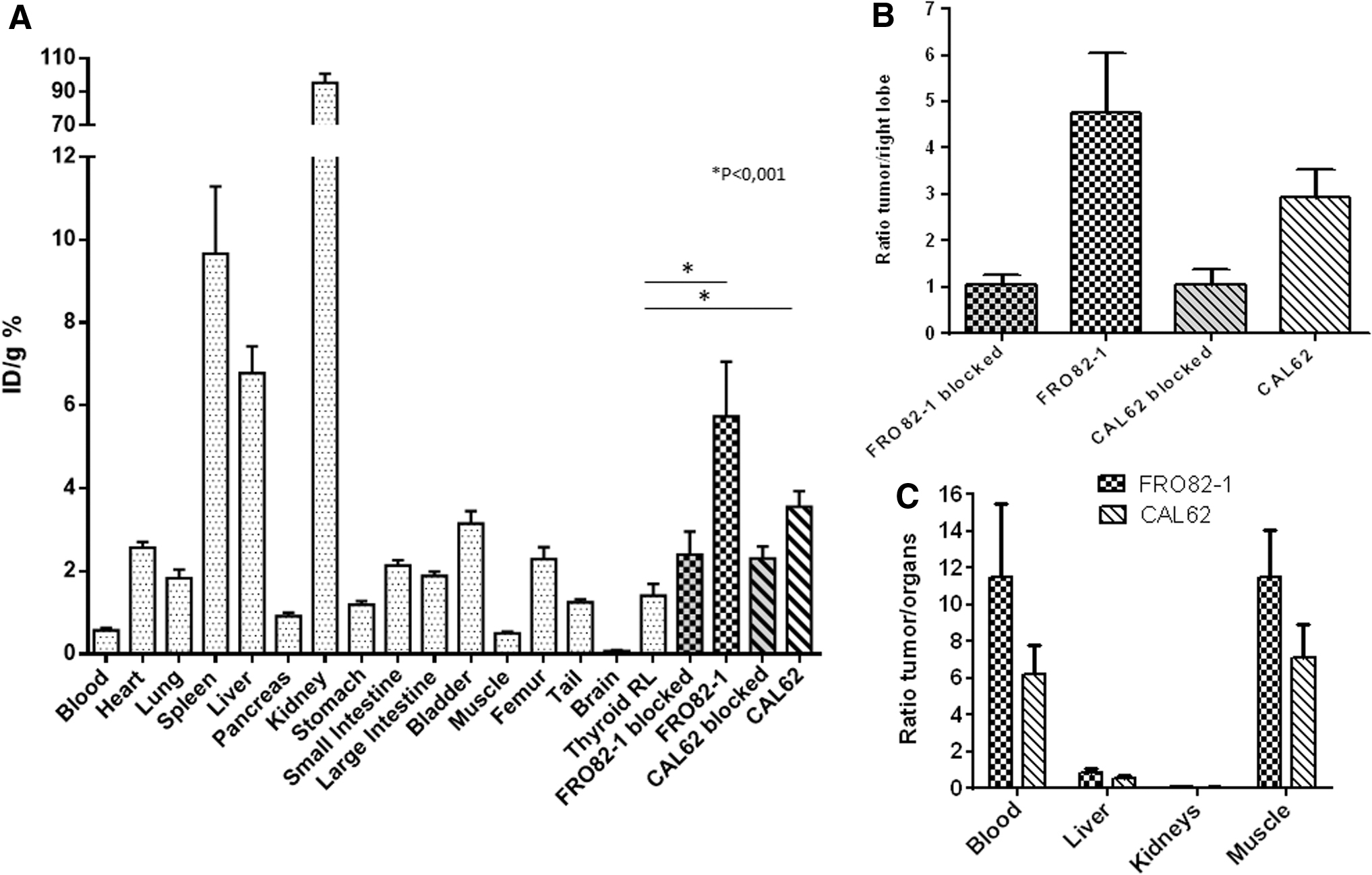
Biodistribution analysis of 89Zr-Dfo-αGal3-Fab-PAS200 in the murine orthotopic tumor xenograft models at 24 hours postinjection. (
In vivo stability assessment of 89Zr-Dfo-αGal3-Fab-PAS200
Analysis of the activity extracted from tumors, blood, and organs revealed a high in vivo stability of this novel αGal3-Fab tracer, indicating negligible amounts of free 89Zr in the radio-TLC (Supplementary Fig. S7A). SDS-PAGE autoradiography under nonreducing conditions allowed the identification of intact 89Zr-Dfo-αGal3-Fab-PAS200 in both tumor and blood. Bands corresponding to molecular weights >100 kDa, which could be attributable to covalent aggregates (possibly mediated by intermolecular disulfide bridges), were found in blood, whereas extracts from liver and kidneys revealed low-molecular-weight bands instead, most likely related to radiotracer metabolites (Supplementary Fig. S7B).
Discussion
The goal of this study was to develop an antibody-derived tracer with favorable affinity and pharmacokinetic characteristics for clinical translation. Using a full-size rat monoclonal anti-Gal3 antibody (M3/38) radiolabeled with 89Zr, we previously achieved a high tumor uptake over five days after injection (11). While demonstrating the potential of Gal3 as a marker for imaging TC, the slow blood clearance, strong liver accumulation, and comparably high radiation doses hampered its potential for clinical translation. Using a F(ab′)2 fragment prepared via proteolytic digestion of the M3/38 mAb, Gal3 targeting was subsequently established as a method for discriminating thyroid tumor nodules from normal thyroid in orthotopic animal models, achieving a high contrast image 48 hours p.i. (12). Now, the development of a recombinant chimeric human/rat Fab with the same specificity for Gal3, which has optimized pharmacokinetics and poses a much lower risk of immunogenicity than a proteolytically prepared animal protein, opens the route toward clinical application.
Gal3 is a member of the β-galactoside binding protein family (27). The 31 kDa glycoprotein has a chimeric structure comprising a C-terminal carbohydrate recognition domain of about 130 residues, which mediates interaction with glycoproteins or -lipids, and a collagen-like, Pro/Gly-rich N-terminal region that is responsible for multimer formation (28). Gal3 is found both intra- and extracellularly, even though it lacks a conventional secretory signal sequence (29). As a multifunctional protein, which is widely expressed by many human cell types, it interacts with a range of intra- as well as extracellular targets and changes their sub- and intercellular localization (30). Due to its selective overexpression in TC, Gal3 represents a potent marker for the visualization and characterization of corresponding tumors, offering a diagnosis independent of the level of NIS expression in TC (31).
To accelerate the translation of targeting Gal3 into clinical application, we here describe the development of an optimized chimeric human/rat Fab, derived from the well-validated hybridoma M3/38 (16), that has high affinity and specificity towards the human Gal3 antigen. Compared with full-size mAbs, Fabs with their smaller size (∼50 vs. 150 kDa) generally show better tissue penetration and faster clearance from plasma, due to efficient kidney filtration and lack of endosomal recycling, which provides crucial advantages for in vivo imaging (13,14). On the other hand, if the clearance from blood is too fast, sufficient accumulation in the tumor may not be reached. As a convenient method to retard rapid renal clearance, we introduced a genetically encoded PAS sequence (13,15,22).
PASylation at the C-terminus of the αGal3-Fab, sterically remote from its paratope, did not hamper antigen affinity, yielding a KD = 1.3 ± 0.1 nM in ELISA and an even lower KD = 0.27 ± 0.1 nM in SPR measurements. Conventional chemical PEGylation of the trastuzumab Fab was reported to negatively affect epitope recognition on the Her2 antigen, which was not observed for the αGal3-Fab (32). Notably, the PASylated chimeric αGal3-Fab exclusively showed immunoreactivity on human tissue sections derived from various malignant thyroid tumors (Fig. 2A–C) if compared with benign thyroid conditions side by side (Fig. 2D–F). This finding is in line with the immunohistochemical staining properties of the parental rat mAb M3/38, which had been extensively tested and characterized in multicenter studies for specific reactivity versus human Gal3 (9,33,34).
The improved antigen affinity (KD of 0.28 ± 0.09 vs. 3.9 ± 0.2 nM for the rat F(ab′)2, both assessed on FRO82-1 cells) and beneficial plasma half-life of our engineered PAS-ylated αGal3-Fab tracer has allowed the visualization of thyroid orthotopic tumors in mice with high sensitivity and at an earlier time point compared with the previously reported Ig reagents (12). The resulting high-sensitivity PET images allowed clear delineation of the tumor in the left thyroid lobe, as confirmed by analysis at necropsy. These characteristics facilitated the ROI analysis of PET images despite the presence of a partial volume effect and the small size of the tumors, leading to a good correlation between in vivo and ex vivo accumulation measurements (R 2 = 0.68; Supplementary Fig. S6).
PET imaging of orthotopic xenograft tumors in mice using the 89Zr-Dfo-αGal3-Fab-PAS200 yielded high target-specific uptake, with signals confined to the left thyroid lobe that contained the tumor tissue and absence of unspecific binding to normal mouse thyroid. Specificity of our innovative probe was further demonstrated by the absence of a tumor signal when the mice were injected with an excess of unlabeled αGal3-Fab-PAS200 and also confirmed by ex vivo accumulation analyses. These findings render 89Zr-Dfo-αGal3-Fab-PAS200 a highly suitable probe for performing PET/CT imaging in patients with thyroid nodules a few hours after tracer injection and, therefore, make it likely to be clinically applicable.
Of note, the proposed diagnostic approach of applying 89Zr-Dfo-αGal3-Fab-PAS200 for the immuno-PET detection of Gal3-positive lesions is not intended for the routine screening of thyroid nodules or for replacing conventional diagnostics workup such as US or FNA cytology (including adjunct molecular tests). Instead, our novel tracer appears suitable to support and improve the accuracy of diagnosis in those thyroid conditions that cannot be easily evaluated with conventional methodology because of: (i) multiple thyroid nodules where clinicians need to decide which nodule to aspirate; (ii) multiple and consecutive Bethesda-1 lesions with “inadequate cytology”; (iii) nodules in mediastinal position that are difficult to access; (iv) suspicious thyroid lesions that are difficult to access due to vascular structures, where FNA biopsy presents a high risk; and (v) micro PTCs (lesions <1 cm, commonly referred to as “occult PTC”), which are currently undetectable preoperatively.
Another condition in which the proposed imaging approach might be useful is recurrence of disease after surgery or the presence of micro-metastases that accumulate radioiodine. In these cases, immuno-PET targeting of Gal3-positive tissue might be beneficial to discriminate neoplastic or hyperproliferative tissue and should help in making a decision about the most appropriate therapeutic intervention (surgery vs. radio-ablation). In fact, recurrences or metastases of DTCs have been reported in patients with high thyroglobulin levels, negative or equivocal 131I whole body scan and/or morphological imaging results (US, magnetic resonance imaging, CT) (35). In particular, after exogenous stimulation by injection of rhTSH, an increased 18F-FDG uptake has been measured (36). However, 18F-FDG is not a specific tracer for TC, because it can be accumulated also by benign lesions due to increased glucose metabolism (37). Since our tracer specifically targets TC cells that overexpress Gal3, independently of their metabolism, stimulation with rhTSH will not be necessary to visualize malignant nodules, metastases or residual tissues. Moreover, a fraction (∼50%) of PDTC and ATC generally retain Gal3 expression but lose the ability to accumulate radioiodine (8,9). In this condition, our novel tracer may be useful eventually to define the extent of the disease. Considering that currently there is no efficient treatment available for PDTC and ATC patients, the highly selective target binding of 89Zr-Dfo-αGal3-Fab-PAS200 opens the potential for new treatments, for example, by replacing 89Zr by a β-emitter such as 177Lu, 90Y or even by a more cytotoxic α-emitter such as 225Ac for radioimmunotherapy. In principle, this theranostic approach could be also applied in the context of DTC that is radioiodine negative.
In conclusion, we have developed a highly specific protein tracer for targeting the human thyroid tumor marker Gal3. Apart from its advantageous functional properties, the simple manufacturing process, yielding a highly defined biopharmaceutical, constitutes an important step toward the development of a specific TC diagnostic agent. Translation of this novel tracer into clinical application should not only help to discriminate between malignant and benign thyroid nodules but also support the monitoring of tumor development, progression, and metastases of other aggressive malignancies where Gal3 plays a pathophysiological role, including melanoma, prostate cancer, and breast cancer (38,39).
Footnotes
Acknowledgments
The authors thank Markus Mittelhäuser (TUM) for animal tracer injection, Giuseppina Pepe (University Sapienza) for support with artwork preparation, and Dr. Martin Schlapschy (TUM) for experimental advice. They would also like to thank Prof. James Nagarajah from the Department of Radiology and Nuclear Medicine of the Radboud University Medical Center for an insightful discussion.
Authors' Contributions
C.D. and A.S. conceived the study. E.P. performed RNA extraction from hybridoma cells, cloning, preparation of plasmid, expression in E. coli, purification and characterization of the αGal3-Fab and αGal3-Fab-PAS200 as well as conjugation with NIRdye and Dfo, and purification and functional characterization of the labeled Fab constructs. F.D.R. performed the orthotopic tumor cell implantation in mice, radiolabeling of Dfo-αGal3-Fab, in vitro experiments, animal PET imaging in vivo, and biodistribution experiments. C.D. and A.S. performed analysis of experimental data. S.R. performed animal PET imaging data acquisition. M.M. performed injection of mice with tracers for FMT and PET imaging. M.S. and W.A.W. provided technical and useful advice for the project. G.S. performed tissue immunostaining and validation of the αGal3-Fab-PAS200 on human tissue sections. A.B. proposed the idea to image thyroid cancer in vivo by targeting galectin-3, performed histological evaluation of normal and neoplastic tissues and provided useful suggestions for the project. E.P., F.D.R., A.S., and C.D. wrote the article. A.S., A.B., C.D., and W.A.W. proofread and revised the article.
Author Disclosure Statement
A.B. has an ownership of a patent related to the use of radiolabeled mAbs to galectin-3 for tumor imaging in vivo (patent no. 1388763, registered on February 20, 2008, Rome, Italy). M.S. reports receiving a commercial research grant from Siemens Medical Solutions and has been acting as consultant for Siemens. A.S. is a shareholder of XL-protein GmbH, the company that commercializes PASylation® technology.
Funding Information
This work was supported by the Deutsche Forschungsgemeinschaft (Grant Nos. DA 1552/2-1 and SK 33/11-1) and the Italian Association for Cancer Research.
Supplementary Material
Supplementary Data
Supplementary Figure S1
Supplementary Figure S2
Supplementary Figure S3
Supplementary Figure S4
Supplementary Figure S5
Supplementary Figure S6
Supplementary Figure S7