
Abstract
Background:
Pediatric papillary thyroid carcinoma (PTC) is a rare malignancy, but with increasing incidence. Pediatric PTCs have distinct clinical and pathological features and even the molecular profile differs from adult PTCs. Somatic point mutations in pediatric PTCs have been previously described and studied, but complex information about fusion genes is lacking. The aim of this study was to identify different fusion genes in a large cohort of pediatric PTCs and to correlate them with clinical and pathological data of patients.
Methods:
The cohort consisted of 93 pediatric PTC patients (6–20 years old). DNA and RNA were extracted from fresh frozen tissue samples, followed by DNA and RNA-targeted next-generation sequencing analyses. Fusion gene-positive samples were verified by real-time polymerase chain reaction.
Results:
A genetic alteration was found in 72/93 (77.4%) pediatric PTC cases. In 52/93 (55.9%) pediatric PTC patients, a fusion gene was detected. Twenty different types of RET, NTRK3, ALK, NTRK1, BRAF, and MET fusions were found, of which five novel, TPR/RET, IKBKG/RET, BBIP1/RET, OPTN/BRAF, and EML4/MET, rearrangements were identified and a CUL1/BRAF rearrangement that has not been previously described in thyroid cancer. Fusion gene-positive PTCs were significantly associated with the mixture of classical and follicular variants of PTC, extrathyroidal extension, higher T classification, lymph node and distant metastases, chronic lymphocytic thyroiditis, and frequent occurrence of psammoma bodies compared with fusion gene-negative PTCs. Fusion-positive patients also received more doses of radioiodine therapy. The most common fusion genes were the RET fusions, followed by NTRK3 fusions. RET fusions were associated with more frequent lymph node and distant metastases and psammoma bodies, and NTRK3 fusions were associated with the follicular variant of PTC.
Conclusions:
Fusion genes were the most common genetic alterations in pediatric PTCs. Fusion gene-positive PTCs were associated with more aggressive disease than fusion gene-negative PTCs.
Introduction
Thyroid cancer is the most common endocrine malignancy in pediatric patients and its incidence is increasing worldwide (1,2). Moreover, thyroid cancer is the second most common cancer among adolescents aged 15–19 years in the United States (1,3). Papillary thyroid carcinomas (PTCs) account for ∼90% of pediatric thyroid carcinomas (4) that differ clinically, pathologically, and genetically from adult PTCs. Pediatric PTCs are usually more advanced at the time of diagnosis with a larger tumor size and higher rate of lymph node metastases and distant metastases occurring mostly in the lungs (5 –7). However, the prognosis of pediatric PTC is excellent (8).
Pediatric PTCs have driver mutations with a different detection rate than in adults. The BRAFV600E mutation is not as common in pediatric PTCs as in adults, and mutations in RAS genes are rare (7,9). RET/PTC rearrangements, the most common being the CCDC6/RET fusion gene (also known as RET/PTC1) in sporadic cohorts and the NCOA4/RET fusion gene (also known as RET/PTC3) in cohorts of radiation-induced PTCs, occur more frequently in pediatric than in adult PTCs (10,11). These two fusion genes have been investigated for more than 20 years in different pediatric cohorts worldwide (10,12 –15) and the association between chromosomal rearrangements and exposure of pediatric patients to ionizing radiation has been reported (10,12). Recently, other types of fusion genes have been found in addition to the RET/PTC rearrangements, but have not been comprehensively studied in a large pediatric cohort. Reported fusions included ETV6/NTRK3 (11), STRN/ALK (16), AKAP9/BRAF (11), AGK/BRAF (17), PAX8/PPARG (18), and others.
In our recent study, we have performed an analysis of genetic alterations in 83 PTCs and 30 benign tumors of pediatric patients. The analysis included HRAS, KRAS, NRAS, BRAF, IDH1, CHEK2, PPM1D, EIF1AX, and EZH1 genes and CCDC6/RET and NCOA4/RET fusion genes (19). Driver mutations were found in 42.2% of PTCs (19). In most PTC samples, a genetic alteration was not detected, thus we decided to extend the analysis to include additional fusion genes.
The aim of the current study was to identify known as well as novel fusion genes in a cohort of 93 pediatric PTC samples. A further goal of the study was to determine any association of the presence of mutations with clinical and pathological features in pediatric patients with PTC.
Materials and Methods
Patients
The cohort consisted of 93 pediatric patients who underwent thyroid surgery from 2003 to 2019 at the Department of Ear, Nose and Throat, 2nd Faculty of Medicine, Charles University and Motol University Hospital in Prague. Informed consent was obtained from all patients or their legal representatives. The study was approved by the Ethics Committee of the Institute of Endocrinology in Prague. Samples were evaluated by an experienced pathologist and classified according to the 7th edition of the AJCC TNM system. Clinical and pathological data were collected from each patient.
DNA and RNA extraction
DNA and RNA were extracted from homogenized, fresh frozen tissues using the AllPrep DNA/RNA/Protein Mini Kit (Qiagen, Venlo, The Netherlands) according to the manufacturer's instructions. DNA and RNA concentrations were measured using a fluorometer (Qubit 2.0; Invitrogen, Carlsbad, CA), purity was evaluated using a spectrophotometer (NanoPhotometer P330; Implen GmbH, München, Germany), and RNA integrity was tested using the Bioanalyzer 2100 and the Agilent RNA 6000 Nano Kit (Agilent Technologies, Santa Clara, CA). All RNA samples used for analyses had an RNA integrity number ≥8.
RNA-targeted sequencing
Detection of known and novel fusion genes was performed by paired-end RNA sequencing using the QIAseq Targeted RNAscan panel (Qiagen, Venlo, The Netherlands) in the first analysis and the FusionPlex Comprehensive Thyroid and Lung panel (ArcherDx, Boulder, CO) in the second analysis.
First, 84 PTC samples were sequenced using the QIAseq Targeted RNAscan panel according to the manufacturer's protocol. Briefly, RNA was reversed transcribed using random primers into double-stranded cDNA whose ends were modified and adenylated, and adapters containing unique molecular tags (UMTs) and sample indexes were ligated. Then, single primer extension was performed, followed by universal polymerase chain reaction (PCR), during which the second sample indexes were added. Purified libraries were quantified using a fluorometer and paired-end sequencing was performed on the HiSeq 2500 sequencer (Illumina, San Diego, CA). Cycles for Read 1 and Read 2 were 231 and 71, respectively. Bioinformatic analysis was performed using the GeneGlobe Data Analysis Center software, version 2.0.2. (Qiagen).
The remaining nine samples that were not sequenced in the first RNA sequencing analysis, five samples with novel or rare fusion gene detected by the first RNA sequencing analysis, and samples that had low complexity, meaning less than 300,000 reads with different UMTs per sample after the first RNA sequencing analysis, were used for the second RNA sequencing analysis using the FusionPlex Comprehensive Thyroid and Lung panel. The libraries were prepared following the manufacturer's instructions similar to the first sequencing run, and paired-end sequencing was performed on the MiSeq sequencer (Illumina). Cycles for Read 1 and Read 2 were 151 and 151, respectively. Bioinformatic analysis was performed using the Archer Analysis software, version 6.0.4 (ArcherDx).
In both analyses, for a gene fusion to be considered as valid, at least five high-quality unique reads had to span the breakpoint and a minimum of three reads had to have a unique start site.
Verification of fusion genes
The presence of fusion genes from RNA sequencing analyses was verified by real-time PCR. RNA (2 μg) was reverse transcribed into cDNA in a final volume of 25 μL using random primers (Promega, Madison, WI) with an annealing temperature of 70°C for 5 minutes, then the AMV reverse transcriptase, RNase inhibitor, and dNTP reagents (Promega) were added and incubated at 25°C for 10 minutes, 37°C for 60 minutes, and 80°C for 5 minutes.
Subsequently, cDNA was diluted five times and amplified with the iQ SYBR Green Supermix (Bio-Rad, Hercules, CA) and gene-specific primers, which were designed using the NCBI Primer BLAST tool (
Real-time PCR was performed as follows using the Light Cycler® 480 system (Roche, Penzberg, Germany): 95°C for 3 minutes, followed by 40 cycles at 95°C for 10 seconds and 60°C for 30 seconds. The melting curve was recorded by holding at 95°C for 10 seconds, cooling to 70°C for 1 minute, and then slowly heating to 95°C (ramp rate of 0.11°C/s) with continuous acquisition of the fluorescence signal. PCRs were performed in duplicate and each experiment included a negative control where RNase-free water was used instead of template cDNA. The evaluation was performed using the Light Cycler® 480 SW 1.5.1. (Roche). Samples positive for CCDC6/RET and NCOA4/RET fusion genes were known from a previous analysis (19).
DNA-targeted sequencing
Detection of point mutations in BRAF (exon 15), HRAS (exons 2 and 3), KRAS (exons 2 and 3), NRAS (exons 2 and 3), and TERT (promoter) genes was performed in all samples as described previously (19). DNA was amplified using gene-specific primers that are available with PCR conditions on request. PCR products were purified using the Agencourt AMPure (Beckman Coulter, Brea, CA), libraries were prepared using the Nextera XT DNA Library Prep Kit (Illumina) according to the manufacturer's protocol and sequenced for 500 cycles by the MiSeq Reagent Kit V2 (Illumina) using the MiSeq sequencer (Illumina). The Integrative Genomics Viewer was used for sequence data visualization.
Statistical analysis
Statistical analysis was performed with Fisher's exact test to compare categorical data and t-test to compare continuous variables. p Value <0.05 was considered as statistically significant.
Results
Study cohort
The cohort consisted of 93 pediatric PTC patients (6–20 years old). The mean age of diagnosis was 14.5 ± 3.4 years and the female-to-male ratio was 2.6:1. A family history of thyroid disorder was present in 30 patients. One patient was diagnosed with Cowden syndrome with germline PTEN mutation. Two patients underwent radiation treatment before the diagnosis of PTC, the first patient was diagnosed with acute lymphoblastic leukemia and the second patient was diagnosed with Hodgkin's lymphoma.
Initial symptoms or signs for detection of PTCs were evaluated in individual patients. In 34 (41.0%) patients, a thyroid nodule was palpated by a family member or by the patient himself. Nineteen (22.9%) patients were followed for endocrine diseases such as goiter, hypothyroidism, or chronic lymphocytic thyroiditis and a thyroid nodule was detected by sonography. In 15 (18.1%) patients, a thyroid nodule was palpated during a pediatrician's examination. In 14 (16.9%) patients, a thyroid nodule was found during examination for another disease, and in only one (1.2%) patient, a thyroid nodule was identified during active screening of a pediatric patient whose father had PTC. In 10 patients, the initial symptoms of PTC were unknown.
Total thyroidectomy (TT) was performed in 82 patients and subtotal thyroidectomy in 11 patients, which was completed to TT in 10 cases. The mean tumor size was 22.1 ± 13.7 mm and 17 patients had microcarcinoma. Among histological variants of PTC, there were 26 (29.9%) cases of the classical variant, 20 (23.0%) cases of a mixture of classical and follicular variants, 29 (33.3%) cases of the follicular variant, 3 (3.4%) cases of the solid variant, 3 (3.4%) cases of a mixture of classical, follicular, and solid variants, 2 (2.3%) cases of the diffuse sclerosing variant, 2 (2.3%) cases of the columnar variant, 1 (1.1%) case of the tall cell variant, and 1 (1.1%) case of the clear cell variant. In six cases, a histological variant was not specified.
Multifocality was observed in 50 (53.8%) cases, extrathyroidal invasion in 38 (40.9%) cases, angioinvasion in 24 (25.8%) cases, lymph node metastases in 58 (62.4%) cases, and distant metastases in 10 (10.8%) cases. Chronic lymphocytic thyroiditis was present in 49 (52.7%) samples, and the frequent occurrence of psammoma bodies was observed in 13 (14%) samples. The specimens with few psammoma bodies (up to 5 bodies per tissue section) were not included.
The median follow-up of the patients was 72 months (range of 2–198 months). Eleven (13.3%) patients had persistent or recurrent disease based on ultrasound or whole-body scintigraphy. Sixteen (19.3%) patients had only biochemical evidence of persistent disease with the basal thyroglobulin blood serum level higher than 1 μg/L or detectable levels of thyroglobulin antibodies. Fifty-six (67.5%) patients had no evidence of disease. One (1.2%) patient, a 7-year-old boy, died due to advanced disease with extensive metastases spread in the lungs before radioiodine (RAI) treatment. Ten patients were not classified due to short-term follow-up (less than one year after surgery). Eighty-four patients received RAI treatment. Eight patients did not receive RAI therapy due to having microcarcinoma (low-risk disease).
Detection of fusion genes
Fusion genes were found in 52 (55.9%) pediatric PTC patients. All fusions included known oncogenes, RET, NTRK1, NTRK3, ALK, BRAF, or MET. Ten different types of RET fusions were identified in 26 patients, four different types of NTRK3 fusions were detected in 14 patients, 1 type of ALK fusion was found in 6 patients, 2 types of NTRK1 fusions were identified in 3 patients, 2 types of BRAF fusions were detected in 2 patients, and 1 type of MET fusion was found in 1 patient (Fig. 1A). A total of 20 types of different fusion genes were identified, of which 11 were interchromosomal and 9 were intrachromosomal rearrangements (Fig. 1B, C). Two coexisting RET fusions were detected in one PTC nodule, the first ACBD5/RET fusion comprised a juxtaposition of exon 11 of the ACBD5 gene and exon 12 of the RET gene, and the second BBIP1/RET fusion was a juxtaposition of exon 1 of the BBIP1 gene and exon 8 of the RET gene.

(
The most common fusion gene was the CCDC6/RET rearrangement found in 13 patients (14%), 1 of whom had an isoform, including also a part of exon 9 of the RET gene (20). Other common rearrangements were ETV6/NTRK3 identified in 10 patients (10.8%), NCOA4/RET and STRN/ALK detected (identical) in 6 patients (6.5%), and RBPMS/NTRK3 found in 2 patients (2.2%). The remaining fusions were not recurrent. Both BRAF fusions with partner genes CUL1 and OPTN were reciprocal. The IRF2BP2/NTRK1 fusion gene had two isoforms, the first isoform was a fusion of exon 1 and the second isoform was of exon 2 of the IRF2BP2 gene with exon 10 of the NTRK1 gene. Each isoform was found in a different patient. No fusion gene-positive patient had a known history of radiation exposure before diagnosis of PTC.
Fusion gene correlations with clinical and pathological data
Clinical and pathological data that correlated with the presence of fusion genes are shown in Figure 2. A more detailed graphical view of age-specific fusion-positive and fusion-negative cases is displayed in Supplementary Figure S1.
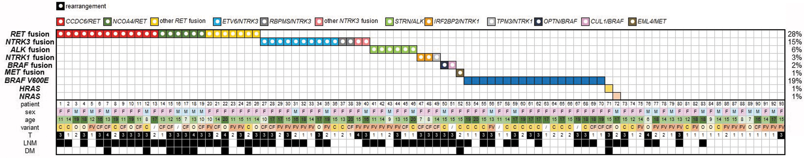
Tile plot of genetic alterations detected in pediatric PTC patients. Clinical and pathological data, including sex, age at diagnosis, histological variant, tumor classification, lymph node metastases, and distant metastases. F, female; M, male; C, classical variant; CF, classical and follicular variants; FV, follicular variant; O, other variant; T, tumor size and extension; LNM, lymph node metastases; DM, distant metastases.
Samples positive for the fusion gene were compared with samples that did not harbor this type of mutation, and the results of the comparison are summarized in Table 1. Fusion-positive samples were significantly associated with the mixture of classical and follicular variants of PTC (p = 0.013), while samples lacking a fusion gene were associated with the classical variant of PTC (p = 0.025). In addition, fusion-positive samples were also significantly associated with extrathyroidal extension (p = 0.012), higher T classification (T3/4) of tumor (p = 0.009), lymph node (p < 0.001) and distant metastases (p = 0.021), chronic lymphocytic thyroiditis (p = 0.001), and frequent occurrence of psammoma bodies (p = 0.004). In addition, patients positive for the fusion gene received more doses of RAI treatment (p = 0.008).
Comparison of Clinical and Pathological Features Between Fusion Gene-Positive and Fusion Gene-Negative Papillary Thyroid Carcinomas and Between RET Fusion and NTRK3 Fusion-Positive Papillary Thyroid Carcinomas
Values highlighted in bold were statistically significant.
Biochemical persistent disease was defined as the presence of a basal thyroglobulin blood serum level higher than 1 μg/L or detectable levels of thyroglobulin antibodies at least one year after surgery. Recurrent/persistent disease was defined as occurrence of a newly identified or persistent structural disease at least one year after surgery.
In three fusion-positive cases, from which two were RET fusion-positive patients, and in three fusion-negative cases, the histological variant was not provided.
In two fusion-positive cases, from which one was an RET fusion-positive patient, and in four fusion-negative cases, RAI therapy records were not provided. One RET fusion-positive patient died before RAI therapy.
Four fusion-positive cases, from which one was an RET fusion-positive patient and one was an NTRK3 fusion-positive patient, and six fusion-negative cases were not classified due to short-term follow-up.
PTC, papillary thyroid carcinoma; RAI, radioiodine; SD, standard deviation.
The samples harboring fusion genes also differed from each other according to the fused oncogene involved. Statistical analysis could only be performed between RET fusion and NTRK3 fusion-positive samples due to the low number of specimens with ALK, NTRK1, BRAF, and MET fusions. The comparison of clinical and pathological data between RET fusion and NTRK3 fusion-positive PTCs is shown in Table 1. RET fusion-positive PTCs were significantly associated with a lower mean age of patients at the time of diagnosis (p = 0.035), lymph node (p = 0.033) and distant metastases (p = 0.020), and frequent occurrence of psammoma bodies (p = 0.006). NTRK3 fusion-positive PTCs were significantly associated with the follicular variant of PTC (p = 0.013). RET fusion-positive tumors were observed in 42.3% (11/26) patients of prepubertal age (up to 12 years) compared with 7.1% (1/14) of NTRK3 fusion-positive patients (p = 0.021) (Fig. 2).
The genetic cause of PTC was detected in all patients with distant metastases (n = 10). In only one case, the point mutation HRASQ61R was found, in the remaining nine cases, a fusion gene was identified (4 × NCOA4/RET, 2 × CCDC6/RET, 1 × RASAL2/RET, 1 × EML4/MET, and 1 × the co-occurrence of ACBD5/RET with BBIP1/RET).
Detection of point mutations
All 93 pediatric PTC samples were also screened for point mutations in BRAF, HRAS, KRAS, NRAS, and TERT genes. In 18 (19%) patients, the BRAFV600E mutation was found, and the HRASQ61R mutation and NRASQ61K mutation were detected identical in one patient (Fig. 1A). No mutation was found in the KRAS gene and in the promoter region of the TERT gene. The HRAS-positive patient underwent radiation treatment for Hodgkin's lymphoma.
The co-occurrence of a fusion gene and somatic point alteration was not identified. Thus, our extended genetic analyses revealed a point mutation or a fusion gene in a total of 72/93 (77.4%) pediatric PTCs. The oncogenic alteration in 21/93 (22.6%) PTCs remained unidentified. Clinical and pathological features of samples are summarized in Supplementary Table S2. Most of these tumors were follicular variants of PTCs, T1/T2 tumor samples. No patient had a recurrent or persistent structural disease, only one patient had a biochemical persistent disease and the other patients remained in remission.
Discussion
An extended investigation of fusion genes in PTC using next-generation sequencing allowed detection of known as well as novel fusion genes (21). Recently, fusion gene studies have been reported in adult patients using comprehensive RNA sequencing panels (22 –24) or studies on a specific fusion gene in adult cohorts have also been performed (25,26). To the best of our knowledge, this is the most comprehensive study of the largest pediatric PTC cohort that has been analyzed using RNA-targeted sequencing.
The total detection rate of fusion genes was 56% and these chromosomal rearrangements were found only in pediatric PTC patients without a known radiation history. The frequency is higher in comparison with other studies of sporadic patients, except the Ukrainian study, in which 64.7% of sporadic patients had the RET/PTC rearrangement (10). Previously reported frequencies ranged from 23.7% to 49% (11,17,27 –29). However, this depends on the number of tested fusion genes or the size of the cohort. Compared with the detection rate of fusion genes in adults, ranging from 7.8% to 15.3% (22 –24), the frequency in pediatric PTC patients is higher. Based on current knowledge, we can only speculate that the more frequent occurrence of fusion genes in pediatric PTC patients may be due to a lower frequency of PTC-associated point mutations whose frequencies increase with aging.
Fusion-positive PTCs in pediatric patients were compared with fusion-negative PTCs and were significantly associated with the mixture of classical and follicular variants of PTC, more aggressive disease (extrathyroidal extension and local and distant metastases), chronic lymphocytic thyroiditis, frequent psammoma bodies, and more doses of RAI treatment. In the literature, this comparison between fusion-positive and fusion-negative PTCs in pediatric patients is lacking. In one study, fusion gene-positive tumors were compared with tumors harboring the BRAFV600E mutation, and those with fusions were larger in size with more frequent diffuse infiltration of the thyroid and lymphatic invasion (29). In another study, tumors harboring the fusion gene or BRAFV600E mutation were grouped together and compared with tumors negative for genetic alterations. Mutation-positive cases had a significantly larger tumor size (17). In the adult PTC cohort, fusion-positive cases were strongly associated with extensive metastases, local invasion, larger tumor size, and higher tumor stage (23).
RET fusions are the most common fusion events in pediatric PTCs. With the detection rate of 28%, this study is consistent with the previously reported frequencies of 24.6–37% (6,17). Ten fusion partner genes with the RET gene were reported. CCDC6/RET and NCOA4/RET rearrangements were recurrent, and SQSTM1/RET, RASAL2/RET, RUFY2/RET, PRKAR1A/RET, and ACBD5/RET fusion genes were not recurrent, but have been previously described in PTC (30 –34). To the best of our knowledge, TPR/RET, BBIP1/RET, and IKBKG/RET rearrangements are novel.
RET fusions were associated with the most aggressive disease (more frequent lymph node and distant metastases), even compared with NTRK3 fusions. The mean age of RET fusion-positive patients was significantly lower than the mean age of NTRK3 fusion-positive patients, and RET fusions were associated with distant metastases compared with NTRK3 fusions. In particular, the NCOA4/RET rearrangement was associated with more aggressive tumor behavior (6), which was also confirmed in our study. RET fusion-positive tumors were significantly associated with frequent psammoma bodies, as previously suggested (29). In another study, psammoma bodies correlated with more aggressive PTC behavior, but not with any particular mutation (35).
NTRK3 fusions were found in 15% of pediatric PTCs, which was comparable with a detection rate of 5.9–22% previously published (14,29,36). Four different fusion partner genes were found and all of these fusions have been previously reported in PTC (22,23,33). The ETV6/NTRK3 fusion gene was the second most common rearrangement in our and another study (36) and was detected in sporadic as well as in radiation-associated PTCs (11).
Compared with RET fusion-positive patients, NTRK3 fusion-positive patients had clinically and pathologically less extensive disease and none of the patients had distant metastases. Furthermore, NTRK3 fusions were significantly more frequent with the follicular variant of PTC, and an association with the histological variant was also observed in other studies (11,26,36,37). The mixture of classical and follicular variants was also found in our study as well as in other studies (22,25). The aggressiveness of NTRK3 fusions is still unclear. NTRK3 fusions have also been published in adults with anaplastic thyroid carcinoma (VIM/NTRK3) and poorly differentiated thyroid carcinoma (ETV6/NTRK3) (16,38).
ALK fusions were detected in 6% of pediatric PTCs that could only be compared with a few studies investigating ALK fusions in pediatric cohorts (16,18,29), of which ALK fusions were detected in only one study with a detection rate of 3/14 (21%) (16). One type of ALK fusion, STRN/ALK, was found in our study. This particular fusion gene has also been described in poorly differentiated and anaplastic thyroid carcinomas (39), and recently, the CCDC6/ALK fusion has been found in medullary thyroid carcinoma in a pediatric patient (40).
NTRK1 fusions are rare. In our study, NTRK1 fusions were found in 3% of cases, TPM3/NTRK1 and two isoforms of IRF2BP2/NTRK1. These fusions have been previously described (22,23,41), and TPM3/NTRK1 has recently been found in RAI-refractory advanced thyroid cancer in an adult woman (33). In other studies, NTRK1 and NTRK3 fusions were frequently evaluated together and not separately as in this study. However, it seems that NTRK1 fusion-positive tumors had different features from NTRK3 fusion-positive tumors. In this study, NTRK1 fusions were detected in three samples, which was an insufficient number to conduct a statistically evaluable comparison. In one study, another type of NTRK1 rearrangement, TFG/NTRK1, was found in an 11-year-old girl with distant metastases in the lungs and a very rapidly growing tumor (42).
Two BRAF fusions were detected in two samples (2%), CUL1/BRAF and OPTN/BRAF. To our knowledge, OPTN/BRAF is a novel fusion and CUL1/BRAF has not been previously described in thyroid cancer, but has been reported in ovarian cancer and melanoma (43,44). For the first time in pediatric PTC, BRAF fusions, AKAP9/BRAF and AGK/BRAF, were identified in radiation-exposed PTCs from Ukraine (11). The worldwide detection rate of AGK/BRAF fusion differs, probably depending on geographic location. The AGK/BRAF fusion was found in 19% of predominantly sporadic PTCs of pediatric patients from Brazil (45) and in 2% of pediatric PTCs from Ukraine (11), while this type of rearrangement was not detected in pediatric patients from the United States or, in our case, from the Czech Republic (16,18,29). Both of the BRAF fusions we found were reciprocal, which was in line with previous reports, where reciprocal BRAF fusions even with other partner genes (AGK and OSBPL9) were detected (11,24).
EML4/MET was the only identified MET fusion; to our knowledge, it is novel and the first detected MET fusion in pediatric PTC. MET fusion (TFG/MET) has been previously described in one adult sample of thyroid cancer (22). A 15-year-old girl positive for EML4/MET fusion had multifocal carcinoma with extrathyroidal extension, angioinvasion, metastases in 18 lymph nodes, and distant metastases to the lung. She received five doses of RAI treatment with a cumulative dose of 700 mCi (25.9 GBq) during a period of four years and now she is in remission.
Fusion genes occur mostly without an additional mutation. RET, NTRK3, ALK, and BRAF fusions were also found in microcarcinomas, suggesting the driver potential of these fusion genes in early stages of PTC development. We detected only one co-occurrence of ACBD5/RET and BBIP1/RET rearrangements in one sample. The co-occurrence of fusion genes with a very unusual high frequency of 11% was reported (17). On the other hand, fusion genes in our study were mutually exclusive with the BRAF or RAS mutation, as suggested by other studies (17,25), except one study describing the co-occurrence of ETV6/NTRK3 and the BRAFV600E mutation (36). In anaplastic thyroid carcinomas, NTRK fusions also co-occurred with TP53 and TERT point mutations (16,38).
Our study has some shortcomings. No functional analyses of novel fusion genes were performed. However, all five novel fusion genes (TPR/RET, IKBKG/RET, BBIP1/RET, OPTN/BRAF, and EML4/MET) contained a known oncogene whose kinase domain was a part of the fusion gene. The TPR, IKBKG, OPTN, and EML4 partner genes encode proteins, which comprise a coiled-coil dimerization domain that was also a part of a particular fusion gene and is required for oncogenic activation. The BBIP1 partner gene encodes a protein of not well-known structure. All partner genes are expressed in the thyroid gland. In other studies, rearrangements containing RET, NTRK3, BRAF, or MET genes were detected and functional analyses were performed. Oncogenic activities of fusion genes were confirmed in all cases (46 –48). Further studies of the fusion gene detection in other pediatric populations are needed to assess correlations between certain fusion genes and clinical–pathological features.
In summary, fusion events were found in 56% of pediatric PTCs, together with point driver mutations in the BRAF and RAS genes, a genetic alteration was detected in 77% of pediatric PTC cases. Fusion gene-positive pediatric PTC cases had more aggressive disease with more frequent extrathyroidal extension and lymph node and distant metastases and consequently received more doses of RAI treatment than patients without fusion genes. Twenty different types of fusion genes were identified, including RET, NTRK3, ALK, NTRK1, BRAF, and MET fusions. Five fusion genes were novel and one fusion gene has not been described in thyroid cancer. Molecular testing for fusion genes could be useful for pediatric patients with PTC for evaluating, prognosis, and identifying targets for targeted treatment in those who fail standard therapy.
Footnotes
Author Disclosure Statement
No competing financial interests exist.
Funding Information
This work was supported by grants from the Ministry of Health of the Czech Republic AZV (16-32665A) and MH CZ—DRO (Institute of Endocrinology—EÚ, 00023761).
Supplementary Material
Supplementary Figure S1
Supplementary Table S1
Supplementary Table S2