
Abstract
Background:
Mutations of thyroid hormone receptor α (TRα1) result in resistance to thyroid hormone (RTHα), exhibiting symptoms of retarded growth, delayed bone maturation, anemia, and severe constipation. Using a mouse model of RTHα (Thra1PV/+ mouse), we aimed at understanding the molecular basis underlying the severe constipation observed in patients.
Methods:
The Thra1PV/+ mouse expresses a strong dominant negative mutant, PV, which has lost T3 binding and transcription activity. Thra1PV/+ mouse faithfully reproduces growth abnormalities and anemia as shown in RTHα patients and therefore is a valid model to examine causes of severe constipation in patients. We used histopathological analysis, confocal fluorescence imaging, transmission electron microscopy (TEM), and gene expression profiles to comprehensively analyze the colonic abnormalities of Thra1PV/+ mouse.
Results:
We found a significant increase in colonic transit time and decrease stool water content in Thra1PV/+ mouse, mimicking constipation as found in patients. Histopathological analysis showed expanded lamina propria filled with interstitium fluid between crypt columns, enlarged muscularis mucosa, and increased content of collagen in expanded submucosa. The TEM analysis revealed shorter muscle fibers with wider gap junctions between muscle cells, fewer caveolae, and hypoplastic interstitial cells of Cajal (ICC) in the rectal smooth muscles of Thra1PV/+ mice. These abnormal histological manifestations suggested defective intercellular transfer of small molecules, electrolytes, and signals for communication among muscles cells, validated by Lucifer Yellow transferring assays. Expression of key smooth muscle contractility regulators, such as calmodulin, myosin light-chain kinase, and phosphorylated myosin light chain, was markedly lower, and c-KIT signaling in ICC was attenuated, resulting in decreased contractility of the rectal smooth muscles of Thra1PV/+ mice. Collectively, these abnormal histopathological alterations and diminished contractility regulators led to the constipation exhibited in patients.
Conclusions:
This is the first demonstration that TRα1 mutants could act to cause abnormal rectum smooth muscle organization, defects in intercellular exchange of small molecules, and decreased expression of contractility regulators to weaken the contractility of rectal smooth muscles. These findings provide new insights into the molecular basis underlying constipation found in RTHα patients.
Introduction
Thyroid hormone receptors (TRs) are critical in mediating the genomic actions of thyroid hormone (T3) in growth, development, and differentiation. Mutations of the THRA gene cause resistance to thyroid hormone (RTHα). 1,2 RTHα patients are characterized by nearly normal thyroid hormone and thyrotropin levels, but they exhibit growth retardation, delayed bone development, anemia, and constipation. 1 –6 The molecular actions of TRα1 mutants in the bone abnormalities, erythroid disorders, and intestine defects have been studied in the Thra1PV/+ mouse, a model of RTHα.
As found in RTHα patients, Thra1PV/+ mice display growth retardation, have abnormal bone development, 7 –9 exhibit anemia, 10,11 and cause epithelial defects in the adult intestine. 12 Collectively, these studies show that the Thra1PV/+ mouse is suitable for further study of other pathological manifestations in patients.
When RTHα patients were first reported, one prominent symptom was severe constipation. Although intestinal defects in Thra1PV/+ mice were reported, 12 it remained unclear how TRα1 mutations altered the activity of the colon, leading to severe constipation. Here, we show that Thra1PV/+ mice exhibit constipation, as evidenced by increased colonic transit time, loss of water content in the stools, and extended colon length. The rectum of Thra1PV/+ mice had abnormal morphological features in the lamina propria, muscularis mucosae, and submucosa.
Transmission electron microscopy (TEM) revealed markedly wider gap-junctions in the smooth muscle cells that impeded intercellular transfer of small molecules. The TEM analysis also showed hypoplastic and reduced number of interstitial cells of Cajal (ICC) to reduce smooth muscle contractility. We further detected decreased expression of key contractility regulators in the rectum of Thra1PV/+ mice. Together, these defects result in impedance of the contractility of smooth muscles to understands how TRα1 mutations act, leading to constipation in RTHα patients.
Materials and Methods
Animals and treatment
All animal studies were performed according to the approved protocols of the National Cancer Institute Animal Care and Use Committee. Generation of Thra1PV/+ mice was previously described. 13 All mice were fed with NIH 31 autoclavable Teklad laboratory animal diets from Envigo (Indianapolis, IN, USA). Further details are described in Supplementary Information.
Transmission electron microscopy
Female wild-type (WT) and Thra1PV/+ mutant mice were used for TEM analysis (mouse age: ∼7.2 months). Mouse colons of ∼1 mm3 were fixed for a minimum of 48 hours at 4°C in 2.5% glutaraldehyde and 1% paraformaldehyde in 0.1 M cacodylate buffer (pH 7.4). The subsequent steps were treated similarly as reported. 14
Isolation of smooth muscle cells from rectum of mice
Rectum was dissected from euthanized female mice WT and mutant mice, aged ∼7.4 months from which smooth muscle cells were isolated as previously described. 15
Lucifer Yellow dye transfer assay
Lucifer Yellow (150 mM) solution prepared as previously described 16 was delivered into a monolayer of smooth muscle cells via the WPI Pneumatic PicoPump (PV 820) on a Leica MZ16. Glass capillaries (TW100F-40) were pulled to inject Lucifer Yellow (∼1.2 nL). Post-injection, confocal fluorescence images were acquired using the Nikon SoRa spinning disk microscope, as described earlier, with the 20 × LambdaS apochromat (N.A. 0.95) water immersion lens, and Tokai Hit Stage Top Incubator to control temperature, humidity, and CO2.
Extended field of view confocal images were collected covering a total area of 9.21 mm2. The tiled images were stitched together into a single large image using the Nikon Elements software (v. 5.3). The stitched images were background subtracted, and the mean fluorescence intensity of Lucifer Yellow positive cells was measured for both mutant and WT cells using the Cellpose 2.0 AI-based image segmentation algorithm with built-in cyto2 model, 17 run through Arivis Vision 4D (v. 3.6) image analysis software (Arivis Imaging, Inc., Boston, MA).
Results
Thra1PV/+ mice exhibit colonic abnormalities mimicking constipation in patients with mutations of TRα1
Figure 1A shows that the body weight of Thra1PV/+ mice was decreased by 43% as compared with WT mice. 13 Evaluation of colonic motility showed that the average colonic transit time for WT mice was 2.58 ± 0.95 minutes (n = 22), and for the Thra1PV/+ mice was 7.97 ± 5.61 minutes (n = 26) (Fig. 1B). The colonic transit time was 3.09-fold longer in Thra1PV/+ mice than in WT mice. Interestingly, the water content was 42% lower in Thra1PV/+ mice than in WT mice (Fig. 1C) and the length of the colon was longer (Fig. 1D-a, with content; D-II-b, content flushed). These observations indicate that Thra1PV/+ mice manifest the constipation phenotype.
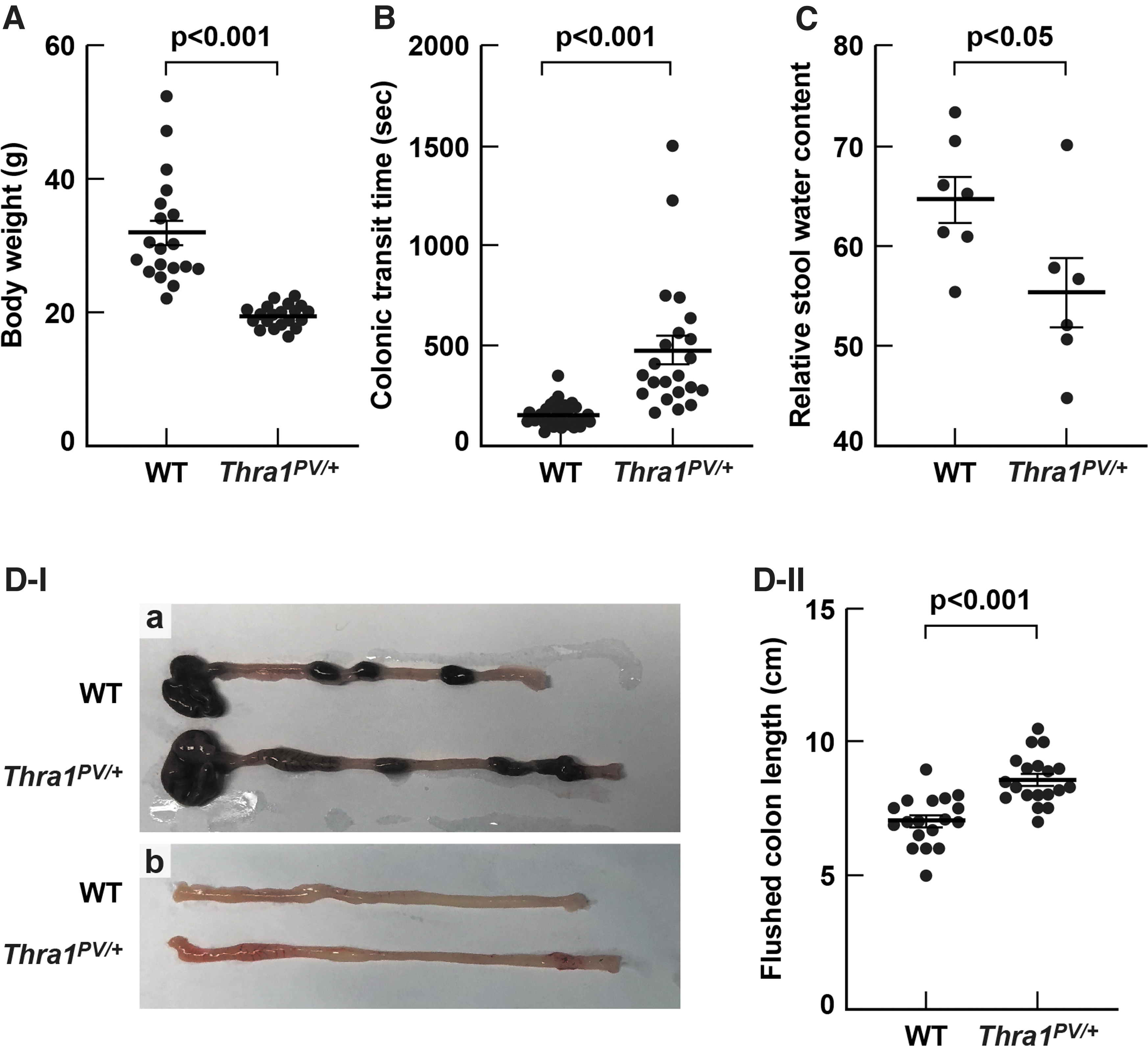
Colonic defects in Thra1PV/+
mice. (
The rectum of Thra1PV/+ mice manifests histopathological defects
Analysis of rectum tissues stained by Periodic acid Schiff (PAS) revealed distinct histological differences between WT and Thra1PV/+ mice. In the WT mice, the colonic crypts were tightly and orderly aligned (Fig. 2A-a). In contrast, in Thra1PV/+ mice, colonic crypts were loosely aligned with enlarged interstitium space around the crypt bases, particularly prominent in the lamina propria (Fig. 2A-b). It is of interest to note that the area of muscularis mucosae was expanded by ∼40% (Fig. 2A-II-b). The submucosa is a fibrous connective tissue layer that surrounds the mucosa and is composed of collagen fibers 18 that can be stained with Masson's Trichrome.
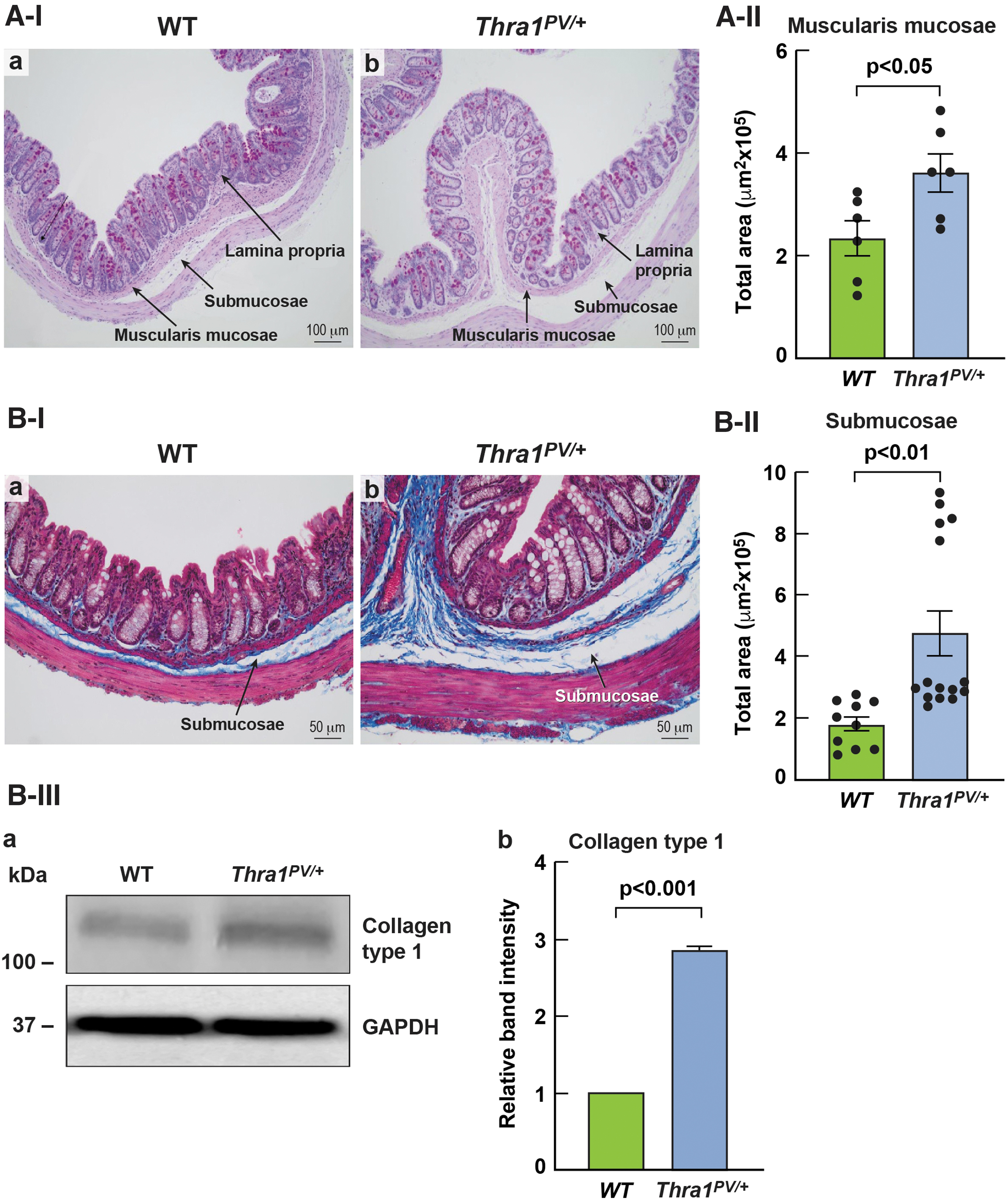
Histopathological abnormalities in the rectum of Thra1PV/+
mice. (
As shown in Figure 2B-I, collagen staining (shown in blue) showed that the submucosa of the rectum in Thra1PV/+ mice (panel b) was more expanded as compared with WT mice (panel b). Measurement of area showed that the submucosae was 1.5-fold larger in Thra1PV/+ mice than in WT mice (Fig. 2B-II). Analysis of the collagen content in the rectum showed that the collagen levels were increased three-fold (Fig. 2B-III-a, -b), supporting that the connective tissues were abnormally expanded, which could derail the transport of fluid and electrolytes in and out of adjacent smooth muscle cells of Thra1PV/+ mice.
One structural element in the smooth muscle that is critical in the intercellular communication is the gap junction. Gap junctions are channels that directly connect the cytoplasm of adjacent cells, allowing the flow of small molecules, ions, and electrical signals to pass through a regulated gate between cells. 19 Gap junctions can be unequivocally identified by TEM. Figure 3 shows the representative TEM images of rectum smooth muscle cells of WT mice and Thra1PV/+ mice at the age of 2.6 months (panels a–d) and 7.2 months (panels e–h).
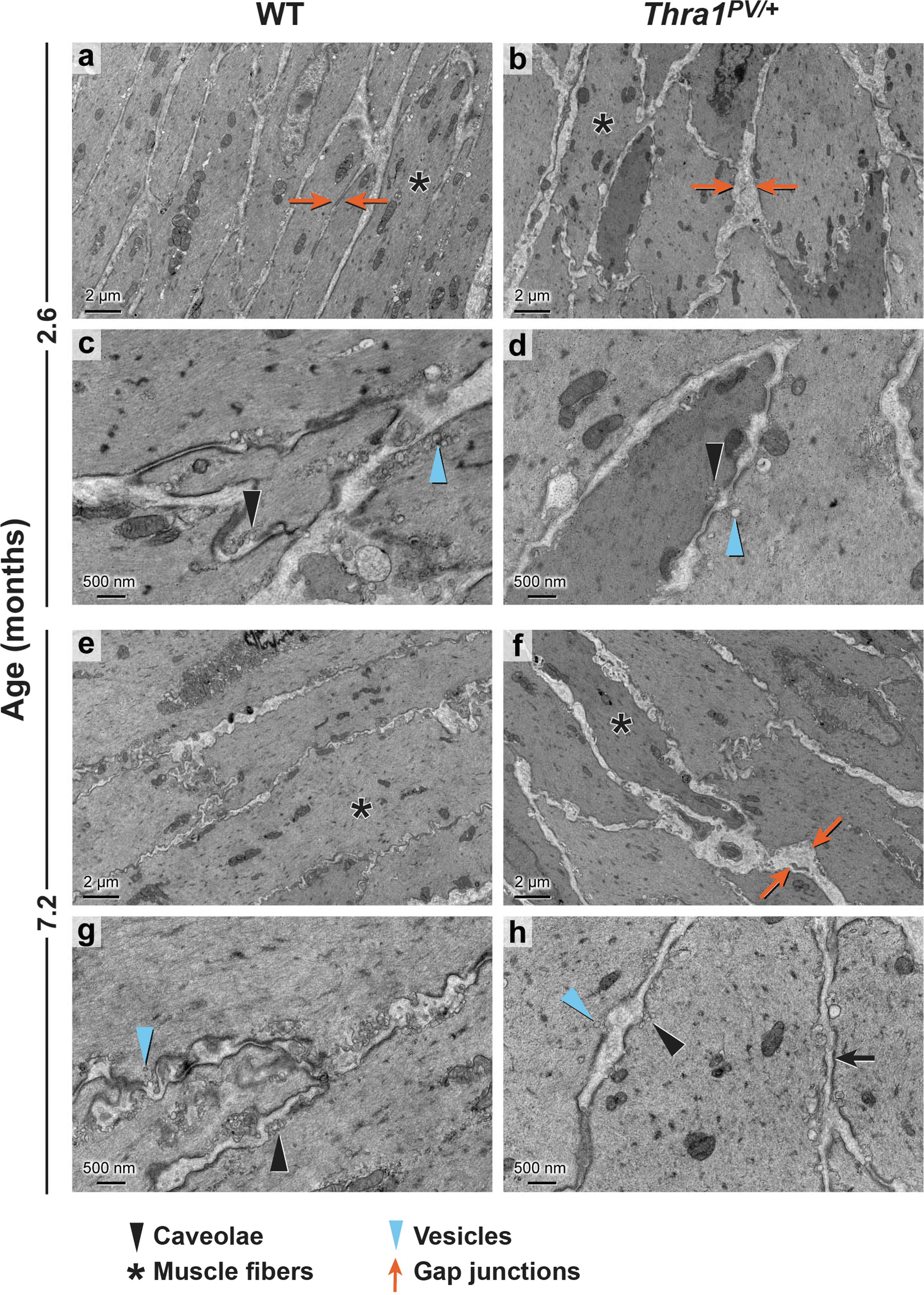
Transmission electron microscopy revealed defects in the smooth muscles of Thra1PV/+
mice. Representative micrographs of WT (n = 4) and Thra1PV/+
mice (n = 4) at the age of 2.6 months (panels
No apparent abnormalities were discernable for nucleus, mitochondria, Golgi apparatus, and ribosomes in the muscle cells of Thra1PV/+ mice. However, at the age of 2.6 months, compared with WT mice, Thra1PV/+ mice exhibited abnormal fibers, frequently twisted (marked by an asterisk* in Fig. 3, at lower magnification of 2 μm, panel b vs. panel a), with wider gaps between muscle cells (red arrows; panel b). As shown in Figure 3, at higher magnification of 500 nm, abundant caveolae (marked by solid arrowhead, panel c) and vesicles (marked by blue arrowheads) were visible in WT mice. In contrast, at the same magnification, fewer caveolae and vesicles were observed in Thra1PV/+ mice (panel d).
Similar shorter fibers with wider gaps (Fig. 3, panel f vs. panel e) and reduced numbers of caveolae, and vesicles were found in the smooth muscle of Thra1PV/+ mice at the older age of 7.2 months (Fig. 3, panel h vs. g). These results indicate that the defects occur at the early age of 2.6 months, persisting into older age.
Gap junction functions are impaired in the rectum smooth muscle of Thra1PV/+ mice
The abnormal structure of gap junctions and other membrane-associated sub-cellular organelles revealed by TEM prompted us to investigate the functional consequences of these changes in Thra1PV/+ mice. To do so, we isolated smooth muscle cells from rectums of WT and Thra1PV/+ mice to assess the ability of intercellular transfer of small molecules. Using confocal imaging, we showed that the smooth muscle cells isolated from rectum of WT and Thra1PV/+ mice exhibited characteristic smooth muscle actin fibers evidenced by immuno-stained actin fibers using anti-α-smooth muscle actin antibodies, establishing the identity of smooth muscle cells (Supplementary Fig. S1A-a, -b).
We further demonstrated no changes in the protein levels of α-smooth muscle actin in the smooth muscle cells isolated from WT and Thra1PV/+ mice (Supplementary Fig. S1B-a, -b). We demonstrated the existence of gap junctions in the isolated rectum smooth muscle cells by a gap junction marker, connexin 43 using super-resolution imaging. Figure 4A-a shows the confocal green fluorescence image for the localization of connexin 43 on the cell membrane of rectal small muscle cells isolated from WT mice.
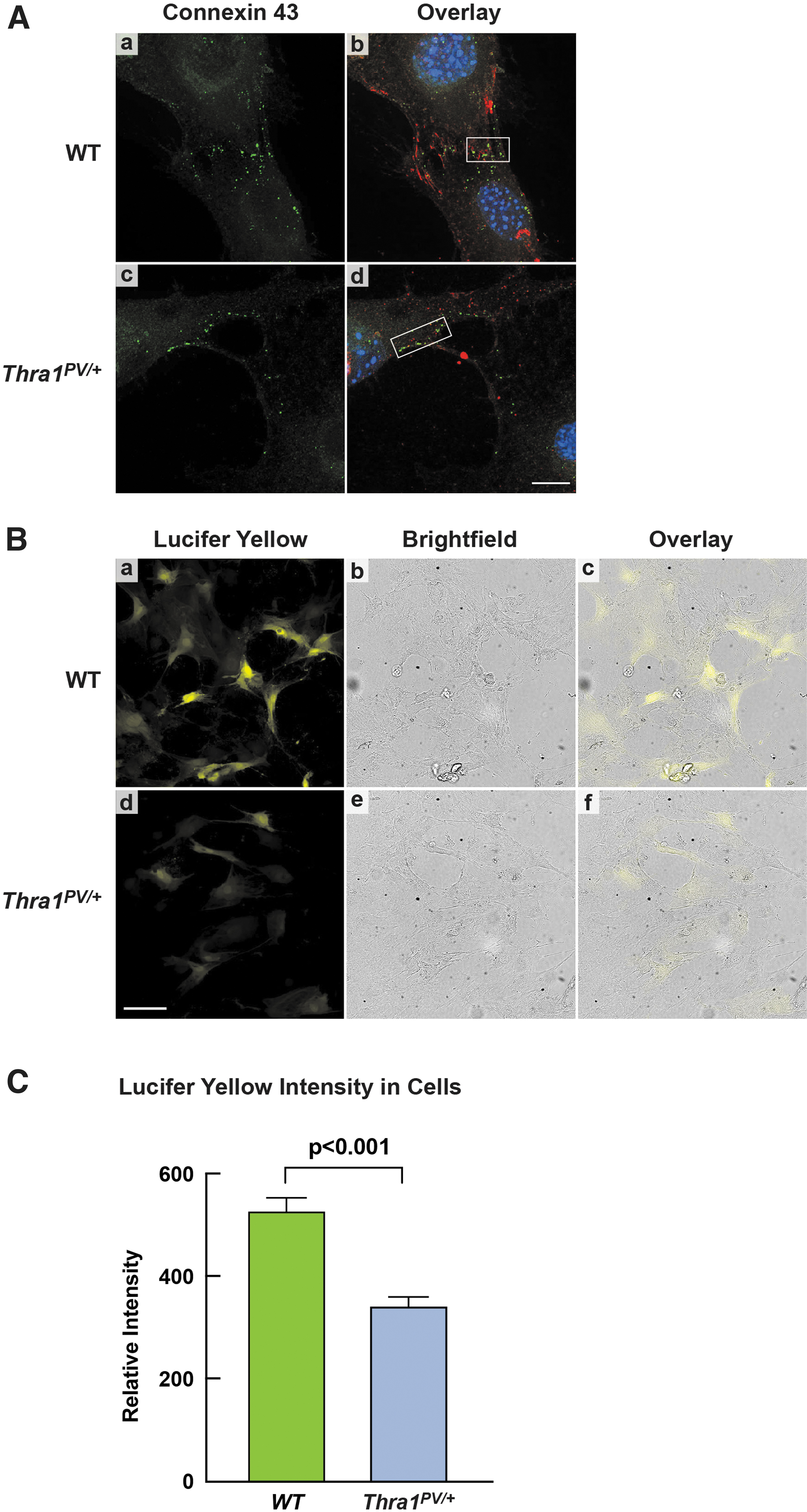
Confocal images of gap junctions and defective intercellular transfer of Lucifer Yellow in the isolated rectal smooth muscle cells of Thra1PV/+
mice. (
The cell boundaries were marked by the MemBrite plasma membrane stained red. Panel b shows the overlay of green fluorescence signals with MemBrite red signals to indicate that connexin 43 was localized on the membrane junctions of the neighboring cells (marked by a white box). We also detected the localization of connexin 43 on the cell membrane of rectum smooth muscle cells of Thra1PV/+ mice (Fig. 4A-c, green fluorescence signals), which were more clearly visualized in the overlaying of green fluorescence signals with the cell membrane marker, red MemBrite (marked by a box in panel d). These findings indicate that the smooth muscle cells isolated from rectums of WT and Thra1PV/+ mice could form gap junctions in vitro.
We next assessed whether the gap junction defects revealed by TEM could affect the intercellular communication by using a fluorescence tracer, Lucifer Yellow. 20,21 Lucifer Yellow has been extensively used to study cell structure and intercellular communication. 22,23 We microinjected identical amounts of Lucifer Yellow into similar numbers of smooth muscle cells isolated from WT and Thra1PV/+ mice. We captured the fluorescence images at an identical time (10 minutes) post micro-injection to allow sufficient time for the tracer dye to transfer into neighboring cells.
As shown in Figure 4B, fewer yellow fluorescent smooth muscle cells were detected in Thra1PV/+ mice than WT mice (panel d vs. panel a). Moreover, the fluorescence intensity in the cells from Thra1PV/+ mice was clearly weaker than that in the WT mice (panels d vs. a). Figure 4B-b and -e show the corresponding bright field to demonstrate that similar number of cells were present in the field imaged by confocal fluorescence microscopy. Panels c and f show the overlay of fluorescence images with the bright field imaging, demonstrating that Lucifer Yellow fluorescence is in the cells that are in contact with the neighboring cells.
To provide quantitative assessment, we measured both the mean intensity of Lucifer Yellow fluorescence per cell and the number of Lucifer Yellow positive cells in an extended field of view area ∼9 mm2. Cells with Lucifer Yellow fluorescence intensity above the background were considered positive. The quantitative data indicated that the mean intensity of Lucifer Yellow fluorescence in the mutant cells was 35% lower than in the WT cells (Fig. 4C). Taken together, these findings indicate that the intercellular communication in the smooth muscles of Thra1PV/+ mice is impaired due to defective gap junctions.
Suppression of smooth muscle contractile regulators in the rectum of Thra1PV/+ mice
The abnormality in the gap junctions and the expanded muscularis mucosa and submucosa suggested that the contractility of smooth muscle in the rectum could be impaired. Figure 5A shows the major regulatory circuit in the control of smooth muscle contractility. Binding of Ca+2 to calmodulin results in conformational change in the calmodulin to interact with myosin light-chain kinase (MLCK), leading to the phosphorylation of myosin light chain (MLC), allowing myosin II to interact with actin to initiate contraction of smooth muscles. The myosin light chain phosphatases (MLCP) acts to dephosphorylate MLC to relax the contractility. Rho kinase (ROCKs) serve as inhibitors to suppress the activity of MLCP for additional regulation of the regulatory circuit. 24,25

Suppression of smooth muscle contractile regulators in the rectum of Thra1PV/+
mice. (
Analysis of the protein levels showed that calmodulin abundance was lower in the rectum of Thra1PV/+ mice (Fig. 5B-I-a and B-II-a). Consistent with lower protein abundance of MLCK (Fig. 5B-I-c and B-II-b), the ratios of p-MLC/total MLC were markedly lower (Fig. 5B-I-d and e, and B-II-c), without changing the protein levels of myosin heavy chain (MYH 11; B-I-g) and α-SMA actin (B-I-i and B-II-e). The protein levels of myosin light chain phosphatase (MYPT1) were lower (B-I-l and B-II-f).
The decreased protein levels of ROCK1 (Rho kinase; B-I-l and B-II-g) were lower, and they allowed MYPT1 to dephosphorylate MLC to further hinder the contractility of the rectum in Thra1PV/+ mice. Taken together, these changes reduced the contractility of smooth muscle cells, which could lead to slow transit time and constipation. Similar regulatory patterns in the isolated smooth muscle cells were observed as in the rectum tissues (Supplementary Fig. S2), indicating that isolated smooth muscle cells retained similar functional characteristics as in the rectum tissues, and they therefore can be used for functional analysis.
Impaired ICC in the rectum of Thra1PV/+ mice
To further explore other mechanisms that could contribute to the constipation observed in patients with mutation of the THRA gene, we evaluated potential defects in rectum ICC of Thra1PV/+ mice. Extensive studies have shown that ICC function as pacemakers to provide a means to coordinate proper propagation of contractions and motility movement. The ultrastructure by TEM (2500 × magnification) shows that ICC in the rectal smooth muscles of Thra1PV/+ mice exhibited marked reduced (hypoplastic) cytoplasm (Fig. 6; panel b, marked by blue arrowheads) as compared with WT smooth muscles (panel a).

Transmission electron microscopy detects hypoplastic ICC in the rectum smooth muscle cells of Thra1PV/+
mice. Representative micrographs of WT (n = 4) and Thra1PV/+
mice (n = 4) at 7.2 months. The magnification of images in panels are marked. The ICC in the rectal smooth muscle cells of Thra1PV/+
mice have significantly reduced (hypoplastic) cytoplasm (panels
All clearly identifiable organelles such as mitochondria appeared normal in size, shape, and structures; however, the cytoplasm appeared to be more condensed in ICC of Thra1PV/+ mice (panels b vs. a, marked by blue arrowheads). The more compacted disorganized mitochondria in ICC of Thra1PV/+ mice could be viewed more clearly at a higher magnification (3000 × ) shown in Figure 6d (marked by yellow arrowheads). The hypoplastic ICC in the rectum smooth muscles of Thra1PV/+ mice suggested the impaired maturation of ICC.
We next ascertained whether the numbers of ICC could be affected in the rectum smooth muscles cells of Thra1PV/+ mice. The c-KIT receptor tyrosine kinase (c-KIT) is known to regulate the development of ICC 26 and has been used as a marker for ICC in the smooth muscle cells. 27 Identification of ICCs was based on c-KIT immunohistochemical staining, localization, and morphologic characteristics of cells by a board-certified veterinary pathologist. The number of c-KIT positive cells was quantified per millimeter within the submucosal region (black arrows, Fig. 7A-I) and in the myenteric plexus (blue arrows, Fig. 7A-I).

Reduced ICC in the rectum submucosa and myenteric plexus of Thra1PV/+
mice. (
Quantitative analysis shows a 70% and 45% reduction of ICC in the submucosa and rectum myentyeric plexus, respectively (Fig. 7A-II, upper panel and lower panel, respectively). The hypoplasia with a reduction in numbers of ICC could lead to weakened motility and slow colonic transit in Thra1PV/+ mice.
The hypoplastic ICC prompted us to ascertain whether the c-KIT signaling is affected in rectum of Thra1PV/+ mice. The c-KIT, a receptor tyrosine kinase and its cognate ligand SCF (stem cell factor), are abundantly expressed in the smooth muscle cells of gastrointestinal tract. 28 The interaction of SCF with c-KIT results in downstream signal transduction to affect proliferation, differentiation, and function of ICC. 27,28 Indeed, we found that protein levels of c-KIT (Fig. 7B-I-a and quantitative data in Fig. 7B-II-a) and SCF (Fig. 7B-I-c and quantitative data in Fig. 7B-II-b) were reduced.
One of the downstream effectors of c-KIT signaling is c-Src, a non-receptor-type tyrosine kinase, which is critical in cell growth, differentiation, migration, and survival. We found that the ratios of p-c-Src (Fig. 7B-I, panel e) as well as the ratios of p-c-Src/total c-Src (Fig, 7B-II-c) were reduced. The findings indicate that TRα1 mutants suppressed the expression of c-KIT and its ligand to attenuate c-KIT signaling via c-Src as one of the downstream pathways to impede the development of ICC.
Discussion
The discovery of patients with mutations of the THRA gene has marked a milestone in understanding the in vivo actions of TRα1 mutants. Significant progress has been made in elucidating how TRα1 mutants cause pathological manifestations of RTHα in the bone and erythropoiesis by using the Thra1PV/+ mouse, a model RTHα.7−11 In the present studies, we provided evidence to show how mutations of TRα1 impacted the morphology and the colonic functions. The colonic transit time was markedly prolonged and accompanied by decreased water content in the feces in Thra1PV/+ mice.
We found enlarged space between the columns of crypts, especially at the junctions with muscularis mucosae. The connective tissue in the submucosa appeared to be loose and expanded with an increased amount of collagen (Fig. 2B-III a and b). The TEM revealed enlarged space in the gap junctions of smooth muscle cells with decreased numbers of caveolae and vesicles, which could adversely affect effective transport of intercellular fluid, small signaling molecules, and electrolytes. Indeed, functional assays by confocal imaging of the intercellular movement of a fluorescence dye (Lucifer Yellow) clearly showed that the intercellular transfer activity of smooth muscle cells was impaired in the rectum muscle cells of Thra1PV/+ mice.
We further discovered that ICC were hypoplasia with reduction in numbers in the rectum smooth muscles of Thra1PV/+ mice and that the expression of key contractile regulators was significantly decreased. Collectively, these morphological defects in the gap junctions, submucosae, muscularis mucosae and ICC, contribute to weakening the colonic activity. These findings have provided new insights into the molecular basis underlying the impeded colonic activity, leading to constipation in patients with mutations of the THRA gene.
The identification of abnormalities in ICC (hypoplastic as well as reduced number) in the rectum smooth muscle cells of Thra1PV/+ mice shed new light on the understanding of the weakened colonic motility in RTHα patients. The ICC, electrically coupled to smooth muscle cells, is responsible to initiate intrinsic pacemaker activity. 25 Gastrointestinal smooth muscle tissues such as the colon possess spontaneous intrinsic pacemaker activity that organizes into phasic contractions, which is the basis for peristaltic activity.
The defective ICC found in the rectum of Thra1PV/+ mice would be less effective to initiate the rhythmicity and contractions of rectum. Together with the wider gap junctions between the smooth muscle cells and the decreased abundance of key smooth muscle contractile regulators, the coordinated movement is further impeded in the rectum of Thra1PV/+ mice. However, although the intrinsic neural plexuses allow a significant degree of autonomy in the gastrointestinal tract functions, it is also known that extrinsic neural inputs from the central nervous system could modulate to further fine tune its activity.
At present, it is unknown how mutations of the THRA gene could impact the basic neural circuitry of the parasympathetic and sympathetic neural inputs to the gastrointestinal tract. The elucidation of the actions of mutant TRα1 on the neural circuitry would await future studies.
It has long been known that constipation or obstipation frequently occurs in hypothyroid patients. 29 However, very few studies have examined how hypothyroidism could cause the constipation in patients. Subsequently, with the creation of mutant mice deficient in the Thrb gene (Thrb−/− mice), the Thra gene (Thra−/− mice), or both the Thrb and Thra genes (Thrb−/−Thra−/− mice), it is possible to assess the role of each TR isoform in the biology of the intestine and colon.
Studies indicated that Thrα−/− mice showed reduced length of the intestine with a greater fragility compared with WT mice. This was also observed in the Thrb−/−Thra−/− mice. On the contrary, no differences between WT and Thrb−/− mice were found, 30,31 indicating the critical role of TRα1 in the development and functions of the intestine. Histological analysis of the proximal and distal parts of the small intestine showed decreased intestinal size and the length of the crypt-villus unit in the Thrα−/− mice compared with WT mice, but no apparent abnormalities were detected in the colons of Thrα−/− mice.
These observations are in contrast with the longer intestine and colon observed in mice expressing dominant negative TRα1 mutant (Thra1PV/+ mice 12 ; and the present studies). Moreover, in the present studies, extensive morphological defects in the lamina propria, muscularis mucosae, submucosae, gap junctions, and ICC, as well as functional colonic deficiencies were detected in Thra1PV/+ mice. These findings indicate that the loss of normal TRα1 function, due to gene knockout and expression of a dominant negative TRα1 mutant, clearly led to distinct functional consequences in the pathology of the intestine and the colon.
The constipation due to hypothyroidism is mediated by insufficient thyroid hormone levels, but with normal functional TRs. The constipation in RTHα is due to the resistance to thyroid hormone in the presence of normal thyroid hormones because of mutated TRα1. These distinct molecular actions in vivo would suggest that the treatment for constipation due to hypothyroidism or RTHα would require the consideration of different therapeutic approaches.
Footnotes
Acknowledgments
The authors thank Christopher King for assistance in the preparation of TEM micrographs. They are grateful to Ross Lake for scanning PAS-stained rectum slides for measurement of areas of muscularis mucosae and submucosae. They thank Mark Willingham for assistance in the insightful interpretation of the histological analysis. They also thank Joelle Mornini, NIH Library, for article editing assistance.
Authors' Contributions
Conception and designs were performed by S.-Y.C. and M.K.; development of methodology and acquisition of data were carried out by M.K., V.H., P.Z., K.B., E.E., W.K.L.D., and S.-Y.C. Analysis and interpretation of data were performed by M.K., S.-Y.C., V.H., P.Z., K.B., E.E., and Y.-J.Z.; writing, review, and/or revision were performed by S.-Y.C. and M.K. Administrative, technical, or material support was organized by S.-Y.C.
Author Disclosure Statement
No competing financial interests exist.
Funding Information
This research is supported by the Intramural Research Program of the Center for Cancer Research, National Cancer Institute, National Institutes of Health (ZIA BC 008752).
Supplementary Material
Supplementary Data
Supplementary Figure S1
Supplementary Figure S2