
Abstract
Background:
Drug resistance is a major obstacle in the treatment of anaplastic thyroid carcinoma (ATC) with the combination of docetaxel (DTX) and doxorubicin (DOX). Excessive intracellular drug efflux is considered a key factor contributing to drug resistance. Therefore, developing drug-loaded microspheres to enhance cellular uptake and inhibit efflux of docetaxel and doxorubicin may be an effective strategy to overcome chemoresistance.
Methods:
Alginate-Doxorubicin@carboxymethyl chitosan-Docetaxel (A-DOX@C-DTX) microspheres were prepared using an electrostatic spraying-assisted microfluidic device. The microspheres were characterized, and the in vitro drug release profiles and extracellular efflux rates were determined. The effects of A-DOX@C-DTX microspheres on drug-resistant ATC cells were evaluated in vitro. In vivo, a subcutaneous tumor model was established in mice to investigate the effects of A-DOX@C-DTX microspheres on tumor growth, as well as the accumulation and release of the materials at the tumor site.
Results:
We successfully prepared aliginate/carboxymethyl chitosan (ALG/CMC) microspheres with a core-shell structure. These microspheres exhibited controllable size, good monodispersity, excellent biocompatibility, high encapsulation efficiency for doxorubicin and docetaxel, and high drug loading capacity. Moreover, the microspheres promoted cellular uptake of doxorubicin and docetaxel while inhibiting their efflux. A-DOX@C-DTX microspheres showed significant inhibition of viability, proliferation, migration, and invasion of drug-resistant ATC cells in vitro. In vivo, A-DOX@C-DTX microspheres suppressed tumor growth, induced tumor tissue necrosis, and promoted tumor cell apoptosis. In addition, the microspheres facilitated the enrichment of active substances in the tumor and their sustained release.
Conclusion:
A-DOX@C-DTX microspheres exhibited superior antitumor effects compared to free drug treatment both in vitro and in vivo, particularly against drug-resistant ATC cells. This improved therapeutic efficacy may be attributed to the enhanced drug uptake and reduced drug efflux in drug-resistant ATC cells facilitated by ALG/CMC microspheres.

Introduction
Anaplastic thyroid carcinoma (ATC) is a rare form of thyroid cancer, accounting for only 1–1.5% of all thyroid cancers. However, owing to its aggressive nature, the mortality rate is nearly 100%, contributing to over 50% of all deaths from thyroid cancer. 1 –3 Chemotherapy has been considered a common and effective treatment modality, and according to American Thyroid Association (ATA) guidelines, the combination of docetaxel (DTX) and doxorubicin (DOX) is a commonly used chemotherapy regimen for ATC. 4,5 However, this treatment often leads to drug resistance, resulting in a worse prognosis. 3,6,7 In addition, ATC cells possess certain stem cell-like characteristics, which may further promote the development of resistance and tumor recurrence. 8 –10 It is currently believed that a key reason for the development of drug resistance is the rapid excessive efflux of drugs from cells, rendering intracellular drug concentrations ineffective. 11 –13 Therefore, achieving controlled release of drugs and increasing drug concentrations at the tumor site may be an effective solution to reduce drug resistance and improve the therapeutic efficacy of ATC. 6,14 –16
In recent years, polymer nanoparticles have been considered as one of the most promising controlled release carriers. 17 –19 Carboxymethyl chitosan (CMC) is an anionic polymer that exhibits high viscosity, low toxicity, excellent biocompatibility, and bio/mucoadhesive properties, making it a potential biomaterial for various biomedical applications. 20,21 However, the rapid degradation rate of CMC limits its use as a drug delivery carrier. 22 Some studies have attempted to develop more optimal drug delivery carriers by combining CMC with other polymers. 22,23 Alginate (ALG) is a natural anionic polymer that possesses gel-forming properties, mucoadhesive properties, low toxicity, biocompatibility, cell affinity, and high capacity for releasing various active substances and has been used in peptide or protein delivery, wound dressings, and cell encapsulation, among others. 23 –30 Furthermore, ALG microparticles are highly bioavailable entities that have small volumes but large surface areas, protecting the core from external influences. 31 However, ALG exhibits slow degradation kinetics. 32 Therefore, we developed ALG-CMC with a core-shell structure to overcome the limitations of ALG and CMC as individual components, aiming to achieve controlled release of drugs while inhibiting drug efflux, thus reversing chemotherapy resistance in anaplastic thyroid carcinoma.
Based on the advantages and specific functionalities of the aforementioned molecules, we designed and synthesized novel microspheres based on CMC, with DTX encapsulated in the core and DOX loaded on the ALG shell, forming core-shell microspheres with CMC-DTX as the core and ALG-DOX as the shell, referred to as Alginate-Doxorubicin@carboxymethyl chitosan-Docetaxel (A-DOX@C-DTX) microspheres. Subsequently, we investigated its effects on anaplastic thyroid carcinoma in both in vitro and in vivo settings.
Methods
Preparation of ALG/CMC core-shell microspheres
The electrospinning apparatus (JDF05, Changsha Nano Apparatus Co., Ltd) was used for microsphere preparation. First, connect the coaxial needle, polytetrafluoroethylene tubing (18S), dispensing needle (18G), and threaded syringe. Then, connect two 10 mL disposable sterile syringes to injection pumps 1 and 2, respectively. Add 5 mL of 1.5% ALG solution to the outer phase syringe and 5 mL of 2% CMC solution to the inner phase syringe. The flow rates of the two syringes are controlled by injection pumps 1 and 2. Once the air is removed from the syringes and tubing by pumps 1/2, this process is paused. Set the voltage to 8–10 kV, inner phase flow rate (Finner) to 0.8 mL/h, outer phase flow rate (Fouter) to 3.6 mL/h, and the collection distance to 3 cm. Then, activate the device to start electrospinning and form microspheres. After observing the liquid starting to eject, collect a small amount of the mixture in a beaker. Once the spraying becomes stable, collect the electrospun microspheres using a Petri dish containing a 2% calcium chloride solution to obtain the final ALG/CMC core-shell microspheres. Wash the collected microspheres thrice with distilled water.
Characterization of ALG/CMC microspheres
The bright-field images of the microcapsules were captured using a DMi8 fluorescence microscope (Leica). The microsphere morphology was characterized using a scanning electron microscope (SEM) (Hitachi) model S-3000N. DOX with red fluorescence was mixed into the ALG solution, and bovine serum albumin-fluorescein isothiocyanate (BSA-FITC) with green fluorescence (SF063, Solarbio) was mixed into the CMC solution. The fluorescence images of the ALG/CMC microspheres were captured using a DMi8 fluorescence microscope. Observing the morphology of microspheres under a microscope, taking photographs, and measuring their diameters, the cytotoxicity of the microspheres was evaluated using a cell counting kit-8 (CCK-8) assay.
Loading DTX and DOX into ALG/CMC microspheres
A measure of 7.5 mg of DOX was mixed with 5 mL of a 1.5% ALG solution, and 10 mg of DTX was mixed with a 2% CMC solution. DOX and DTX were loaded into ALG and CMC, respectively. The preparation of A-DOX@C-DTX microspheres followed the same steps as described in “Characteristics of A-DOX@C-DTX Microspheres.”
The encapsulation efficiency and drug loading of A-DOX@C-DTX microspheres were determined using high-performance liquid chromatography (HPLC). The detailed methodology can be found in the Supplementary Data S1.
In vitro drug release
The release of DOX and DTX was determined using HPLC. The detailed methodology can be found in the Supplementary Data S1
In vitro cytotoxicity assays
Cell culture
Human ATC cells, BHT101 (undifferentiated), were obtained from iCell Bioscience Inc. The cells were cultured at 37°C under 5% CO2 using BHT101 cell-specific culture medium (iCell-h432-001b, iCell Bioscience Inc). Initially, BHT101 cells were seeded in culture medium containing 50 μM of DOX and DTX and incubated for 24 hours to allow drug exposure. After drug treatment, the culture medium was replaced with fresh medium, and the cells were allowed to recover and resume growth. The cells were cultured in this concentration of drugs for 1–2 weeks until stable growth was observed. Subsequently, the cells were subjected to the next round of screening at a higher concentration. This process was repeated gradually increasing the concentrations of DOX and DTX. After 3 months of culture, the BHT101/R resistant strain was obtained.
CCK-8
BHT101 and BHT101/R cells were seeded in a 96-well plate and incubated until reaching approximately 70% confluence. ALG/CMC microspheres, free DOX/DTX, or A-DOX@C-DTX microspheres were added to the cells and incubated for 24 or 48 hours. After removing the drug-containing culture medium, fresh medium containing 10% CCK-8 solution (C0037, Beyotime) was added to the wells. The plate was then placed back in a 37°C CO2 incubator and incubated for an additional 1.5 hours. The absorbance of each well was measured at 450 nm wavelength.
Flow cytometry for cellular uptake and efflux
Cellular uptake or efflux of DOX (or DTX) was assessed using flow cytometry. The detailed methodology can be found in the Supplementary Data S1.
Clonogenic assay
The detailed methodology can be found in the Supplementary Data S1.
Transwell assay
The detailed methodology can be found in the Supplementary Data S1.
Flow cytometry apoptosis assay
After treating BHT101 cells and BHT101/R cells with DOX/DTX, ALG/CMC, or A-DOX@C-DTX microspheres for 48 hours, staining was performed using the FITC Annexin V Apoptosis Detection Kit I (556547, BD Pharmingen) following the instructions. Cell apoptosis rates were detected using a flow cytometer.
In vivo antitumor activity
Animal model
All experimental protocols were reviewed and approved by the Wenzhou Medical University Experimental Animal Ethics Committee (Ethics Approval Number: wydw2024-0016).
Twenty-two 4- to 5-week-old male Specific Pathogen-Free-grade BALB/C-nu/nu mice were purchased from SiPeiFu (Beijing) Biotechnology Co., Ltd. A subcutaneous injection of 100 µL of BHT101/R cell suspension with a density of 5 × 10⁶ cells/mL was administered to the mice. After 1 week, most of the mice had tumor volumes exceeding 50 mm3.
At this point, 16 mice were randomly divided into the following four groups: control group, DOX/DTX group, ALG/CMC group, and A-DOX@C-DTX microspheres group, with four mice in each group. DOX/DTX, ALG/CMC, and A-DOX@C-DTX microspheres were injected into the tumors of the mice in their respective groups. In the groups receiving free DOX/DTX and A-DOX@C-DTX microspheres loaded with DOX/DTX, the dosage of DOX was equivalent to 5 mg/kg, and the dosage of DTX was equivalent to 3.6 mg/kg, based on the encapsulation efficiency of ALG and CMC in the A-DOX@C-DTX microspheres. The mice were treated every 6 days for a total of 4 doses. During the treatment period, tumor volume was measured every 3 days. At the end of the experiment, the mice were euthanized, and photographs of the tumors were taken, followed by measurement of tumor volume and weight.
In vivo imaging of mice
The remaining six mice were randomly divided into two groups: Free 1,1'-dioctadecyl-3,3,3',3'-tetramethylindotricarbocyanine iodide (DiR) group and ALG/CMC@DiR group. As previously described, a subcutaneous injection of BHT101/R cell suspension was administered to establish a subcutaneous tumor model in mice. After 1 week, the Free DiR group was injected with free DiR dye (AAT-22070, Amylet Scientific) into the tumor, while the ALG/CMC@DiR group was injected with ALG/CMC microspheres encapsulating DiR into the tumor. In vivo imaging using the IVIS Lumina Series III (PerkinElmer) was conducted to observe the fluorescence intensity of DiR in the tumor, which reflects the accumulation of the material and the release of the encapsulated substance at the tumor site.
Hematoxylin–eosin staining
Freshly collected tumor tissue was fixed in 4% paraformaldehyde. The sections were stained using the standard hematoxylin–eosin (H&E) staining protocol. Once the staining was completed, neutral resin was immediately used to mount the slides. After air-drying the slides, they were observed and photographed under a microscope.
Immunohistochemistry
The preparation of paraffin sections followed the same steps as described in 3.14. Blocking solution was added to the tissues and incubated at room temperature for 1 hour. After blocking, the tissues were incubated overnight at 4°C with anti-Ki67 antibody (9449T, Cell Signaling Technology). The following day, SignalStain® Boost Detection Reagent (#8125, Cell Signaling Technology), was added and incubated at room temperature for 1 hour. Visualization was performed using the SignalStain® DAB Substrate Kit (#8059, Cell Signaling Technology). Neutral resin was added, and the slides were observed and captured under a microscope.
Immunofluorescence
The preparation of paraffin sections followed the same steps as described in 3.14. TUNEL Cell Apoptosis Detection Kit (C1086, Beyotime) was used to detect TUNEL-positive expression.
Data analysis
Experimental data collected from at least three independent replicates were imported into GraphPad Prism 8 software for statistical analysis. The measured data were presented as mean ± standard deviation. Two-group comparisons were analyzed using the t-test, while comparisons among multiple groups were analyzed using one-way analysis of variance. A two-way analysis of variance was performed to compare the effects of 0, 10, 20, 40, 60, 80, and 100 μM DOX or DTX treatment for 48 hours on the viability of BHT101 cells and BHT101/R cells. A p-value less than 0.05 was considered statistically significant.
Results and Discussion
Characterization of synthesized ALG/CMC microspheres
Microfluidics is an advanced technology that allows precise fluid control at the microscale. 33 –36 Hydrogel microspheres prepared using microfluidic techniques exhibit uniform size distribution, making them suitable for low-dose injections. Owing to their larger surface area, these microspheres possess enhanced adsorption capacity, making them excellent candidates as drug delivery carriers. 37 –41 Figure 1A illustrates the synthesis of ALG/CMC microspheres using the electrostatic spray-assisted microfluidic device. The optical microscope image in Figure 1B shows the obtained ALG particles, which exhibit stable dispersion and a spherical shape. The average particle size of the synthesized ALG microspheres was measured as 270.33 ± 2.98 μm (Fig. 1J). Further synthesis of ALG/CMC microspheres with an ALG shell and CMC core is depicted in Figure 1B. The average particle size of the synthesized ALG/CMC microspheres was determined to be 289.73 ± 4.46 μm (Fig. 1J). SEM was used to characterize the microstructure of the ALG/CMC microspheres. Figure 1D presents an SEM image of a single ALG/CMC microsphere, while Figure 1E and Figure 1F show localized views of ALG/CMC microspheres at different magnifications. The ALG/CMC microspheres demonstrated a spherical core-shell structure, with a rough outer surface and distinctive folding. To further confirm the core-shell structure, DOX and BSA-FITC were separately loaded into ALG and CMC, and the resulting red and green fluorescence were observed under a fluorescence microscope, as shown in Figure 1D–G. The ALG shell exhibited red fluorescence from ALG-DOX, while the CMC core emitted green fluorescence. Importantly, blank ALG/CMC microspheres showed no significant impact on the viability of normal human thyroid cells (Nthy-ori3-1) and ATC cells (BHT101) at different time points (24 and 48 hours) and concentrations (0, 50, 100, 200, 400, 600, 800, and 1000 μg/mL), demonstrating excellent biocompatibility of the microspheres (Supplementary Fig. S1). These results confirm the successful synthesis of ALG/CMC microspheres with a core-shell structure. The microspheres exhibit controllable size, good monodispersity, and excellent biocompatibility, and their rough surface structure facilitates interactions with cells.
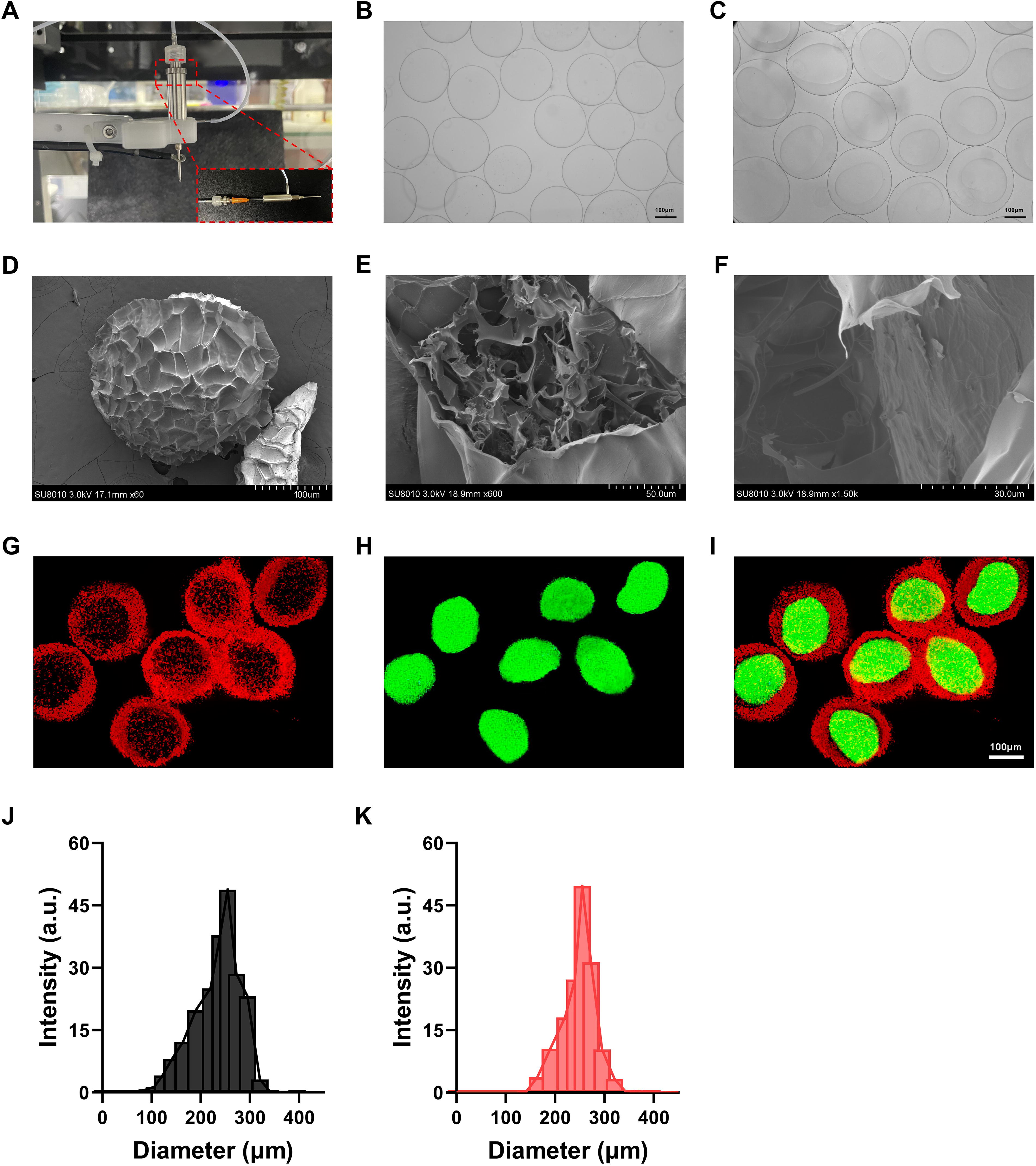
Characterization of alginate/carboxymethyl chitosan (ALG/CMC) microspheres.
Characteristics of A-DOX@C-DTX microspheres
We loaded DOX into the ALG shell and DTX into the CMC core, resulting in the synthesis of coloaded A-DOX/C-DTX microspheres. The encapsulation efficiency of DOX was determined to be 64.5%, with a drug loading of 2.5% (Supplementary Fig. S2A, B). The encapsulation efficiency of DTX was found to be 35.5%, with a drug loading of 1.8% (Supplementary Fig. S2C, D). The efficacy of chemotherapy poses a major challenge in clinical cancer treatment. 42 Owing to the narrow therapeutic window of most chemotherapy drugs, there is a small difference between the required dosage for achieving therapeutic effects and the dosage that induces toxic reactions. 42,43 However, free chemotherapy drugs often exhibit sudden and short-term release, which significantly affects the effectiveness of chemotherapy. Therefore, drug delivery carriers are needed to achieve slow and sustained release of chemotherapy drugs. 44 The biocompatible materials ALG and CMC used in this study possess good gelation properties, biocompatibility, and mucoadhesion. ALG, as the outer shell, has a large surface area and slow degradation kinetics, facilitating drug loading and sustained drug release. 21,31,32 Consistent with our expectations, free DOX and DTX exhibited burst release within the first 10 hours, with approximately 70% of the drugs being released, while the release of A-DOX@C-DTX microspheres was slower and more sustained (Fig. 2A, B). Furthermore, owing to the “particle-in-particle” construction of A-DOX@C-DTX microspheres, DTX is encapsulated within the CMC core, and ALG acts as an effective physical barrier on the outer layer, resulting in a progressive and sustained release of DTX.
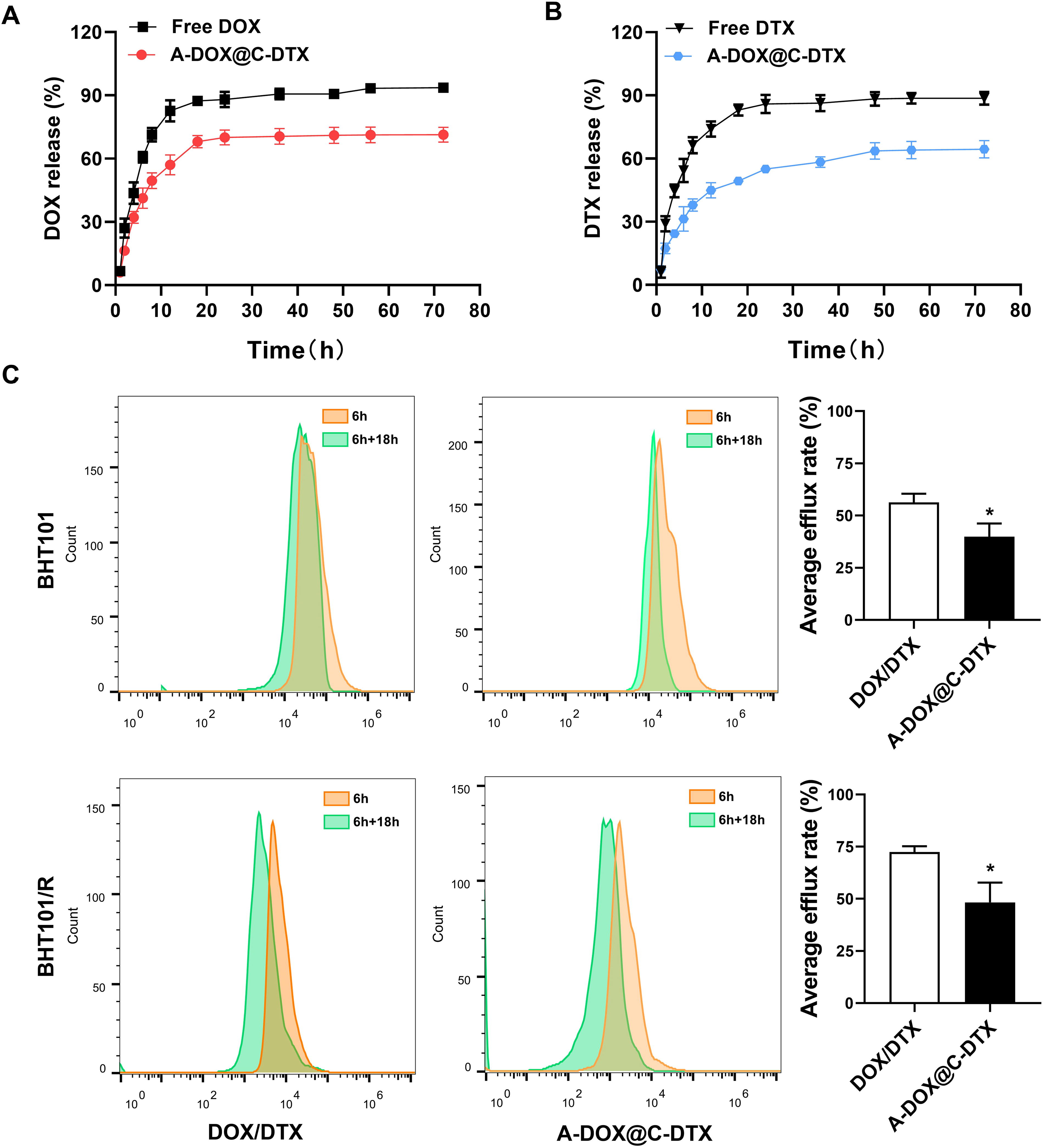
In vitro release profiles and cellular uptake and efflux of alginate-doxorubicin@carboxymethyl chitosan-docetaxel (A-DOX@C-DTX) microspheres.
Cell uptake and efflux were measured using flow cytometry, as shown in Figure 2C. The efflux rates of DTX/DOX and A-DOX@C-DTX in BHT101 cells were 56.30% and 39.91%, respectively. In BHT101/R cells, the efflux rates were 72.48% and 48.32%, respectively. ALG/CMC encapsulation significantly reduced the efflux of chemotherapy drugs in ATC cells, particularly in drug-resistant ATC, which may contribute to enhancing the sensitivity of ATC to chemotherapy drugs.
Inhibition of proliferation, migration, and invasion and promotion of apoptosis in resistant ATC cells by A-DOX@C-DTX microspheres
We investigated the effects of A-DOX@C-DTX microspheres on undifferentiated thyroid carcinoma cells in vitro. The viability of BHT101 cells treated with 0, 10, 20, 40, 60, 80, and 100 μM DOX or DTX for 48 hours, as well as BHT101/R cells treated with 0, 10, 20, 40, 60, 80, 100, 120, 140, and 160 μM DOX or DTX for 48 hours, was measured using the CCK-8 assay. The statistical results indicate that when the concentration of DOX is ≥60 μM, the viability of BHT101/R cells is significantly higher than that of BHT101 cells. Similarly, when the concentration of DTX is ≥80 μM, the viability of BHT101/R cells is significantly higher than that of BHT101 cells. This suggests that drug-resistant ATC cells exhibit a significant decrease in sensitivity to DOX/DTX. The IC50 values of DOX and DTX in BHT101 cells were determined to be 71.0 and 75.3, respectively. After acquiring drug resistance, the IC50 values increased to 140.0 and 142.5 for DOX and DTX, respectively, resulting in resistance indices of 2.0 and 1.9, indicating successful establishment of the drug-resistant cell line (Supplementary Fig. S3A, B). We ultimately selected a concentration of 50 μM DOX and 36 μM DTX for a 48-hour treatment of the cells in subsequent in vitro experiments.
First, the sensitivity difference of BHT101 cells and BHT101/R cells to DOX/DTX was assessed using the CCK-8 assay. The results showed that compared to BHT101/R cells, BHT101 cells were more sensitive to DOX/DTX, indicating the successful establishment of a drug-resistant ATC cell line. In contrast, under the treatment of A-DOX/C-DTX microspheres, there was no significant difference in the viability of BHT101 cells and BHT101/R cells, suggesting that A-DOX/C-DTX microspheres can enhance the sensitivity of drug-resistant ATC cells to DOX/DTX (Supplementary Fig. S3C, D). The CCK-8 assay was performed again to evaluate the impact of A-DOX@C-DTX microspheres on the viability of BHT101 and BHT101/R cells. The results showed that treatment with DOX/DTX or A-DOX@C-DTX microspheres significantly inhibited the viability of both BHT101 and BHT101/R cells, with A-DOX@C-DTX microsphere exhibiting the strongest effect (Fig. 3A, B). In the colony formation assay, treatment with DOX/DTX or A-DOX@C-DTX microspheres significantly suppressed the colony formation of both BHT101 and BHT101/R cells. Moreover, compared with the DOX/DTX treatment group, A-DOX@C-DTX microspheres further reduced the number of colonies formed by BHT101 and resistant BHT101/R cells, indicating inhibited cell proliferation (Fig. 3C–E).
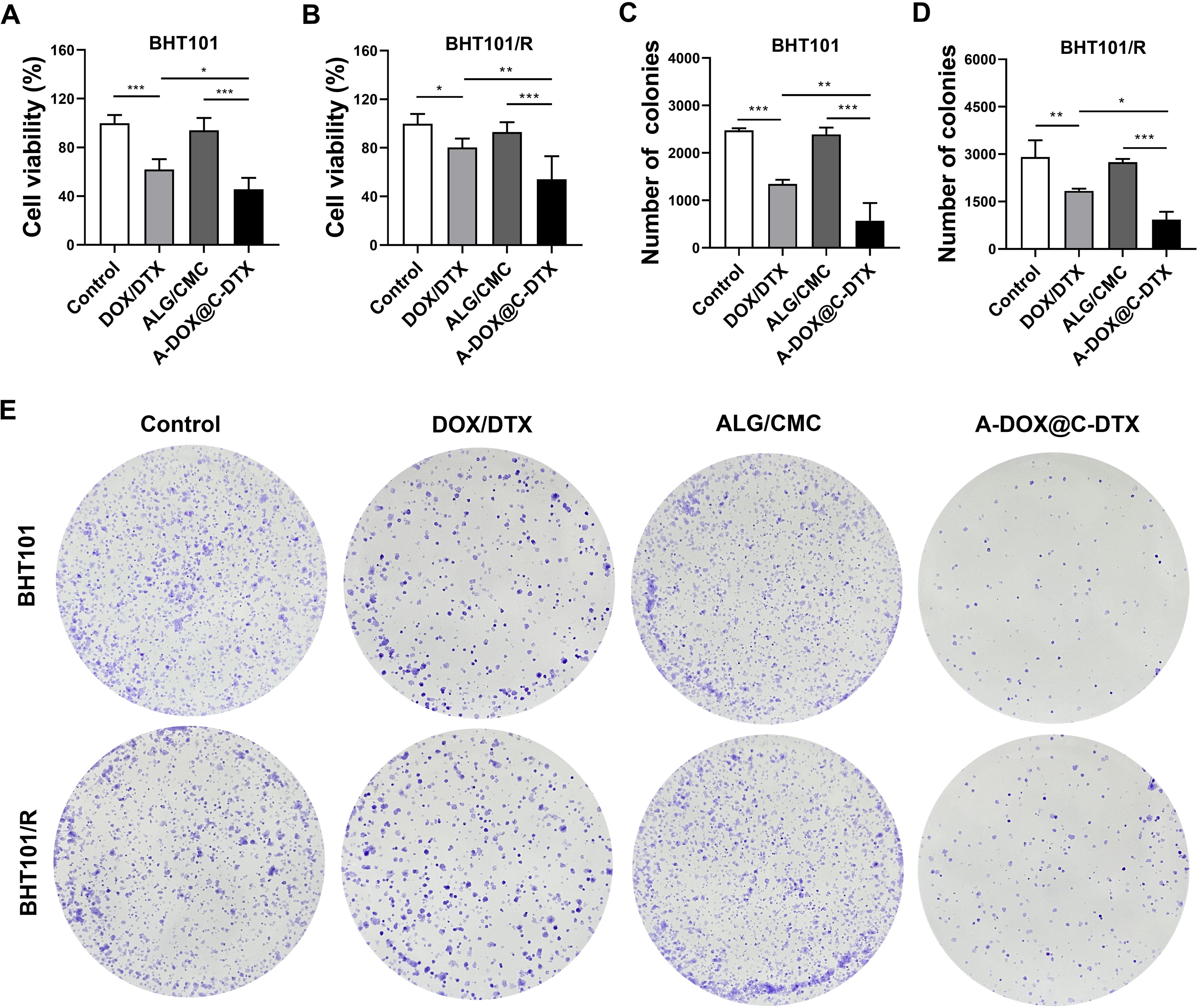
Effects of A-DOX@C-DTX microspheres on the viability and proliferation of BHT101 and BHT101/R cells.
Transwell assay was used to assess changes in cell migration and invasion abilities, and the results are shown in Figure 4. Treatment with DOX/DTX and A-DOX@C-DTX microspheres inhibited the migration and invasion of both BHT101 and BHT101/R cells. Furthermore, compared with the DOX/DTX treatment group, A-DOX@C-DTX microsphere treatment further suppressed the migration and invasion abilities of BHT101 and BHT101/R cells.
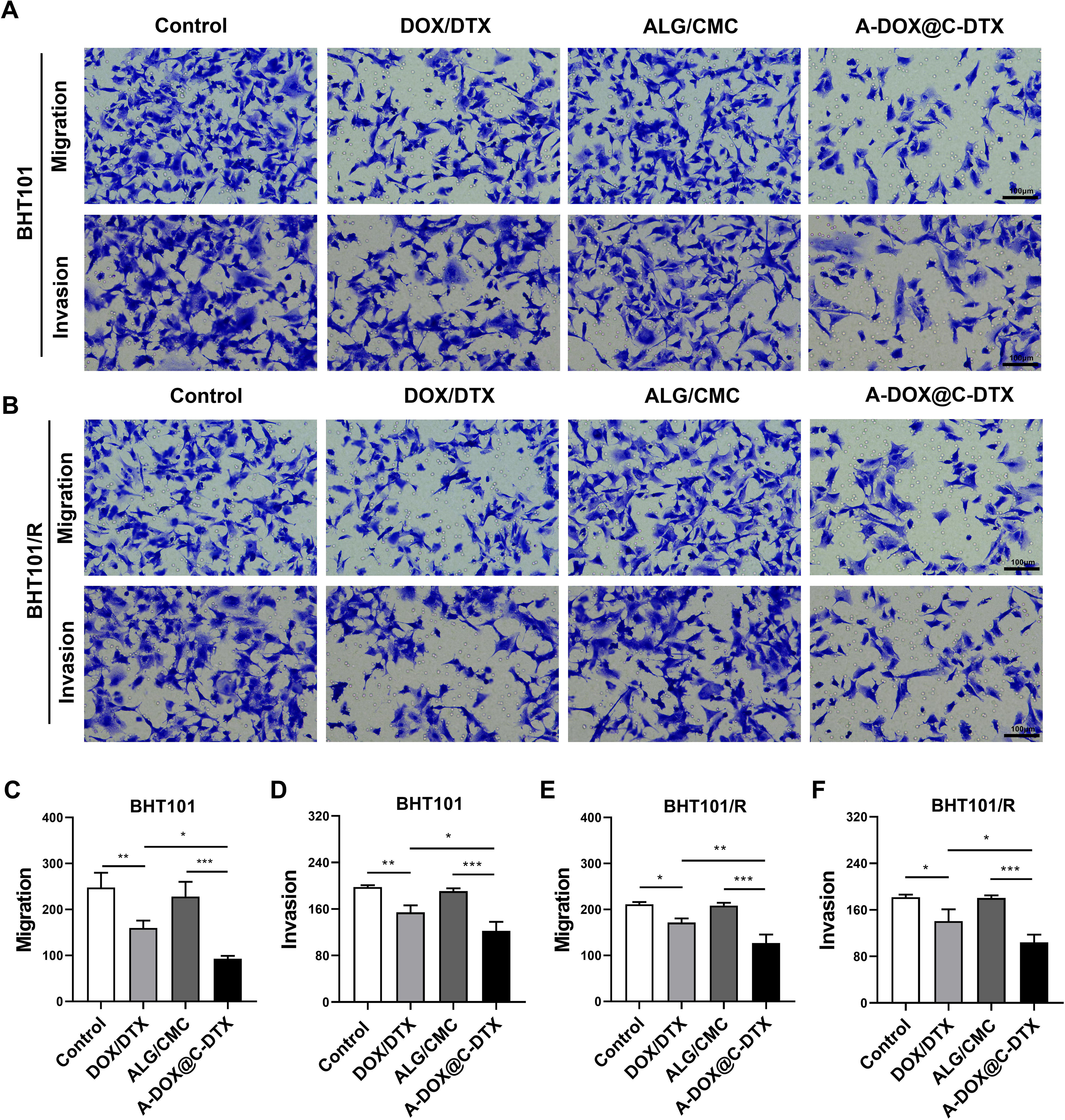
Effects of A-DOX@C-DTX microspheres on the migration and invasion abilities of BHT101 and BHT101/R cells.
The impact of A-DOX@C-DTX microspheres on cell apoptosis is depicted in Figure 5A–D. DOX/DTX treatment induces apoptosis in both BHT101 and BHT101/R cells. However, when encapsulated in ALG/CMC microspheres, the ratio of apoptotic cells further increases in BHT101 and BHT101/R cells. These findings demonstrate that ALG/CMC microsphere encapsulation enhances the sensitivity of resistant ATC cells to DOX/DTX. It achieves this by suppressing cell proliferation, migration, and invasion while promoting apoptosis, thus overcoming drug resistance. Based on the aforementioned outcomes, it can be concluded that DOX/DTX loaded into ALG/CMC microspheres exhibits improved efficacy in inhibiting or even eliminating resistant ATC cells. This effect could be attributed to enhanced internalization of DOX/DTX within resistant ATC cells. 45,46
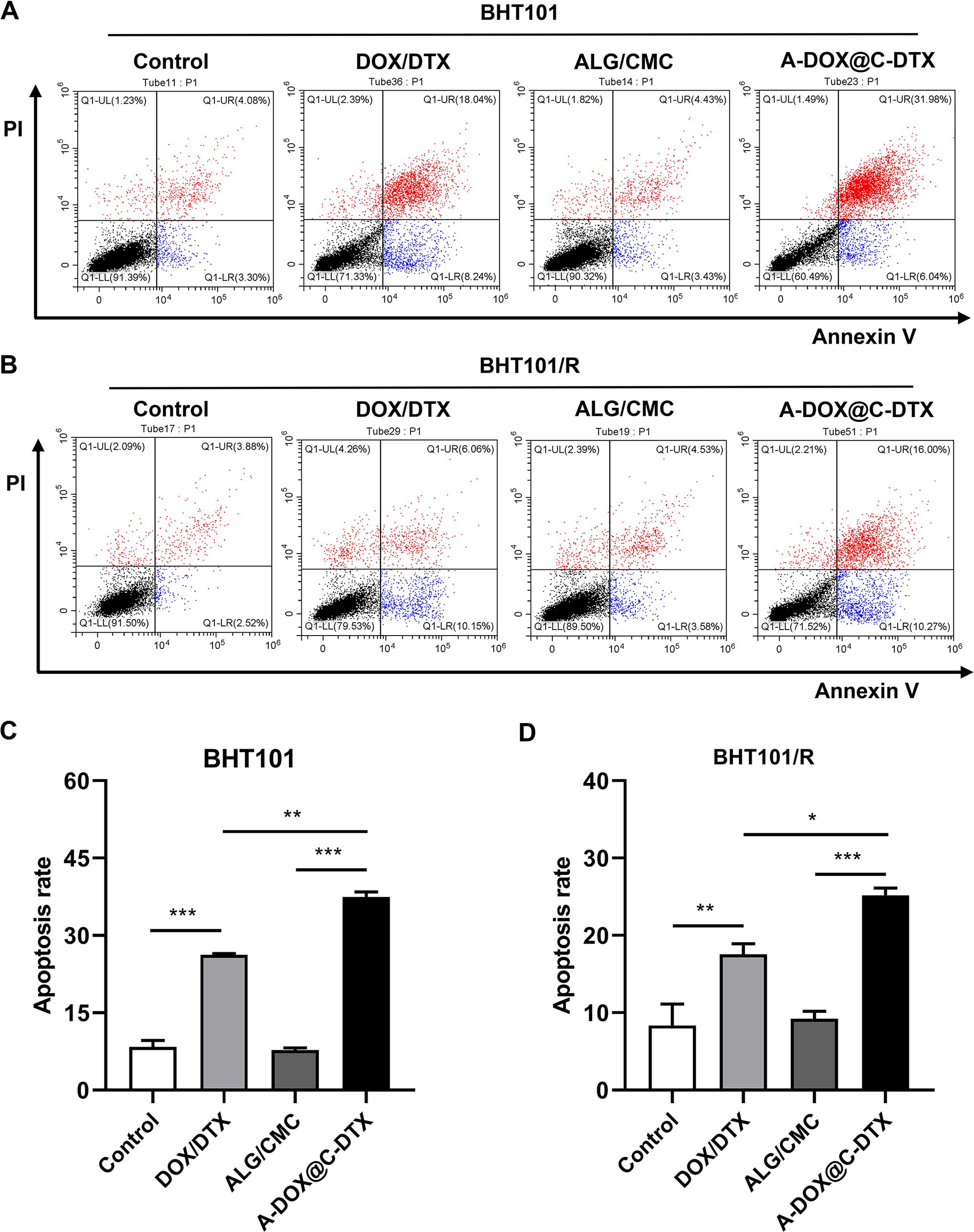
Effects of A-DOX@C-DTX microspheres on apoptosis in BHT101 and BHT101/R cells.
ALG/CMC microspheres promote sustained release of encapsulated agents at the tumor site
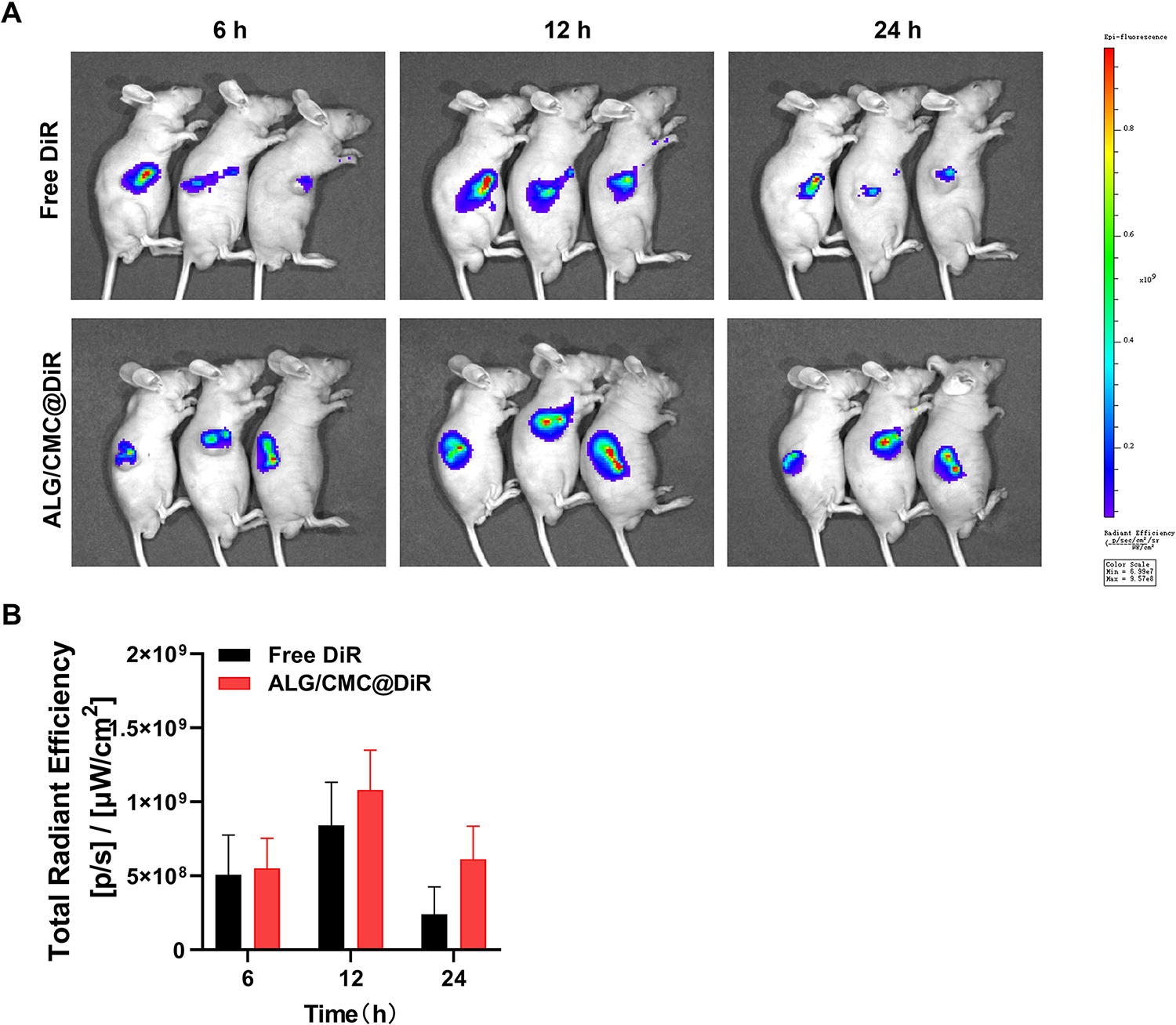
In vivo imaging (Fig. 6A–B) was used to examine the accumulation of the materials at the tumor site and the release of the encapsulated agents. Both the DiR and ALG/CMC@DIR treatment groups exhibited increasing near-infrared signals at the tumor site over time, followed by attenuation. However, compared with the DiR group, the A-DOX@C-DTX group showed significantly stronger fluorescence signals at the tumor site at the same time points, with a slower decay. This indicates that the microspheres can effectively concentrate the active substances in the tumor and achieve sustained release.
Effects of ALG/CMC microspheres on the release of encapsulated agents.
Inhibition of tumor growth by A-DOX@C-DTX microspheres
To further validate the impact of A-DOX@C-DTX microspheres on chemo-resistant ATC in vivo, a mouse tumor model was established by subcutaneously injecting chemo-resistant ATC cells. During the treatment period, the body weight of mice in each group was monitored. No significant weight loss was observed, indicating minimal side effects and excellent biocompatibility of the materials (Supplementary Fig. S4). At the end of the experiment, the mice were euthanized, and tumor photographs were taken to measure tumor volume and weight. The results showed that treatment with DOX/DTX led to a decrease in tumor volume and weight, demonstrating a certain level of inhibition of tumor growth. However, treatment with A-DOX@C-DTX microspheres exhibited a remarkable tumor-suppressive effect (Fig. 7A–C). Figure 7D shows the results of H&E staining, indicating that the tumor tissue morphology was normal in the Control group and ALG/CMC group. However, the other treatment groups exhibited varying degrees of nuclear damage and morphological changes in tumor tissue, with the A-DOX@C-DTX microsphere treatment group showing the largest area of necrosis. Ki67 protein is highly expressed in cancer cells and is a commonly used tumor marker to evaluate cell proliferation activity. The expression levels of Ki67 in each group are shown in Figure 7E, with the Control group and ALG/CMC group showing the highest Ki67 expression levels, indicating rapid tumor growth and high malignancy. The DOX/DTX treatment group showed a slight decrease in Ki67 expression levels, while the A-DOX@C-DTX microsphere treatment group exhibited the most significant decrease in Ki67 expression levels. TUNEL staining (Fig. 7F) was used to detect apoptosis in tumor cells in each group. The Control group and ALG/CMC group showed the lowest levels of TUNEL-positive staining, indicating normal tumor cell growth. The DOX/DTX treatment group showed an increase in TUNEL-positive staining, indicating some tumor cell destruction. The A-DOX@C-DTX microsphere treatment group showed a more significant increase in TUNEL-positive staining, indicating the best antitumor effect. These results demonstrate that ALG/CMC-encapsulated DOX/DTX is superior to free DOX/DTX treatment and exhibits excellent antitumor activity.

Effects of A-DOX@C-DTX microspheres on mouse tumors.
Discussion
To combat ATC and prolong patient survival, clinical practice typically involves a combination of surgery, radiotherapy, and chemotherapy. However, there is often a discrepancy in the size of the treatment area between surgery and radiation therapy, leading to damage to surrounding tissues and organs. 1,47 Chemotherapy has long been considered an effective treatment modality, but the emergence of drug resistance severely impacts its efficacy. Currently, strategies to overcome resistance and avoid monotherapy-induced drug resistance have been proposed and discussed, including combination therapies with radiation, chemotherapy, or targeted drugs. 48,49 The use of biomaterials for the delivery of chemotherapy drugs has been extensively studied in cancer treatment, but there is limited research on the application of biomaterials in combating ATC. Based on this, our study developed a novel drug-loaded microsphere combining biomaterials and chemotherapy drugs. This microsphere can simultaneously encapsulate docetaxel and doxorubicin, achieving co-encapsulation and spatiotemporal release of the two drugs. It also enhances cellular uptake of the drugs and inhibits drug efflux, thereby reversing chemotherapy resistance.
The efficacy of chemotherapy poses a major challenge in clinical cancer treatment. 42 The combination of biomaterials and chemotherapy drugs plays a significant role in improving cancer treatment efficacy. Combined therapy using biomaterials and chemotherapy drugs has been shown to enhance tumor control and reduce adverse reactions by improving drug payload, pharmacokinetics, and targeted delivery. 50,51 Combination therapy appears to be the standard approach in traditional chemotherapy to overcome cross-resistance and achieve synergistic therapeutic outcomes without significantly increasing toxicity. In general, monotherapy using a single drug is insufficient to induce tumor regression. Through combination chemotherapy, the synergistic effects of two or more drugs targeting different disease pathways, genes, or cell cycle checkpoints can be utilized to increase the chances of eradicating cancer. 52 ATC patients typically receive two or more drugs, such as taxanes (paclitaxel, DTX) combined with cisplatin or DOX. 4,53,54 Docetaxel is a mitosis-specific drug, while doxorubicin is a cell cycle-nonspecific drug. The combination of the two drugs exhibits synergistic effects and has a stronger tumor-inhibiting efficacy. 54,55 However, most current research on biomaterials focuses on delivering single drugs, severely limiting the therapeutic efficacy for ATC. Therefore, we developed a core-shell microsphere using electrospinning-assisted microfluidic technology, with ALG as the shell loaded with doxorubicin DOX and CMC as the core loaded with DTX. This combination therapy utilizing biomaterials and chemotherapy drugs offers advantages such as the use of lower doses of chemotherapy drugs, elimination of adverse cytotoxic effects, and improved efficacy, making it a promising approach to enhance cancer treatment outcomes. 56 In addition, the ALG/CMC core-shell microspheres can carry multiple chemotherapy drugs in a layered manner, allowing for optimized synergistic drug ratios within a single carrier. 17 This combination therapy approach is not only applicable to ATC but also provides a reference for the treatment of other diseases.
Previous studies have reported the potential of ALG/CMC as a scaffold in dental, orthopedic, and chronic wound applications because of its good biocompatibility and slow degradation rate, receiving consistent praise. 23,57,58 Inspired by these studies, this research utilized ALG/CMC microspheres coloaded with DOX/DTX for ATC treatment, demonstrating superior antitumor effects both in vitro and in vivo compared with free DOX/DTX treatment. In addition, it should be noted that in in vitro experiments, the sustained release property of our microspheres may potentially limit the in vitro efficacy of the drug to some extent.
It has been reported that reduced drug uptake and increased efflux are important factors contributing to drug resistance in tumors. 42,59 Therefore, enhancing cellular drug uptake and inhibiting drug efflux may partially reverse chemotherapy resistance. In fact, previous studies have demonstrated the effectiveness of this approach. 46 For example, bovine serum albumin-doxorubicin-mesoporous organosilica nanoparticles (BSA-DOX-MONPs) developed by Han et al. were able to reverse chemotherapy resistance in ATC cells by increasing drug uptake and inhibiting drug efflux. 46 In this study, we also observed similar functionality in A-DOX@C-DTX microspheres, which may contribute to reversing chemotherapy resistance in ATC cells to DOX and DTX. However, the molecular mechanism underlying the microspheres’ inhibition of drug efflux remains to be further investigated. In addition, it is worth mentioning that, compared to previously developed ATC chemotherapy drug delivery systems, 46 our ALG/CMC microspheres support the co-encapsulation and spatiotemporal release of DOX and DTX, offering great potential as a drug delivery system for combined chemotherapy applications.
In summary, the combination therapy utilizing biomaterial microspheres ALG/CMC for layered delivery of chemotherapy drugs (DOX and DTX) is a suitable alternative approach for ATC treatment.
Conclusion
In this study, we successfully constructed ALG/CMC core-shell microspheres capable of efficient coloading of DOX and DTX, enabling simultaneous and sustained release of the two drugs. The synthesized A-DOX@C-DTX microspheres exhibited superior antitumor effects compared to free drug treatment both in vitro and in vivo, particularly against drug-resistant ATC cells. This improved therapeutic efficacy may be attributed to the enhanced drug uptake and reduced drug efflux in drug-resistant ATC cells facilitated by ALG/CMC microspheres.
Footnotes
Authors’ Contributions
Y.Z.: Methodology (lead); writing—original draft (lead); formal analysis (lead); and writing—reviewing and editing (equal). F.Y.: Investigation (lead); writing—original draft (equal); data curation (lead); and writing—reviewing and editing (lead). H.Z.: Software (lead) and writing—reviewing and editing (supporting). S.L.: Conceptualization (lead); writing—original draft (supporting); project administration (lead); and supervision (lead).
Data Availability
The data used to support the findings of this study are available from the corresponding author upon request.
Author Disclosure Statement
All the authors declare that there are no conflicts of interest.
Funding Information
Y.Z. has received funding from the
Supplementary Material
Supplementary Figure S1
Supplementary Figure S2
Supplementary Figure S3
Supplementary Figure S4
Supplementary Data S1