
Abstract
Introduction
According to the National Hospital Ambulatory Medicare Care Survey, there were approximately 124 million emergency department (ED) visits in the United States in 2008. 1 Initial visits involving care for lacerations in the ED occur at a rate of almost 75/10,000. 2 When people experience a skin laceration, one of the major questions is whether ED care is required, specifically, whether a laceration repair is necessary. Some lacerations are large and clearly need repair, whereas others are small and can be managed at home. A large middle category laceration is one where some patients may have trouble with self-triage. These lacerations require evaluation by an experienced provider to determine management. This is typically done with an in-person medical examination in an ED or other urgent care setting.
Advances in digitally based camera technology and their ubiquity in new generation mobile phones may help play a role in the triage of lacerations. This would involve a patient taking a picture of the laceration with a mobile phone and sending it to a provider. Being able to obtain a medical opinion prior to entering the healthcare system may improve the care of these cases because it could lead to avoidable ED visits and lower healthcare costs. Previous studies have assessed the use of telemedicine for chronic wound diagnosis and management, as well as remote dermatology consultation, and found it to be effective. However, no studies to our knowledge have explored the utility of patient-generated mobile phone camera images to help with the triage of lacerations or with recommendations for patients when they return for follow-up care in an ED setting.
The goal of this study is to assess the quality of patient-generated mobile phone images and determine the agreement between using mobile phone–generated images versus in-person assessments for laceration management in the ED.
Subjects and Methods
Subjects, Site, and Sampling Method
This was a prospective study conducted in an urban, academic ED in Washington, DC, with an annual volume of more than 70,000 visits in 2010. The ED has a 4-year residency program and is also staffed by physician assistants and rotating medical students. Medical students, physician assistants, and residents are supervised by ED attending physicians 24 h/day. Routine laceration care is preferentially performed by physician assistants; however, sometimes ED residents, medical students, attending physicians, or consulting service physicians also may repair lacerations when the physician assistants are unavailable.
Research assistants were available in the ED approximately 12 h/day on weekdays to enroll patients in the study, so the sampling method can be described as a convenience sample. When research assistants were present in the ED, potential subjects were patients identified by their chief complaint as having a laceration. Exclusion criteria included lacerations in the perineum/genital areas, wounds requiring immediate and emergent care (i.e., a major hemorrhage or severely injured patient), patients who refused participation, and patients who did not have a mobile phone with a camera. Each subject gave informed consent to be enrolled; child assent forms were provided for minors, and their guardians were required to provide informed consent. No medical students were used to enroll patients.
Procedures
Eligible patients were approached by a research assistant for enrollment in the study prior to any provider evaluation. Subjects obtained four pictures of their laceration with their mobile phone camera and then e-mailed or text-messaged it to a dedicated account. Patients were instructed precisely how to take up the four photographs: one close up, one at 2–3 feet, one from the left, and one from the right (Fig. 1). In some cases, a disposable ruler was used for scale next to the laceration. All photographs were taken while the patient was in a triage room or treatment room. Patients were also given the option of having images taken by a family member or friend. Subjects completed a questionnaire that included a history of their laceration, documenting when and how it occurred and where it was located on the body.
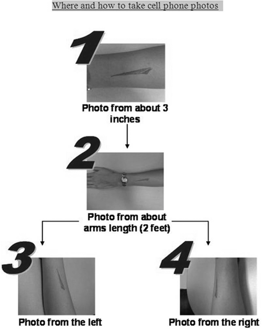
Instructions given to emergency department patients on where and how to take mobile phone camera pictures of their laceration.
Prior to any in-person evaluation, an ED attending physician, physician assistant, or ED resident evaluated the questionnaire and mobile phone images. The healthcare professional then completed a worksheet to assess the quality of the mobile phone images using a 10-point Likert scale and to document a diagnosis and management plan. Then the same provider performed an in-person examination and documented his or her diagnosis and management plan again. The patient was also asked if his or her mobile phone–based management plan was different from the in-person management plan and why.
Data Analysis
The population was described using standard descriptive statistics. Agreement was calculated three ways. The first was the percentage of cases where there was complete agreement (i.e., both the mobile phone and in-person assessments for laceration repair were identical). The second was the percentage of cases where there was agreement or where the in-person evaluation was not to repair the wound. This was intended to calculate the percentage where there was no undertriage, which we think would be the major safety issue with this technology (i.e., telling a patient that it does not need repair when, in actuality, it does after an in-person evaluation). The third was a κ statistic, which is a statistical measure of inter-rater agreement from 0 to 1. It is typically interpreted such that values less than 0 are “no agreement,” 0–0.20 as “slight,” 0.21–0.40 as “fair,” 041–0.60 as “moderate,” 0.61–0.80 as “substantial,” and 0.81–1 as “almost perfect” agreement.
Results
In total, 106 patients were approached for enrollment. Of those, 12 patients were excluded because forms were not filled out correctly, so that 94 paired assessments were available for inclusion in the study. Sixty-three (67%) ultimately received a laceration repair in the ED.
Of the 94 patients included for the analysis, 10% were younger than 18 years old, 18% were between 18 and 24 years old, 30% were between 25 and 35 years old, 22% were between 36 and 50 years old, 16% were between 51 and 65 years old, and 3% were older than 65 years of age. Thirty-four percent of enrolled patients were female.
The most common body areas with lacerations were the hand (36%), followed by the head/face (23%). Mechanisms of injury were most commonly described by patients as a cut (51%). In the majority of cases, the laceration was <3 h old (65%) (Table 1). On a scale of 1 to 10, with 1 being the poorest (minimum) and 10 being the best (highest), the median score for image quality rated by ED clinicians was 6, with an interquartile range of 4–8.
Demographics of the Study Group Seen in an Emergency Department for a Laceration Viewed with a Mobile Phone Camera Paired with In-Person Evaluation
There was complete agreement in terms of the laceration management in 81 of 94 cases (87%), with a κ statistic of 0.65 (moderate agreement). A total of 89 of the 94 (95%) cases had either complete agreement or were not undertriaged. Of the 13 cases where there was a discrepancy, in 6 it was due to poor image quality, in 3 the image was adequate but did not properly represent the problem, in 3 there was other history of findings that altered care, and in 1 the image looked worse than the actual injury in person.
Limitations
This study was limited in that it was a small study conducted in a single ED, which limits the external validity of these findings. It was also conducted only in patients who had triaged themselves or were triaged to the ED for care, so this may have biased toward more severe lacerations. There was also a variety of mobile phone cameras used for this study; certain mobile phone camera images may be better than others, which may explain some of the differences in image quality. Finally, there may be variation in provider opinions about whether certain marginal lacerations need to be repaired. However, by pairing assessments within providers, we were able to control for differences in decision-making. But in reality, it is possible that a provider's opinion from reviewing a cell phone image may differ from an in-person evaluation in the same way that in-person evaluations may differ between providers. We also had providers assess image quality using a Likert scale, which has not been directly validated, but we think has good face validity.
Discussion
We found that there was a high rate of agreement in management decisions about repairing lacerations using a history and four pictures obtained by the patient using a mobile phone camera. When considering the safety of this program, we felt the most detrimental outcome would be advising a patient based on a mobile phone image to not come to the ED (i.e., undertriage), while in actuality a sutured repair was needed. Using these criteria, only 1 in 20 lacerations was undertriaged, and, in the majority of cases, it was due to poor image quality. In a real-life setting where management decisions were being made, the clinician could potentially ask a patient to obtain additional images.
Now that mobile phone cameras have become common in the United States and around the world, several recent studies have focused on mobile phones as an emerging telemedicine technology. Plastic surgeons and dermatologists using mobile phone camera images found a 75% and 94% concordance between multiple remote physicians' assessments of acute extremity wounds and leg ulcers, respectively. 3,4 There was similar agreement between phone-based and in-person assessments for extremity wounds in using mobile phone images. 5 Plastic surgery attending physicians found a high concordance between in-person wound assessment and remote photograph-based wound assessment using 3.3 megapixel digital camera images captured by plastic surgery residents. 6 Similarly, photograph-based assessment of pressure ulcers has been shown to be 89% concordant with in-person assessments. 7 Compared with our study, we found similar agreement in wound assessments; however, previous studies used physician-generated images, whereas our study used patient- or other layperson-generated images at the primary photographer.
Image quality was moderate in this study but also highly variable, showing that most patients are capable of taking adequate images for wound assessment. But not infrequently, poor images are generated, indicating that, in a real-life setting, additional images may need to be obtained. It is also possible that with better mobile phone camera technologies becoming standard and more general familiarity with using mobile phone cameras that the quality of unsupervised photos could improve.
Conclusions
There is moderate agreement between mobile phone and in-person evaluations of lacerations on the decision to repair. Images obtained by patients are of highly variable quality, which may be a key limitation. Mobile phone camera images may be useful to assess lacerations without a clinician assessment; however, additional larger studies are needed to assess safety, outcomes, and cost impacts of a program prior to widespread implementation.
Footnotes
Disclosure Statement
No competing financial interests.