
Abstract

Introduction
Ensuring telemedicine tools reach those with the greatest need is not as simple as delivering devices to a remote clinic—there is often the issue of power availability and device compatibility. One tool that is quickly becoming omnipresent even in the most hard-to-reach places is the mobile phone and more specifically the smartphone. Microscopes, ultrasound, and other point-of-care devices can utilize a smartphone for processing power or become components that can directly function with a smartphone.
Why Mobile Phones?
According to the International Telecommunication Union, as of October 2010, 76.2% of the world's population had mobile cellular phone subscriptions or about 5.3 billion people. Even in developing countries, the number is impressive, with 67.6% penetration. This number drives much of the development of mobile phones (m-phones) or smartphones in use of point-of-care diagnostics. Aydogan Ozcan, Ph.D., Associate Professor of Electrical Engineering at the University of California—Los Angeles (UCLA), says “With 5 billion cell phone subscribers, this drives costs enormously low and the profit margins become fantastic for all these advanced technologies. There's a lot of profit, and that's why the complex technologies are becoming part of a bigger and bigger system that we couldn't have imagined before.”
The sheer ubiquity of m-phones has helped drive down both the costs of the chips and processors used in phones as well as the costs of the devices themselves. Also, the technology itself has become powerful enough to drive telemedicine and POC diagnostic devices. Sailesh Chutani, PhD, CEO and Cofounder of Mobisante, Inc. (Redmond, WA), says, “If you didn't have the CPUs, you couldn't do the real-time imaging. You need enough horsepower, and since the development of Qualcomm chips, that became practical.”
Although m-phones and cellular towers are commonplace throughout the world, a regular, reliable, functioning power grid is not. There are numerous low-resource settings globally, often remote, which are fairly close to cellular antennas but do not have a power grid; they do, however, have access to cell phone charging stations that draw electricity from generators or solar or wind power. These elements have come together to make the m-phone the logical nexus for mobile point-of-care diagnostic technology, especially in low-resource areas.
Telemicroscope via Lens Technology
UCLA's Dr. Ozcan is the head of a team that has developed a powerful microscope that utilizes the processing power of an m-phone with a modification of the lenses. In fact, the device Ozcan's team has developed is a “lens-less” microscope. Lenses, Ozcan explains, have been performing an “analog computation on light” for decades, which explains why lens grinders have worked to magnify the image to better see with the human eye. He describes it as “an analog computer.”
By utilizing digital imaging technology, it is possible to duplicate the same results under certain conditions. Ozcan says, “Then the microscope becomes an analog computer without any lenses. That makes the microscope much simpler, cost effective, and lightweight.”
Essentially their device detects shadows of the cells it is examining. Because a cell—or bacteria—is semitransparent, if light is cast through it and scattered, it is possible to detect these holographic shadows of the cells using the retinas of the m-phone. The digital shadow images are processed using the graphics-processing power of the phone to reconstruct the image. Oczan says, “Essentially, if your cell phone has a camera on it, we remove the lens and bring the cells very close to it so the cells cast their shadows. We capture the images, and then we construct those. If your cell phone doesn't have a camera in it, we have different versions, which are like your laptop having a webcam. Or we can attach one to your USB port and convert your laptop into a webcam” (Fig. 1).
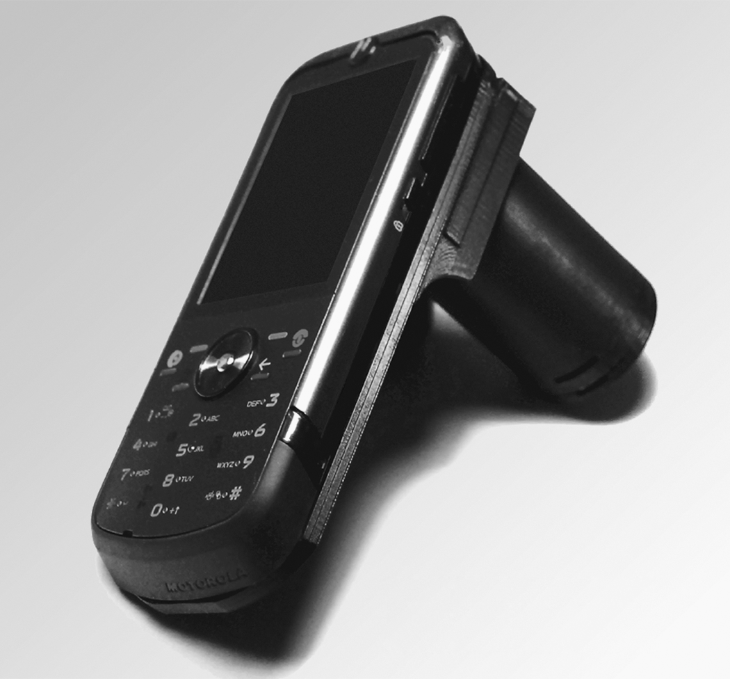
An image of the lens-free microscope and cell phone developed by Aydogan Ozcan's group at UCLA. Courtesy of Ozcan Research Group at UCLA.
Resolution for the microscope is submicron, less than one millionth of a meter, roughly equivalent to the image seen through a 10× microscope lens. This makes it useful for viewing parasites such as malaria or bacteria. The technology itself is very similar to a phase-contrast microscope, which is very good for looking at objects surrounded by water.
Although Ozcan's team does not yet have a commercial product available, he notes that the most expensive part of the technology is the sensor, which costs approximately $15. The added advantage of the processor being part of a mobile phone, of course, is the potential for telemedicine, all within a portable, hand-held form factor.
“If you're targeting global health, some of your limitations are related to bandwidth—it's still expensive, so you can't count on it to be free or low cost,” Ozcan says. “Globally, the handsets are as good as we have in the U.S. [though] some of the innovations we come up with here can't be immediately translated to the global health settings, like Africa.”
He notes that, for instance, in some resource-poor settings, smartphones are likely to be a second-generation Blackberry, which can cause a gap in terms of development and utilization. Still, with a typical microscope costing in the thousands of dollars and relying on electricity to function, it is easy to see how a phone-powered microscope could be useful in the field. Ozcan's group is currently working on a smartphone-based fluorescent microscope (Fig. 2).
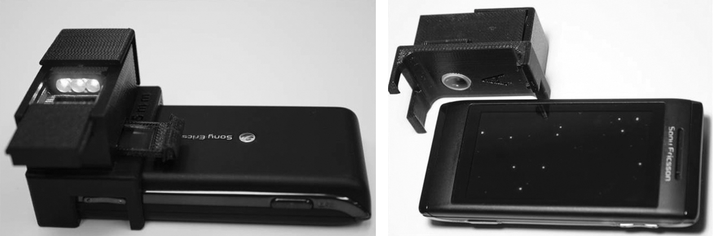
Images of a wide-field fluorescent microscope on a cell phone under development by Ozcan's group. Courtesy of Ozcan Research Group at UCLA.
The Microfluidics Microscope
Although Ozcan's group is using “lens-less lenses” for their technology, there are other approaches. Changhuei Yang, Ph.D., Professor of Electrical Engineering and Bioengineering at the California Institute of Technology's Biophotonics Laboratory, is using microfluidics technology to develop a smartphone-based microscope. Yang calls the device a “subpixel resolving optofluidic microscope” (SROFM), or “optofluidic microscope” for short. That is to say, it is a combination of optics and microfluidics, microfluidics being the study of small, typically submillimeter fluids.
Yang says, “The strategy we are applying is basically the fact that most samples you want to image are in fluid form. So instead of a slide you inject it into a chip that would run the fluid across a scanning system to do imaging. This is such that you can manufacture these systems very easily and end up with the complete microscope on the chip that costs only a couple of dollars.”
This system involves a microscope system that is plugged into the smartphone. It uses the phone's computational ability for processing, but the scanning itself is performed by the chip. Again, Yang says that their first uses are for malaria cell analysis and other waterborne parasites. Current resolution of SROFM is 0.75 μm, roughly comparable to a 10× light microscope lens (Fig. 3).

A diagram of subpixel resolving optofluidic microscope (SROFM). A sample is sent through the chip, smaller than a quarter. The phone's processor creates and analyzes a holographic image of the specimen. Courtesy of Guoan Zheng, Ph.D., Caltech Biophotonics Laboratory.
Integrated Camera and Multimedia Messaging Services
Livia Bellina, M.D., is a pathologist currently working with St. Vincent Hospital in Dinajpur, Bangladesh. Affiliated with Azienda Sanitaria Locale 6 in Palermo, Italy, Bellina is using telemicroscopy to train rural health workers and is affiliated with Mobile Diagnosis and Medical Exchange, Medting. Mobile Diagnosis utilizes the Web-based platform of Medting to share medical images and videos with medical professionals worldwide for education and clinical diagnosis.
Bellina's approach is very different and basic—essentially she simply places the camera from a smartphone against the ocular of a common optical microscope and takes a photograph. Then, using multimedia messaging services (MMS), which is text messaging that can send images, she sends the images to distant reference centers for diagnosis and second opinions (Figs. 4 and 5). Bellina outlined her approach in a Diagnostic Pathology article titled “Mobile cell-phones (M-phones) in telemicroscopy: Increasing connectivity of isolated laboratories” (Bellina L and Missoni E. 2009; 4:19). She responded to questions for this article via e-mail from Bangladesh.
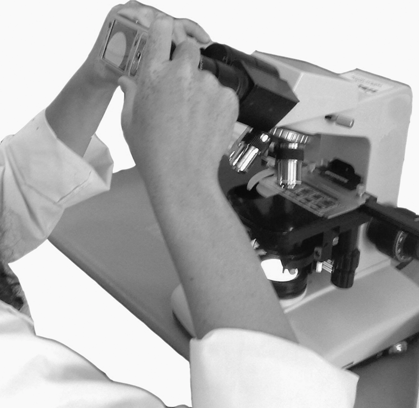
A mobile phone with a digital camera is held to the ocular of a standard light microscope to capture an image. Courtesy of Dr. Livia Bellina, M.D.
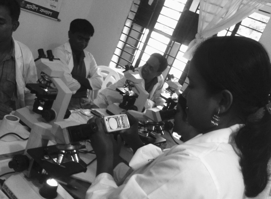
Healthcare workers in Bangladesh using mobile phones to capture images through standard light microscopes. Courtesy of Dr. Livia Bellina, M.D.
Bellina notes that in the wake of the global financial crisis (often dubbed “the great recession” in the United States), the global market decreased in general, but the mobile connection market increased, especially in the developing world. “The global market is looking at low-cost technologies for developing countries. This is the future of commerce.” She says that relatively simple technologies such as smartphones, which are often prepaid for longer terms of service, have “dual results: to improve rural, low-resource setting healthcare and to improve the global economy.”
She finds m-phones work best in the health field for sending text, capturing and sending diagnostic images via MMS, and where feasible, for quick, low-cost telemedicine. She cites telediagnosis, second opinions, monitoring for disease, training in low-resource settings, and the doctor–patient relationship as being the most likely uses of smartphone-based medicine in the global health market. “I think the future of telemedicine is mobile medicine, so the future for rural community education will be mobile training and m-learning.”
Bellina notes in particular that digital capture through a regular microscope has uses for dermatology and parasite identification, although there is also a need for ultrasound. The two limitations she has found working in Bangladesh and Uganda revolve around government-controlled broadband—often only lower-resolution images can be sent, resulting in unusable image quality—and the relative lack of training and education that many local, rural health workers have. “A transmission of images can be made, but health workers have to know the basic diagnostics.”
Telephonic Ultrasound
Although a number of researchers and consortiums are working to develop “Lab on a Chip” technology that can be powered by an m-phone, Mobisante, Inc., is the first company to receive FDA approval for its smartphone-based ultrasound imaging system, MobiUS. The entire product comes as a package, which includes the probe or transducer linked to a smartphone that has the software that runs the systems. Because it utilizes a smartphone, it then has Wi-Fi or cellular capability for sending data for archival purposes, telediagnosis, or a second opinion (Fig. 6).
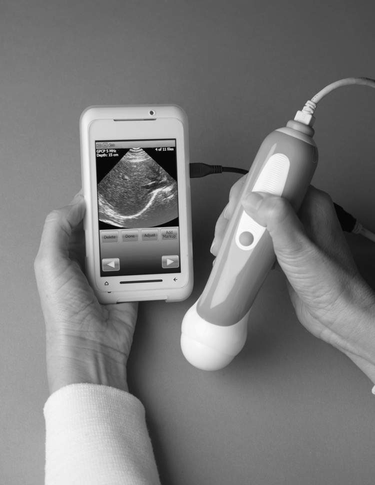
The Mobisante, Inc., MobiUS, a portable ultrasound device that uses a Microsoft Windows-based smartphone for processing and image recover. Courtesy of Mobisante, Inc.
Sailesh Chutani, Ph.D., CEO and Cofounder of Mobisante, says “What we give you is the complete system. We give you the phone, we give you the transducer, and everything is preinstalled and preconfigured.”
At the moment, the phone utilizes a Windows Mobile operating system. This is perhaps not surprising in that Chutani was formerly a senior director at Microsoft in Windows Mobile. Chutani says “What we need long-term, as certain platforms get standardized, is support for the phone from a technical perspective. We need to be able to draw power for the transducer from the phone. That basically doesn't exist today on Android or iPhone, although we're in conversations with Android and I think it'll come around pretty quickly. When it does, we'll probably have it for Android phones.” They currently have no plans to use the iPhone for this technology.
Chutani believes that MobiUS is just the start of what he thinks will revolutionize global healthcare and mobile telemedicine. “We're talking about a true point-of-care diagnostic platform at the heart of which you'll have a smartphone, and around it you'll have a set of peripherals, sensors, or smartchips that essentially will enable healthcare workers to work with a very powerful set of devices.”
He further points out that he expects that these point-of-care diagnostic devices will be powerful enough and inexpensive enough that most of the frontline work can be done on them. “That's the first line of defense. That's our vision and we're looking at medical imaging next and, in fact, the cell phone cameras as a second sensor in our platform for dermatology.”
The device as currently marketed in total costs about $7,500 for the United States, although Chutani indicates that Mobisante has a roadmap to reduce that price dramatically. “The cost of the device is one thing, but because we have connectivity it gives you the ability to centralize the higher expense costs, working to where the cost per independent scan is one dollar or less, which appears to be the threshold around the world that people are willing to pay.” They also have plans to utilize cloud computing, which should help drive down the cost per scan.
Down the Road
It can be rather startling to look at a typical smartphone and realize that even a decade ago you could not reproduce the computing power for the price and size you are holding in your hand. Go back two decades and it is the stuff of science fiction. Now an iPhone or Android phone has almost the power of a laptop computer for a lower price and comes equipped with a cellular phone and an 8.0 megapixel digital camera, often with high-resolution video capabilities. With all this power at our fingertips, Chutani asked the question: Why are some of the organizations that fund public health initiatives sitting on the sidelines? Why isn't there more excitement in the public health community to get this out there, to do a large-scale deployment and test this out?
Part of the answer may be that it is just too new. And perhaps it is just a matter of time. Chutani says, “We've taken the regulatory and reimbursement issues out, but these things just take time for people's imaginations to catch up.”