
Abstract
Introduction
Tele-ultrasound has emerged as an effective method of providing sonographic services to remote areas. 1 –6 Tele-ultrasound electronically transmits images from one location to another in order to obtain expert interpretation from a consulting radiologist. The radiologist's interpretation is returned, typically via e-mail, to the referring physician to confirm or refute the original ultrasound diagnosis. The main use of telemedicine has been to allow isolated healthcare practitioners to contact consulting specialists in order to obtain a second opinion. 7,8
Antarctica is the highest (the average elevation is 2,300 m), coldest (mean temperatures range from 0°C to −70°C), driest, windiest, and most isolated continent on Earth. Medical practice in such conditions constitutes an extreme form of remote care where telemedicine links are a necessity. 1,9 Many Antarctic stations do not utilize ultrasound. In 2001, two Acuson Aspen (Oceanside, CA) digital ultrasound scanners capable of real-time and store-and-forward transmission of ultrasound imaging were deployed to the two largest U.S. Antarctic Program (USAP) research facilities: McMurdo Station, a coastal facility on the Ross Sea, and the Amundsen-Scott South Pole Station, at the geographic South Pole. It was anticipated that the introduction of ultrasound technology would increase diagnostic accuracy and would reduce the number of medical evacuations. 10
To date, there has been no evaluation of the impact of ultrasound examination on medical events in the Antarctic. Additionally, the importance of tele-ultrasound for clinical management in Antarctica has not yet been assessed. Given the operational impact of a medical evacuation from South Pole or McMurdo Station to a tertiary-care center, avoiding a misdiagnosis is essential. Although McMurdo Station may occasionally have a trained sonographer on site during the summer season, during the winter season (and throughout the year at the South Pole), exams are acquired and interpreted by physician operators with basic ultrasound skills. Examining the accuracy of this data acquisition and interpretation is an essential consideration for precise diagnoses. 6 Accordingly, the purpose of this investigation was to review the medical events requiring ultrasound examination and to determine the importance of ultrasound in clinical management in Antarctica. The secondary purpose was to review the accuracy of ultrasound acquisition and interpretation by remote technicians and physicians. We hypothesized that expert interpretation of tele-ultrasound scans by a consulting radiologist leads to improved diagnosis and patient care as well as reductions in intercontinental aeromedical evacuations.
Subjects and Methods
A retrospective study of patients referred for ultrasound examination at McMurdo Research Station and the Amundsen-Scott South Pole Station was conducted. The clinical population consisted of ambulatory USAP employees or participants of its contracting agencies who presented to the Medical Clinic at McMurdo or South Pole Station and were deemed, by the attending physician, to have a medical condition that required ultrasound imaging. Because of the remote nature of Antarctic deployment, all subjects had undergone a prescreening health examination and laboratory testing to qualify for Antarctic duty. Data collection began in October 2002, the beginning of the Austral summer, and ended in October 2003. Ultrasound examinations were conducted using an Acuson Aspen ultrasound unit (Fig. 1).
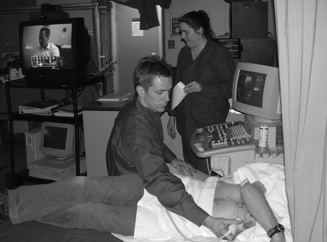
Real-time tele-ultrasound scanning at McMurdo Station.
Ultrasound Acquisition
Ultrasound images were acquired by four individuals: one certified ultrasound technician, two medical doctors (with MD degrees), and one physician assistant. Both MDs received basic ultrasound acquisition training prior to deployment. The ultrasound exams were divided into primary and follow-up exams. The primary exam was the first ultrasound to be conducted for a particular patient problem, whereas the follow-up exam was the second ultrasound exam conducted per patient problem. The ultrasound studies were categorized according to organ system: (i) abdominal, (ii) gynecologic, (iii) genitourinary, (iv) cardiac, (v) venous, (vi) endocrine, and (vii) musculoskeletal.
Tele-Ultrasound Transmission
For ultrasound store-and-forward interpretation, still and cine images were recorded in a Digital Imaging and Communications in Medicine format and stored on the Acuson's internal hard drive. The ultrasound files were then transmitted via the T-1 satellite communications link, from either McMurdo or South Pole Station to the general radiology reading room of Diversified Radiology Associates at Presbyterian Saint Luke's Hospital, Denver, CO. A standard radiology report was generated once the images were read by one of the attending radiologists. The report was then forwarded by e-mail to the McMurdo or South Pole medical account, where it was accessible by the station's physician. Real-time ultrasound sessions between the Antarctic Stations and diversified radiology were conducted at 384 kilobits per second.
Ultrasound Interpretation
For store-and-forward exams, patients were scanned initially by the remote sonographer, MDs, or physician assistant who, on completing the examination, documented their major and minor findings in writing and also gave a final overall impression. Each case was then reviewed and interpreted by a radiologist or cardiologist in the Continental United States (CONUS). The CONUS physicians in turn documented their major and minor findings and gave a final interpretation. With the exception of three echocardiograms, all ultrasound exams were then read by one of three certified radiologists from Diversified Radiology Associates. The echocardiograms were read by two cardiologists from the University of Texas Medical Branch at Galveston.
Analysis of Image Quality
The quality of the primary ultrasound exam was classified according to the radiologist's interpretation. 11 “Diagnostic” exams were of sufficient quality that the radiologist could make a diagnosis with confidence. In the case of “partially diagnostic” exams, a diagnosis was given with reservations. “Nondiagnostic” exams lacked sufficient data or were of poor quality to make a definitive diagnosis. The follow-up exams were omitted from quality analysis to avoid a learning effect bias. Similarly, real-time ultrasound exams were excluded as they were interpreted in real time, and image acquisition could be directed by the radiologist.
Analysis of Diagnostic Interpretation
Analysis of diagnostic data received from the reading radiologist was classified as either “concordant” or “discordant.” 11 “Concordant” readings were defined as true-positive or true-negative readings. Two further subclassifications of the concordant category included (1) “minor variation,” where the physician or sonographer identified the abnormality but did not appreciate the full extent of the injury or illness or a second separate minor injury was missed, and (2) “gave other significant information” and reflected results that were not expected by the referring physician either with respect to the pathology in question or on new findings identified by the radiologist. “Discordant” readings reflected false-negative or false-positive interpretations in which the physician or sonographer did not appreciate the presence of an injury or illness or else reported pathology that was refuted by the radiologist's interpretation (Table 1).
Classification of Effects on Diagnosis
Analysis of Clinical Management
Data on the effects of tele-ultrasound on clinical management were obtained by review of the patient's medical chart. The medical charts for all USAP participants were housed at the Raytheon Polar Services Company headquarters in Denver. Charts were examined to determine the patient's initial treatment prior to receipt of the ultrasound report and the treatment administered after the date the ultrasound report was received. The following categories were assessed to determine what effects tele-ultrasound had on treatment: (a) “no effect” on treatment; (b) “prevented an unnecessary treatment”; (c) “quality of the treatment was changed”; (d) “new treatment”; and (e) “medical evacuation was prevented”; this was determined by comparing the symptoms and presumed diagnosis with the course of action in similar cases prior to the introduction of ultrasound imaging. Two physicians reviewed these data for consistency. The last three categories were as follows: (f) “contributed to a medical evacuation”; (g) “resulted in additional specialist consultation,” that is, when consultation occurred with a second specialist, in addition to the radiologist; and (h) resulted in “additional testing.”
Results
In total, 66 ultrasound exams were conducted on 49 individual patients between October 2002 and October 2003. Data were collected on 100% of the ultrasound exams.
Ultrasound Acquisition
Four individuals conducted the store-and-forward exams at McMurdo and South Pole Stations (Table 2). Sonographer A and MD-B conducted all exams at McMurdo Station with the exception of one conducted by the physician assistant. At South Pole Station, MD-C conducted all exams. Sonographer A was a certified ultrasound technician, MD-B was an emergency room physician, and MD-C was an internist. MD-B, MD-C, and the physician assistant had received basic ultrasound skills training.
Ultrasonographers
Data are number (%).
Tele-Ultrasound Transmission
Sixty-two ultrasound exams (94%) were interpreted as store-and-forward exams, whereas four (6%) were interpreted in “real-time.” Table 3 illustrates the gender breakdown of ultrasound exams at South Pole Station and McMurdo Stations for both summer and winter seasons.
Ultrasound Exams at McMurdo and South Pole Stations
Ultrasound Interpretation
With the exception of three echocardiograms, all ultrasound exams were read by certified radiologists from Diversified Radiology Associates. The echocardiograms were read by two cardiologists from the University of Texas Medical Branch at Galveston.
Exam Type
Primary ultrasound exams accounted for 53 (80%) of the exams, whereas 13 (20%) constituted follow-up ultrasound examinations. Abdominal, genitourinary, and gynecology ultrasounds accounted for 63.6% of primary ultrasound exams (n=42). Ultrasound exam of the deep venous system was the next most frequent study type (12.1%, n=8), followed by dermatology (9.1%, n=6), cardiac (7.6%, n=5), endocrine (6.1%, n=4), and musculoskeletal (1.5%, n=1). With respect to follow-up scans, gynecologic exams were the most frequent with five exams (35.7%), followed by abdominal (three, 21.4%), and two each for venous (25%) and endocrine (50%) and one cardiac (20%). Thirty-nine ultrasound exams (59.1%) demonstrated pathology. Of the four exams conducted in “real-time,” two were cardiac (to rule out pericardial tamponade and to assess extent of inferior wall infarction), another was venous (to rule out deep vein thrombosis), and one was abdominal (to rule out gangrenous gallbladder).
Image Quality
Diagnostic quality data were collected on 52 primary ultrasound exams. Ninety-five percent of the sonographer's exams were diagnostic. Only 4.8% of the sonographer's exams were partially diagnostic, and none was nondiagnostic. In comparison, 77.4% of exams by the MDs were diagnostic, 16.1% were partially diagnostic, and 6.5% were nondiagnostic (Table 4).
Diagnostic Quality of Ultrasound Exams
Diagnostic Interpretation
In examining the overall diagnostic effects, the four “real-time” ultrasound exams were omitted from analysis because of the immediate feedback received from the reading radiologist during these exams. Table 5 illustrates the breakdown of diagnostic interpretation. Of the 62 store-and-forward exams, 88.7% had concordant readings. Of the concordant readings, 16.4% yielded “minor diagnostic variations,” whereas 18.2% “gave other significant information.” There was a greater number of discordant readings among the MDs (n=6) in comparison with the sonographer (n=1).
Diagnostic Interpretation of Ultrasound
Clinical Management
Patient management was affected in 51 cases (96.2%) (Table 6). The most common effect was in “causing a new treatment.” It is important that ultrasound examination prevented aeromedical evacuation in 25.8% of cases. These data were determined by the ability or inability to definitively treat the assumed diagnosis at either the South Pole or the McMurdo Station Medical Clinic. For example, a patient with lower quadrant abdominal pain prior to the introduction of ultrasound imaging would have been evacuated (Table 7).
Effect of Ultrasound on Clinical Management
Case Breakdown of Ultrasound Exams Averting Medical Evacuation
Discussion
The present investigation examined the impact of tele-ultrasound on the diagnosis and treatment of patients at two USAP research stations. To our knowledge this is the first study to review medical events requiring ultrasound examination and to assess the importance of ultrasound in the clinical management of patients in Antarctica.
Tele-Ultrasound Transmission
The majority of ultrasound exams were interpreted in the “store-and-forward” format (94%), with only 6% conducted in real time. These results suggest that in similar remote, prescreened populations, the majority of ultrasound examinations may be conducted in a store-and-forward format, assuming a similar or higher level of operator expertise. Store-and-forward format does not impact operational bandwidth in the same manner as a real-time guided ultrasound session, which, in some cases, may take up to one hour to complete. 12,13 These aspects are important for both South Pole and McMurdo Station, where operational bandwidth is limited and is required for the transmission of scientific data. 14,15 Furthermore, because both stations operate on “New Zealand” time, real-time consultations with the CONUS can be problematic with time zone differences of 18–20 h. The store-and-forward format is also convenient for the referring physician and the consulting radiologist, who is not constrained by the demands of being available at a specified time. Real-time availability is more challenging at the South Pole Station, which is limited to a 12-h window of daily satellite connectivity. Furthermore, this window processes 2 h earlier each month. Consequently, real-time links require significant coordination.
Exam Type
The greatest number of ultrasound exams were equally distributed among the abdominal, genitourinary, and gynecology categories. Because of the multiple organ systems in each of these ultrasound categories and the prevalence of nonspecific clinical symptomology, it is not surprising that these three categories represent the majority of ultrasound exams. Consequently, ultrasound training programs for remote-duty physicians should include a specific focus on these anatomical regions.
In total, 13 follow-up ultrasound exams were conducted. In 6 of these cases, the second ultrasound was to follow the evolution of pathology diagnosed on the primary exam. In 2 cases it was to confirm treatment effects following diagnosis made on the primary exam. This number of follow-up exams demonstrates that ultrasound is a useful tool in remote environments with limited medical technology to confirm or refute the presence of pathology and to follow treatment effects or disease progression. 16
Image Quality
Overall, 84.6% of the primary ultrasound exams were diagnostic. A further 11.5% were partially diagnostic. It is not surprising that the sonographer had a higher rate of diagnostic exams (95%) compared with the MDs (77.4%). This is comparable to a previous study of a diagnostic ultrasound service for general practitioners, which found that 85% of the store-and-forward images obtained by general practitioners were of diagnostic quality. 17
Diagnostic Interpretation
Fifty-five (88.7%) of the store-and-forward exams had concordant readings. Only seven exams (11.3%) had discordant readings. This is an excellent rate of concordance given that non-radiologist physicians conducted 59.6% (n=31) of the ultrasound exams. This result demonstrates the utility of ultrasound among these non-experts. However, there remains room for improvement given that six of the seven discordant readings were conducted by the station physicians. This illustrates the importance of tele-ultrasound and the value of radiological consultation, especially in patients with a new onset of illness or injury. 18 The number of ultrasound exams with minor diagnostic variations and those that gave significant other information were similar between the physicians and the sonographer.
Clinical Management
A surprising finding was the magnitude to which ultrasound examination affected intercontinental aeromedical evacuation. Given the operational impact of an evacuation from South Pole or McMurdo Station to New Zealand, a distance of up to 5,600 km, a 25.8% reduction in aeromedical evacuations in a single year is significant. For example, between 2000 and 2003, three winter evacuations occurred at South Pole Station at a cost of between $1.0 and $1.5 million (R.S., data from Portage River Locums). During the 2001–2002 summer season alone, there were 62 medical evacuations and patient transports from South Pole and McMurdo Station at a cost of up to $5,000 per patient transport. 19 The conditions most frequently ruled out in the present study that would have otherwise required medical evacuation were appendicitis, cholelithiasis, renal lithiasis, pregnancy beyond 8 weeks of gestation, and venous thromboembolic disease. Each one of these conditions has profound implications and potentially adverse consequences in isolated populations without access to tertiary-level healthcare. 7,20
Limitations
Several novel tele-ultrasound technologies may provide additional advantages in the remote Antarctic environment. For instance, Arbeille et al. 21 demonstrated that robotized tele-ultrasound was able to visualize the main abdominal organs in 87% of cases, allowing an expert in clinical ultrasound to guide a robotic arm to acquire images. Furthermore, three-dimensional ultrasound image reconstruction, a relatively recent addition to ultrasound, may remove the need for real-time radiological expertise to guide precise probe angle and position for diagnostic-quality images. 8 Nevertheless, this investigation suggests that even basic ultrasound imaging and tele-ultrasound are valuable additions to remote medical care for isolated populations with limited access to tertiary-healthcare facilities.
Conclusions
This study demonstrated that ultrasound is a useful and repeatable imaging modality in remote environments with limited diagnostic technology. Tele-ultrasound allowed remote physicians to confirm or refute the presence of clinical pathology and to follow treatment effects or disease progression. The present study also indicates that remote-duty physicians should receive broad ultrasound training that includes abdominal, genitourinary, gynecology, cardiac, and venous imaging. Finally, our findings indicate that diagnostic ultrasound should be available at all Antarctic Stations for medical care and to mitigate intercontinental evacuations.
Footnotes
Acknowledgments
We would like to thank Raytheon Polar Services Company and the National Science Foundation Office of Polar Programs.
Disclosure Statement
No competing financial interests exist.