
Abstract
Introduction
In most developed countries, the prevalence of dental caries declined during the last three decades of the 20th century and at the beginning of the 21st century 1 ; however, it remains the most common oral disorder and the cause of tooth loss in adolescents.
Institutionalized adolescents usually have little access to preventive or therapeutic dental services, 2 and studies have reported that the oral health status of this group is worse than that of the general population. 3 –6 This situation is aggravated in part by the fact that not all correctional centers have a structure for dental care, so some incarcerated adolescents must be transferred for treatment. The waiting time depends on the availability of professionals, and safe transport continues to be a fundamental concern for officers. 2
Digital imaging, long suggested as a strategy for identifying oral conditions, could be used to deliver clinical diagnostic services to remote areas devoid of dental specialists or even lacking general dental practitioners for referral and treatment recommendations. 7 However, teledentistry has been infrequently used as a method of screening, diagnosis, consultation, and referral in dental practice. 8
Some studies have demonstrated that digital images have potential in screening dental conditions, especially in the early diagnosis of dental caries. This method has been described as effective in detecting caries in preschool children. 8 The presence of calculus, gingivitis, and dental fractures, as well as malocclusions, has also been diagnosed remotely in children, using accessible and low-cost technologies. 9
A system of telemedicine consultation, developed at a correctional center in an attempt to reduce the cost of conventional dental appointments, showed that teleconsultations were received with enthusiasm by authorities at the center. Cost is also a factor because inmates' transportation to healthcare facilities is often expensive. It is cheaper and more accessible to transport data from place to place than to move doctors or patients. 10
Establishing diagnostic methods that allow a previous screening of dental caries is a good strategy to set priorities and minimize spending for transporting inmate population. Teledentistry can be an alternative to solve this problem, as it has been considered a practical and potentially cost-effective method of providing dental healthcare, increasing the possibility of early diagnosis and preventive treatment that can significantly reduce the frequency and severity of oral disease 8,11 –13 ; however, it is important to know the validity of this diagnostic tool.
The aim of this study was to determine whether a store-and-forward teledentistry system is a valid method of screening for dental caries in a cohort of juvenile offenders, based on digital photographs.
Subjects and Methods
Sample Selection
This study was conducted as a cross-sectional census that evaluated a cohort of 102 male juvenile offenders ranging from 15 to 19 years of age, who were undergoing social and educational measures of deprivation of liberty at a juvenile detention facility, CENSE São Francisco, in the city of Piraquara, state of Paraná, southern Brazil. The entire inmate population was invited to participate during March–July 2010. Those who voluntarily agreed to participate were informed about the study objectives and procedures. Only one adolescent did not cooperate and thus was excluded from the sample. The research was approved by the Universidade Federal do Paraná Research Ethics Committee (protocol number CEP/SD 836.171.09.11) and by the Paraná State Ethics Committee of Children and Youth Department (protocol number 022/2009). The informed consent terms were signed by the institution's director, as the inmates were under state responsibility.
Calibration Procedure
The clinical examiner and the distant consultants had been previously trained to assess the decayed, missing, and filled teeth (DMFT) index, and agreement was measured by the kappa coefficient. A PhD professor of pediatric dentistry with extensive experience acted as the examination referee.
Data Collection
Prior to the dental examination, a registration form addressing such sociodemographic factors, health-related aspects, and dental experience, with items on marital status, formal schooling, work activities, reported discomfort in teeth and mouth, and use of tobacco or other drugs, was filled out. The short form of the Oral Health Impact Profile, cross-culturally adapted and validated for Brazilian Portuguese, was also administered through interviews by a single, trained researcher to measure the impact on oral health related to quality of life (data not shown).
Clinical examinations were conducted by one of the researchers, using standard dental equipment, a light reflector, an air syringe as needed, a No. 5 plane mirror, a standard probe (Trinity, São Paulo, Brazil), and gauze pads. The examiner determined the DMFT index according to World Health Organization criteria 1 and recorded the results on an individualized caries exam form.
The oral condition of adolescents was documented using a professional digital camera (EOS 300 Rebel; Canon, Tokyo, Japan) with a 100/35-mm macro lens and a Canon circular flash system. After correct positioning of the adolescent, the teeth were dried, and the cheeks and lips were held apart using plastic dental retractors to allow better visualization of the teeth and intraoral structures.
At least five intraoral photographs of each participant were taken: one frontal, two lateral (right and left), and two occlusal images (upper and lower) (Figs. 1 –5). The images were 220–800 KB in size, with a minimum resolution of 600 dpi. Clinical images were saved in JPEG format, and the remote assessment of caries was made using these files. The participants could not be identified by these images, as all photographic records were restricted to oral tissues.
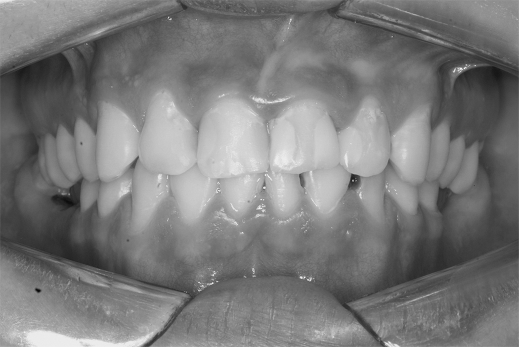
Intraoral frontal photograph. In this image can be seen the labial surfaces of the restored maxillary incisors and the buccal surfaces of the sound maxillary canines.
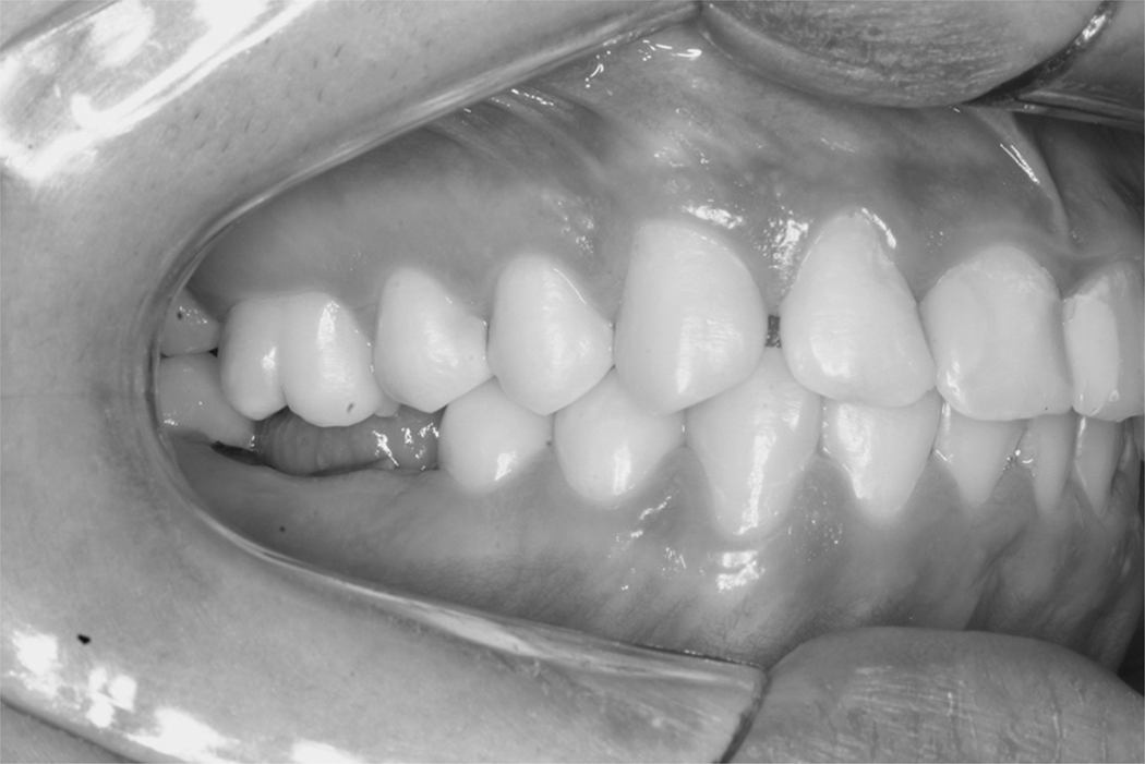
Intraoral right-side photograph. It is possible to observe the residual roots of the mandibular right first molar, the restored maxillary incisors, and the sound mandibular right incisors.
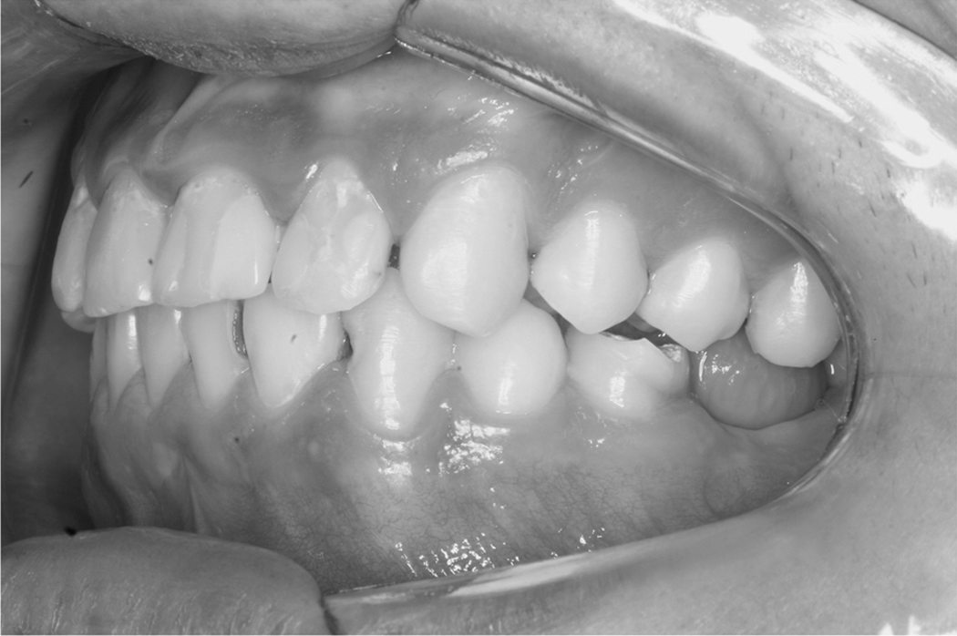
Intraoral left-side photograph. Note a missing tooth in the mandibular arch (left first molar) and some decayed teeth (mandibular left incisors and canine).
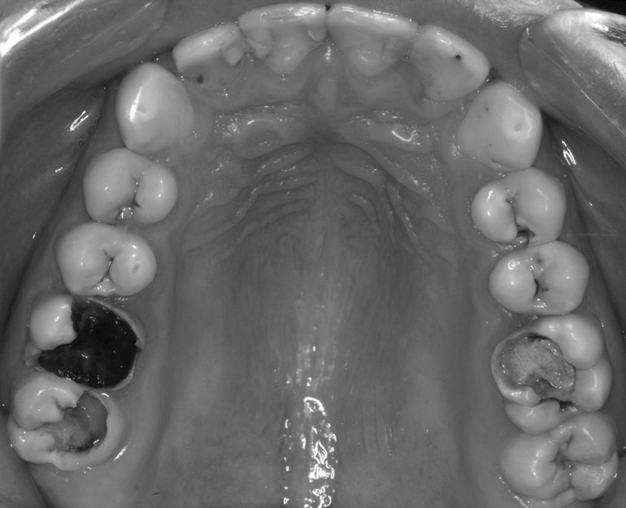
Intraoral upper occlusal photograph. Varying degrees of caries development can be observed, requiring different types of treatment.

Intraoral lower occlusal photograph. It is possible to see the missing mandibular left first molar, decayed left incisors and canine, restored left second molar, residual roots of the right first molar, and decayed right second molar.
Data Transmission
After the oral inspection, performed in person by the on-site examiner, two other different distant examiners evaluated the images. For the distant diagnosis, each photograph was assigned a numerical code to prevent subject identification. Two methods of image transmission were used. In the first method, the images were uploaded to a file-sharing service (
Statistical Analysis
The DMFT index components were analyzed through descriptive statistics. Agreement between the traditional examination and the distant consultants was measured by the kappa coefficient of agreement, considering each tooth. Sensitivity, specificity, accuracy, positive predictive values, and negative predictive values were also calculated. To calculate sensibility and specificity, the cut point adopted was the presence or absence of untreated caries. Filled and missing teeth were excluded from the analysis.
Statistical analysis was performed using the Statistical Package for the Social Sciences software (version 15.0; SPSS Inc., Chicago, IL).
Results
This study evaluated 102 male juvenile offenders having an average age of 16.84 years (standard deviation=0.941).
Almost 15% of the subjects had never seen a dentist before admission to the juvenile detention facility, and more than 60% of those who had access to routine dental care used public health services.
Calibration
Results of the inter and intra-examiner calibrations ranged from “strong” to “almost perfect” (κ=0.78 to κ=0.86) according to the classification proposed by Landis and Koch. 14
Dmft Index
For the traditional clinical examination, the mean DMFT index for this sample was 5.9, ranging from 0 to 21 (standard deviation=4.49), wherein 93.1% of the adolescents had at least one carious lesion, of which 77.5% were untreated. The study evaluated a total of 3,264 teeth, 600 of which were counted as decayed, filled, or missing. The frequency of each component is shown in Table 1.
Frequency of Decayed, Missing, and Filled Teeth Index Components, According to the 1999 World Health Organization Criteria, for Adolescent Inmates Between 15 and 19 Years Old in Curitiba, Brazil (n=102)
DMFT, decayed, missing, and filled teeth; WHO, World Health Organization.
Interexaminer Agreement and Accuracy Tests
Kappa statistics were applied to the data with the purpose of determining interexaminer agreement beyond that expected through chance. The results ranged from “strong” to “almost perfect” and are shown in Table 2.
Interexaminer Agreement for Decayed, Missing, and Filled Teeth Index Considering Clinical and Distant Examinations
According to Landis and Koch. 14
Sensitivity, specificity, accuracy, positive predictive values, and negative predictive values were calculated between each distant consultant and the clinical examiner separately and in parallel, considering both distant consultants together (a tooth was considered decayed when at least one dentist diagnosed the lesion), with the purpose of improving telediagnosis sensitivity (Table 3). In all cases, specificity was higher than sensitivity, ranging from 97% to 98%.
Sensitivity, Specificity, Accuracy, Positive Predictive Values and Negative Predictive Values for Comparisons Between Traditional Clinical Examination (Gold Standard) and Teledentistry Evaluations, Individually and in Parallel
A, accuracy; NPV, negative predictive value; PPV, positive predictive value; Se, sensitivity; Sp, specificity.
Discussion
In this research, data obtained on dental caries through remote diagnosis were similar to those obtained by traditional visual inspection. This finding can be especially useful in telehealth activities through the use of relatively low-cost tools such as digital cameras and personal computers with Internet-based connections.
In the present investigation more than 93% of the sample had at least one instance of caries. Although the mean DMFT index of the inmates was similar to that found in the general population, these youths usually have more decayed teeth requiring treatment, which burdens the public health system. 2,4,6,15
Despite its specificities, the study group is an example of the applicability of this technology to other selected populations because the screening of patients requiring immediate intervention for caries treatment could reduce the human, social, and economic cost of the diagnostic procedure, allowing better allocation of resources and increasing the quality of individuals' health.
Other investigators have studied teledentistry in various clinical situations, such as caries-risk screening in kindergartens, 16 screening patients for dental emergencies, 17 expert opinion in cases of oral and maxillofacial surgery, 18 and oral mucosal diseases, 19 –21 as well as supervision of orthodontic treatment 11 and implant planning. 22 The literature also supports the idea that teledental consultations are as reliable as those conducted by traditional methods. 7,10,11,16,20,21
In the present study, agreement between the clinical examiner and Consultant 1 was considered “almost perfect,” that between the clinical examiner and Consultant 2 was “strong,” and that between both distant consultants was also “almost perfect.” Specificity was fairly satisfactory even when consultants were considered individually.
Regarding dental caries diagnosis in public health services, specific tests are useful in reducing avoidable costs, due to additional diagnostic procedures and unnecessary treatment. Combining the results of both consultants in parallel made it possible to increase sensitivity of telediagnosis, indicating, as expected, that two distant consultants together are more likely to identify untreated carious lesions.
A pilot study compared data from traditional visual screenings with those from digital images transmitted using teledentistry. 7 In the first phase, 137 children were examined in person by a dental technician and an assistant to obtain initial data. The indices used were the DMFT index and deciduous decayed, missing, and filled teeth. Two months later, a second test was performed using an intraoral camera on 27 children randomly selected. Three dental technician students and a dentist conducted the teleconsultations. The images were interpreted by the participants of the first stage, using the same indexes. The system was used both to achieve full diagnosis and to screen patients. Kappa statistics indicated a perfect agreement (κ=1.0) in three categories, which were primary teeth to be extracted, permanent missing teeth, and permanent filled teeth. For both the primary and permanent teeth in the decay groups, the kappa statistics showed moderate agreement (κ=0.58 and κ=0.50, respectively), and for the group in which primary teeth were to be filled, the agreement was very good (κ=0.93). Despite the fact that agreement between the assessments had been evaluated by kappa statistics, sensitivity and specificity were not calculated. However, these data might help validate the method.
Another investigation assessed the feasibility and reliability of teledentistry in dental caries screening in a convenience sample of 50 preschool children between 4 and 6 years. 16 A calibrated dental examiner evaluated children enrolled in an inner-city childcare center. Following the oral examination, images of the children's teeth were obtained by a trained telehealth assistant, and the recorded digital images were transmitted via the Internet to a computer at the remote dental site. After a 2-week washout period, the images were read by the same examiner. Agreement between traditional dental examinations and teledentistry examinations was considered good (κ=0.61). The sensitivity of the teledentistry examination was 100%, and specificity was 81%. In this study, the criterion for dichotomization was the presence or absence of previous caries experience, whether treated or not. The sensitivity value of 100% can be explained, in part, because missing and filled teeth were included in the analysis, which are more easily identified even if done remotely.
In the present research, the cut point adopted was the presence or absence of untreated dental caries lesions. Filled and missing teeth were excluded from the analysis. This definition was adopted because untreated caries has a greater impact on the patient and the healthcare system. As the criteria were different, comparing other sensitivity and specificity values with the results obtained by the present study is difficult.
The presence of calculus, gingivitis, and dental fractures, as well as malocclusions, was also diagnosed remotely in preschool children. 9 The results indicated sensitivity between 94% and 100% and specificity between 52% and 100%. The positive predictive value was between 67% and 100%; the negative predictive value was between 94% and 100%, suggesting that remote diagnosis of children's dental problems based on photographs constitutes a valid resource to exclude referred children to a dentist for treatment, but further studies should be carried out to bolster the validity of this procedure of referring children for the same treatment.
As can be seen, there is much variation in kappa values, and, according to Patterson and Botchway, 7 this feature may be due to the difficulty of diagnosing caries from images; without the use of explorers, no tactile information can be transmitted. The agreement described in their investigation was greater in sound, missing, and filled teeth. As expected, less agreement was observed in the detection of decay, considering the difficulties of caries diagnosis even in vivo. On the other hand, Kopycka-Kedzierawski et al. 16 affirmed that, most likely, the disagreement on caries diagnosis between teledentistry imaging and the traditional oral examination was attributable to the greater spectral sensitivity and illumination of the oral cavity by the intraoral camera, in that imaging of a child's mouth provides high-quality pictures for discrete evaluation.
An item of discussion in teleconsultations is the feasibility of digital images for oral diagnosis. If the images transmitted via teledentistry correspond to those provided in person, consultations between specialists in central locations and healthcare workers in remote areas can be integrated. Clinical photography needs at least 1 million pixels and 24-bit color. 23 The camera used in this study produces images of more than 1 million pixels (1,536×1,024), has the recommended color specifications, and is considered more than adequate for producing good-quality images.
If there is concern about the loss of detailed diagnostic information when using just photographs in a clinical setting, both digital radiographs and standard films can be easily transmitted to increase diagnostic confidence. 7
In this study, only photographs were used for teledentistry evaluation. In some cases, it was difficult to identify incipient caries occurrences and differentiate them from darkened enamel defects. When composites were present and depending on the lighting, identifying filled teeth was a challenge. The store-and-forward teledentistry model is not recommended for emergency cases because in these situations treatment should be performed as soon as possible.
Another limitation is that training someone to make good-quality photographs using a professional camera is time consuming, whereas if nonprofessional digital cameras, or even smartphone cameras are used, this training might be reduced. Further studies should be undertaken to evaluate the quality of images captured by these portable—and more commonly used—devices by clinicians and other healthcare staff. Moreover, the selected sample might not represent the entire population of inmate adolescents in Brazil, and the descriptive analysis of epidemiological caries should be accepted with caution.
Despite these limitations, this survey indicated that teledentistry can be considered a feasible approach to screening and referral of inmate adolescents for dental treatment, saving economic and human resources. The tools to be used in other studies depend on the purpose of each investigation and the available technology. In this research, simple technologies such as digital cameras and electronic transmission were used with good results, demonstrating that the benefits of teledentistry can be experienced even without expensive resources.
Footnotes
Disclosure Statement
No competing financial interests exist.